
Source- textbook of oral pathology Shafers and Google images
Source- textbook of oral pathology Shafers and Google images


STUDY NOTES ⚕️
Cancrum oris is a special type of gangrene.
Causes:
🔍 Malnourishment.
🔍 Major diseases like diptheria, Whooping cough, measles, kala azar, typhoid, etc.
🔍 These factors lead to invasion of opportunistic organisms like the Vincent’s organisms – Borrelia vincentii and fusiforms causing ulcerations, erosions and eventually fibrosis.
Clinical Presentation:
👩⚕️ It is an extensive ulcerative disease of the cheek mucosa usually occuring in malnourished children.
👨⚕️ As the disease progresses, there may be complete reduction of the cheek thickness.
Treatment:
💊 Ryle’s tube feeding.
💊 Improve the nutrition.
💊 Appropriate antibiotics: Metronidazole 400mg thrice daily for 7-10 days.
💊 Surgical reconstruction of the cheek.
Complications:
⚠️ Fibrosis leading to restricted movement of the jaw.
⚠️ Sepsis, toxaemia and death.
SOURCE: Manipal Manual of Surgery (3rd edition)
~Sunantha✍️


Written by -Sanjana Agrawal Dentowesome 2020
Source -Borley textbook

STUDY NOTES ⚕️
Glassgow Coma Score is used to assess the level of consciousness properly instead of using vague terms like semi-conscious, obtundant, etc.
Hence it is widely used thereby avoiding observer errors in the observation patients.
NEUROLOGICAL ASSESSMENT USING GLASSGOW COMA SCALE:
1️⃣ Eyes Open:
Spontaneously – 4
To speech – 3
To pain – 2
None – 1
2️⃣ Best Verbal Response:
Oriented – 5
Confused – 4
Inappropriate words – 3
Incomprehensible sounds – 2
None – 1
3️⃣ Best Motor Response:
Obeys commands – 6
Localises pain – 5
Withdrawal to pain – 4
Flexion to pain – 3
Extension to pain – 2 (due to raised intracranial pressure)
None – 1
~*~
⭐ Maximum score is 15.
⭐ Minimum score is 3.
⚠️ If a patient has a score of 7 or less than 7, then he/she is said to be in coma.
SOURCE: Manipal Manual of Surgery (3rd edition)
~Sunantha✍️

AMALGOMER Technology is the latest innovation in restorative dentistry. For the first time the strength of a classic amalgam restorative has been combined with the aesthetics and the many other advantages of Glass Ionomers.
In short AMALGOMER is the world’s first GIC to pass the ISO strength test requirements for amalgam (ISO1559:2001) as well as that of the GIC standard (ISO9917:1991).
Features:
Amalgomer CR High Strength Posterior GI
AMALGOMER CR High Strength Posterior GI Restorative offers a wide range of features:
1) Ceramic Reinforcement
2) Exceptionally low wear
3) High Radiopacity
4) Excellent for core build ups
5) High strength, exceeds 300MPa compressive strength
6) Universal tooth shade or white
7) Natural Adhesion to tooth structure
Amalgomer High Strength Anterior GI
AMALGOMER High Strength Anterior GI Restorative offers a wide range of features:
Amalgomer Light Cure Varnish
A light curable varnish for protection of AMALGOMER and AMALGOMER CR restorations. This protects the restoration against moisture. This can be used with any GI restorative.
Amalgomer Conditioner
Dentine conditioner for use prior to placing AMALGOMER and AMALGOMER CR restorations.
Amalgomer mixing spatulas
Flexible plastic glass ionomer mixing spatulas.
Dr Iswarya V
General Practitioner,
Trivandrum.
Reference : Amalgomer official product website

Role of Platelet – Rich Fibrin (PRF) in dentistry
It is a natural Fibrin-based biomaterial prepared from autologous blood and is clinically used to deliver growth factors in high concentration to the site having a bone defect or requiring augmentation.
It is introduced by Dr. Choukroun. et. Al, 2000. It is a second generation platelet concentrate that contains platelets and growth factors, prepared from self blood devoid of anticoagulant or other artificial modifiers.
Preparation
10ml of human blood is taken in a test-tube without any anticoagulant and is centrifuged in a tabletop centrifuge machine for 12 minutes at 2500RPM or 10 minutes at 3000RPM.
After centrifugation, the three components in the blood are localised in the test tube.
Fibrin clot is extracted from the test-tube with a pair of sterile forceps and PRF is obtained by removing the red clot from its lower end.
Applications
Advantages
Disadvantages
Contraindications
Should not be performed on patients with:
Dr Iswarya V
General Practitioner,
Trivandrum
Reference : Oxford Clinical Dentistry


Dr. Mehnaz Memon🖊
References: Davidson’s Principles and Practice of Medicine Textbook
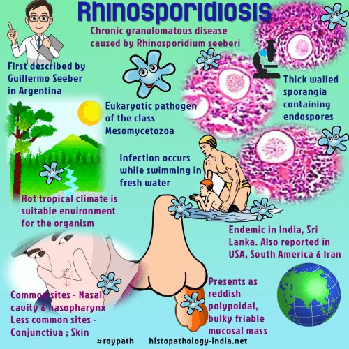
(i) Rhinosporidiosis is a chronic granulomatous disease characterised by formation of friable polyps, usually confined to the nose, mouth or eye.

(i) Causative agent is Rhinosporidium seeberi.
(iii) More than 80% cases are reported in India and Sri Lanka.
(iv) The mode of infection is not known but most infections occur in males who have frequent contact with stagnant water or aquatic life.
ORAL MANIFESTATIONS
•Oronasopharyngeal lesions appear as soft red polypoid growth which spread to pharynx and larynx.
• These lesions often contain mucoid discharge and are vascular.
LAB DIAGNOSIS
• The fungus has not been cultivated.
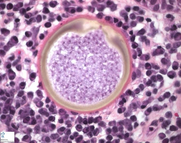
• Diagnosis depends on the demonstration of sporangia
•Tissue sections stained with H & E stain show large number of endospores within the sporangia embedded in a stroma of connective tissue and capillaries.
The sporangium (10-200 um) contains thousands of endospores (6-7 µm in diameter)

Source – textbook of microbiology for dental students c p baveja and Google images

Have you ever dreamt of having wonderful lips as you see in ads?
Are you worried about how your lips do appear?
YES. Find out the route to ‘pretty lips’ – Lip Fillers
Lips and eyes make up the important parts of face and determine to an extent, the beauty of an individual. Lips form a major aspect of cosmetic treatment.
Factors to consider for lip augmentation :
Injectable intradermal Fillers are commonly used for lip augmentation. These can be done fully in a single session. The procedure seem difficult and challenging but can be done in a safe, effective and as an in-office procedure by a skilled professional. It is advisable to consult a plastic surgeon before any cosmetic procedure.
There are many types of intradermal Fillers, the most common being products that contain substances similar to hyaluronic acid. Allergic reactions are unlikely as these are biocompatible.
Benefits:
Points to ponder:
Dr Iswarya V
General Practitioner,
Trivandrum
