Extraoral Force in Orthodontics: A Closer Look at Class II, Division 1 Malocclusions

Orthodontic philosophies, much like those in medicine, tend to swing with the pendulum of trends and innovations. In the medical field, we’ve seen treatments rise and fall in popularity—antihistamines were once heralded as a cure-all, and antibiotics became the go-to for nearly every ailment. Similarly, in orthodontics, we’ve witnessed an evolving landscape of treatments and tools: non-extraction versus extraction approaches, debates over which teeth to extract, and a constant shift between fixed and removable appliances. Each innovation, from square tubes to round tubes and from labial to lingual appliances, has had its moment in the spotlight.

In this article we will explore: What is the role of the extra-oral appliance? Where does it find use? What are its limitations? How valid are the multiplicity of claims made for it?

Investigating the Facts: A Study of 150 Cases

In a detailed study of 150 Class II, Division 1 malocclusions, headplates and plaster casts were analyzed to assess the role of extraoral force. Among these cases, 107 exhibited normal mandibular arch form, tooth size, and basal bone relationships. These findings suggest that in many cases, the mandibular arch is not the primary culprit in malocclusion; rather, the anteroposterior discrepancy lies in the maxilla. This raises an important question: Should orthodontic therapy target the maxilla while leaving the mandibular arch undisturbed?

The clinical reality supports this approach. Prolonged Class II therapy directed at the mandibular arch often results in unwanted tipping or forward sliding of the lower teeth. By focusing forces on the maxilla, we may achieve better results, including improved tooth interdigitation, reduced overbite and overjet, and restored muscle function and facial aesthetics.

The Debate Around Extraoral Force

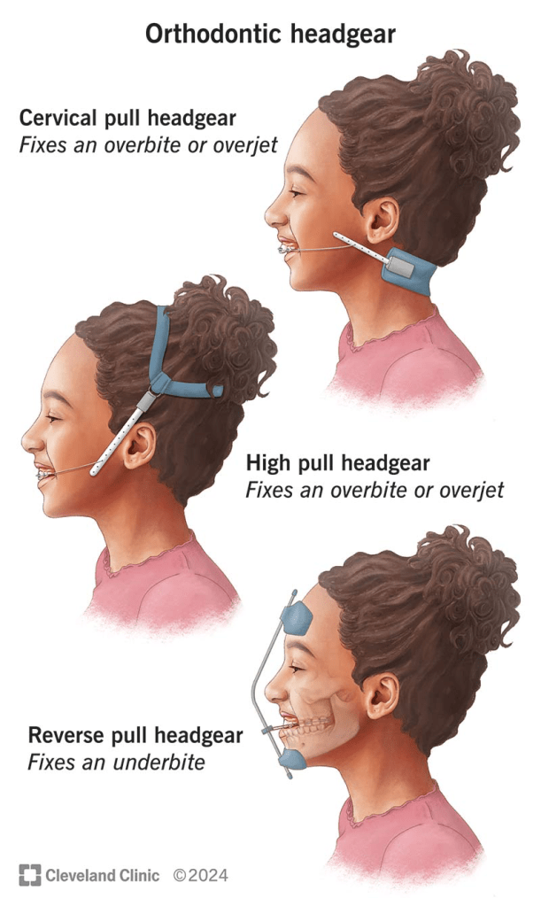
The literature on extraoral force is filled with conflicting claims. Some argue that it restricts maxillary growth, while others suggest it only affects alveolar growth. There are debates about whether it moves teeth bodily or merely tips them, and whether it allows the mandible to grow forward or simply frees occlusal interferences. Even the choice of appliance—headgear versus cervical bands—sparks disagreement.

To bring order to the conflicting claims about extraoral appliances, we must approach the topic with objectivity. What truly happens in a controlled group of cases? Which cases benefit most from extraoral force, and where does it fall short? By critically evaluating both successes and failures, we can better understand the indications, contraindications, and unanswered questions surrounding this treatment modality.

The appliance used consisted of molar bands, an .045 stainless steel labial arch wire with vertical spring loops at the molars, and continuous loops at the lateral canine embrasures to receive the cervical gear. The cervical gear featured a metal tube with a continuous internal spring to provide distal motivating force. In select cases, incisors were banded at certain stages of therapy.

Patients were categorized into three age groups to analyze outcomes based on developmental stages:

  • Deciduous dentition: 3 to 6 years
  • Mixed dentition: 7 to 10 years
  • Permanent dentition: 11 to 19 years

This stratification allowed for a nuanced understanding of how age and dentition stage influenced treatment outcomes.

The study revealed several key insights, supplemented by observations from routine practice where extraoral anchorage was employed in diverse scenarios. These included:

  • Bolstering anchorage during full edgewise therapy
  • Closing spaces created by distal movement of anterior teeth
  • Uprighting individual teeth
  • Serving as an active retainer

The study confirmed that Class II, Division 1 cases vary significantly, even when focusing on three core characteristics:

  1. Maxillomandibular basal relationship
  2. Overjet
  3. Overbite

The severity of discrepancies across these factors, combined with patient-specific variables such as morphogenetic patterns, motivation, cooperation, and growth during therapy, made the prognosis unpredictable. Success or failure was influenced by the degree of deviation from the norm in each factor and the interplay between them.

Can extraoral force alone, directed against the maxilla, correct Class II, Division 1 malocclusions?

The goal of establishing normal tooth interdigitation, eliminating excessive overbite and overjet, and restoring muscle function and appearance is ambitious. Achieving these outcomes universally is contingent on numerous factors:

  • Hereditary patterns
  • Age and sex of the patient
  • Presence or absence of third molars
  • Growth increments during treatment
  • Patient cooperation

Deciduous Dentition Group (3 to 6 years)

  • Sample Size: 14 cases, all selected for their severity, characterized by significant basal dysplasias.
  • Outcomes:
    • Successful correction: Achieved in 3 cases.
    • Partial improvement: 3 cases showed near-successful results.
    • Residual Class II relationship: Persisted in over half the cases, though to a lesser degree.
    • Basal adjustment: Anteroposterior basal adjustment was observed in 11 out of 14 cases.
    • Muscle function: Most patients exhibited improved muscle tone and function, along with a reduction in abnormal muscle habits.
    • Overjet correction: Often led to excessive lingual tipping of maxillary incisors, especially in cases without pre-existing spacing.
    • Overbite correction: The least satisfactory aspect of treatment.

Mixed Dentition Group (7 to 10 years)

  • Sample Size: 50 cases (34 girls, 16 boys).
  • Outcomes:
    • Normal molar relationship: Achieved in 29 cases, though not always accompanied by normal canine relationships.
    • Overjet correction: Similar to the deciduous group, excessive lingual inclination of maxillary incisors was noted in some cases.
    • Vertical correction: More pronounced and successful compared to the deciduous group.
    • Severe discrepancies: Cases with the greatest deviation from normal in basal relationship, overbite, and overjet showed the least favorable results.

Case Examples

  1. Patient A.L.
    • Presented with severe basal malrelationship, marked overjet, and normal overbite.
    • Outcome: Immediate and gratifying response due to anterior spacing and lack of excessive overbite.
  2. Patient J.K.
    • Presented with a similar profile but without anterior spacing.
    • Outcome: Removal of maxillary second molars facilitated mesiodistal adjustment, resulting in successful correction across all parameters.

Permanent Dentition Cases

  • Sample Size: 36 cases (19 boys, 17 girls)
  • Growth Correlation: A clear link was observed between the pubertal growth spurt and positive response to mechanotherapy.
  • Outcomes:
    • 25 patients responded well enough to eliminate Class II characteristics, achieving normal interdigitation and improved aesthetics.
    • Success was highly dependent on a combination of favorable growth, patient cooperation, and other individual factors.

Can Extraoral Force Achieve Bodily Distal Movement of Maxillary Teeth?

The ability of extraoral force to influence maxillary growth, move teeth bodily distal, or merely tip them distally has been a subject of debate.

Maxillary Growth

  • Observation: There is no evidence that maxillary growth, as governed by sutures, is significantly affected by extraoral force. Claims of growth inhibition require substantiation, which is currently lacking.
  • Alveolar Growth: However, maxillary alveolar growth can be influenced. Changes in the anteroposterior apical base relationship are among the most significant findings, as demonstrated by cases like Patient A.M.

Distal Movement of Maxillary First Molars

  • Controversy: The possibility of bodily distal movement of maxillary first molars remains contentious. While some authorities categorically deny this, evidence from the study suggests otherwise:
    • Cases Supporting Movement:
      • Bodily distal movement has been observed in some cases, though it is not the norm.
      • Occasionally, this movement occurs unpredictably or can be facilitated by the removal of maxillary second molars during active treatment (Figs. 8 and 9).
    • Normal Path Restriction: In most cases, extraoral force restrains the maxillary first molar from moving forward along its natural path or tips it distally.

Challenges with Tipping

  • Excessive Distal Tipping: One drawback of extraoral appliances is the tendency for excessive distal tipping of maxillary first molars.
  • Mitigation Strategies:
    • Allowing maxillary second molars to erupt before treatment.
    • Removing maxillary second molars during treatment.
    • Using bands or Rocky Mountain-type crowns on second deciduous molars instead of first permanent molars in the mixed dentition stage.
    • Employing a headcap instead of cervical gear, as the headcap is associated with reduced tipping tendencies.

Does Extraoral Force Tip Maxillary Incisors Lingually, Moving Apices Labially?

Yes, extraoral force can cause lingual tipping of the maxillary incisors, with their apices potentially moving labially. This effect is a notable concern in orthodontic treatment, particularly in cases with significant basal discrepancies.

Lingual Tipping of Maxillary Incisors:

  • Lingual tipping is a frequent outcome when extraoral force is applied, especially in attempts to correct overjet in cases with marked maxillomandibular basal dysplasia.
  • This tipping often results from the inability to fully eliminate the basal malrelationship.

Overjet Correction Challenges:

  • Correcting overjet in the presence of basal discrepancies often necessitates:
    • Excessive lingual inclination of maxillary incisors.
    • Excessive labial inclination of mandibular incisors.
    • A combination of both adjustments.
  • These compromises are sometimes unavoidable to achieve acceptable occlusal and esthetic outcomes.
  • Between the two options, lingual tipping of maxillary incisors is considered the lesser compromise compared to labial tipping of mandibular incisors.

Does Extraoral Force, Directed Against the Maxillary First Molar, Impact Maxillary Second or Third Molars?

The impact of extraoral force on the maxillary second and third molars cannot be definitively answered with a simple “yes” or “no.” However, clinical observations and studies provide insights into potential effects:

Temporary Impact on Second Molars:

  • Excessive distal tipping of the maxillary first molars due to extraoral force can temporarily affect the eruption path of the maxillary second molars.
  • Once the distal force is removed, the first molars typically upright themselves, allowing the second molars to erupt.

Crossbite and Eruption Issues:

  • In some cases, maxillary second molars have been observed to erupt buccally, resulting in crossbite.
  • While it is not definitively proven that this is caused by extraoral force, there is a strong likelihood of a connection.

Non-Eruption Cases:

  • Four documented cases showed non-eruption of maxillary second molars following extraoral mechanotherapy.
  • This suggests that extraoral force may sometimes inhibit the eruption of the second molars, likely due to changes in the eruption path or space limitations.

Impact on Third Molars:

  • The diversion of the second molar’s eruption path could also influence the eruption of the maxillary third molars, though this requires further investigation.

Space Limitation in the Alveolar Trough:

  • Observations indicate that the alveolar trough may have limited capacity. If space is consumed by distal movement or tipping of the first molars, it may affect the eruption and alignment of second and third molars.

Growth and Timing in Class II Correction

  1. Importance of Growth:
    • Growth is a critical factor in addressing Class II discrepancies. Successful treatment often relies on leveraging the pubertal growth spurt to maximize skeletal and dental changes.
    • The maxillary alveolodental complex can be restrained during growth, allowing for a more favorable adjustment of the anteroposterior relationship with minimal reliance on tooth movement.
  2. Optimal Age for Treatment:
    • Girls: Best results observed between 10 to 13 years.
    • Boys: Optimal outcomes seen between 12 to 17 years.
    • Exceptional cases, such as a 19-year-old boy with significant mandibular growth during a late growth spurt, demonstrate the variability of growth potential.
  3. Uncertainty of Growth:
    • While growth is pivotal, its predictability remains a challenge. The degree of mandibular growth and its impact on correcting Class II malocclusions vary significantly between individuals.

Unilateral Response to Extraoral Force

  1. Observation of Unilateral Effects:
    • In some cases, unilateral response to extraoral force was noted, particularly in the canine region. This posed challenges in achieving bilateral symmetry.
  2. Contributing Factors:
    • Sleeping Position: Patients reported consistently sleeping on one side, which appeared to correlate with reduced movement on that side.
    • Chewing Habits: Favoring one side during eating may also contribute to unilateral response, though this remains inconclusive.
  3. Management Strategies:
    • In some cases, a lower lingual appliance was used to provide additional elastic traction, helping address asymmetry. However, unilateral response persisted in certain cases.

Challenges in Achieving Complete Correction

  1. Residual Discrepancies:
    • Even with significant improvement in overjet and molar relationships, Class II characteristics in some segments, particularly the buccal region, may remain unresolved
  2. Future Considerations:
    • The causes of unilateral response and incomplete correction remain areas for further research and clinical focus. Factors such as patient compliance, growth variability, and appliance design must be studied in greater detail.

Does Extraoral Force Free Occlusal Interferences, Stimulate Forward Mandibular Positioning, or Promote Mandibular Growth?

The effects of extraoral force on occlusal interferences, mandibular positioning, and growth remain a topic of debate. The current evidence provides insights but lacks conclusive proof for some claims.

Freeing Occlusal Interferences:

  • Extraoral force can alter inclined plane relationships between maxillary and mandibular teeth.
  • In cases of mandibular overclosure caused by occlusal interference, combined extraoral force and bite plate therapy can effectively eliminate functional retrusion.
  • However, functional retrusions are less frequent and less severe than previously believed.

Stimulating Forward Mandibular Positioning:

  • Claims that extraoral force promotes forward mandibular positioning via a neurogenic reflex posture mechanism lack robust evidence.
  • While such repositioning cannot be categorically dismissed, it has not been consistently demonstrated under controlled, biometric conditions.

Stimulating Mandibular Growth:

  • There is no conclusive evidence that extraoral force or any orthodontic appliance can stimulate mandibular growth beyond the individual’s inherent morphogenetic pattern.
  • Apparent acceleration or increased growth rates reported in some studies (e.g., guide planes) have not been reliably duplicated in controlled experiments, such as those conducted at Northwestern University.

Class II to Class I Transformation:

  • Eliminating distal displacement through extraoral force does not result in the transformation of a Class II malocclusion into a Class I malocclusion.
  • The role of growth and morphogenetic patterns remains the primary determinant of mandibular development.

Challenges and Limitations

  • Need for Controlled Studies:
    • Many claims regarding mandibular growth stimulation and repositioning remain anecdotal or based on uncontrolled studies. Rigorous biometric analyses are necessary to substantiate such claims.
  • Physiological Variability:
    • Individual growth patterns, genetic predispositions, and environmental factors contribute to the variability in response to orthodontic treatment.
  • Role of Functional Appliances:
    • While functional appliances may influence mandibular posture temporarily, their long-term impact on growth remains uncertain.

The Role of Lip Growth in Orthodontic Treatment Planning: Insights for Orthodontic Students

Understanding the growth patterns of the maxillary and mandibular lips is essential for effective orthodontic treatment planning. As orthodontic students, recognizing the interplay between facial soft tissues and the underlying hard tissues, particularly during the critical growth period from 8 to 18 years, can significantly influence treatment outcomes. This blog post delves into a lip growth and its implications for orthodontic therapy, providing data-driven insights and clinical guidelines.

Key Findings on Lip Growth

1. Maxillary Lip Length

Maxillary Lip Length Growth (mm)Age 8Age 18Percentage IncreaseLargest growth occurred between 
Males17.7321.5321.43%Ages 10 and 16.
Females17.7319.8812.11%Ages 10 and 14.

2. Maxillary Lip Thickness

Maxillary Lip Thickness Growth (mm)Age 8Age 18Percentage Increase
Males10.7715.7646.33%Growth was continuous, with the largest increase between ages 12 and 16.
Females10.9012.5014.68%Growth occurred primarily between ages 10 and 14.

3. Mandibular Lip Length

Mandibular Lip Length Growth (mm)Age 8Age 18Percentage Increase
Males19.1426.5338.56%Largest increase occurred between ages 12 and 16.
Females19.1422.7318.65%Growth occurred primarily between ages 10 and 16.

4. Mandibular Lip Thickness

  • Males: Increased steadily from ages 8 to 16, with a plateau between ages 16 and 18.
    • Largest increases occurred between ages 14 and 16.
  • Females: Growth was less pronounced, with significant increases only between ages 10 and 14.

Clinical Implications for Orthodontic Treatment

1. Sexual Dimorphism in Lip Growth

  • Males experience greater increases in both lip length and thickness compared to females.
  • Females show more limited growth, particularly in lip thickness during puberty.

2. Extraction Therapy Considerations

  • Female Patients: Limited lip thickening during puberty makes the effects of extraction therapy more noticeable, especially in patients with straight or concave profiles. Treatment plans should be approached with caution to avoid adverse effects on facial esthetics.
  • Male Patients: Greater lip thickening provides more flexibility for extractions without significantly impacting facial profile fullness.

Maxillary Lip Thickness Growth Rates

  • Males:
    • 0.5 mm/year (ages 8–12).
    • 0.7 mm/year (ages 12–16).
  • Females:
    • 0.5 mm/year (ages 10–12).
    • 0.3 mm/year (ages 12–14).

Conclusion

Understanding the growth patterns of maxillary and mandibular lips is essential for predicting soft-tissue responses and planning effective orthodontic treatments. Sexual dimorphism plays a significant role, with males experiencing greater growth in both lip length and thickness compared to females. These differences must be factored into treatment decisions, particularly for extraction therapy, to ensure optimal esthetic and functional outcomes. As orthodontic students, incorporating these insights into clinical practice will enhance your ability to deliver patient-centered care.

Comparing Forsus FRD and PowerScope: A Cephalometric Analysis

Introduction
Malocclusion, characterized by changes in teeth positioning and skeletal growth, represents a global public health concern.

  • Historical Context: Correction attempts date back to at least 1000 BC.
  • Prevalence: Class II malocclusion affects one-third of the population and is categorized as skeletal or dental in origin.
  • Etiology:
    • Only 20% of Class II Division 1 cases are due to maxillary protrusion.
    • The majority stem from mandibular retrusion.

This prevalence has led to the development of functional appliances aimed at stimulating mandibular growth. These devices are divided into removable and fixed functional appliances.

Fixed Functional Appliances

  • Types:
    • Rigid: Herbst appliance, MARA (Mandibular Advancement Repositioning Appliance).
    • Flexible: Jasper Jumper, Scandee tubular jumpers.
    • Hybrid: Forsus Fatigue Resistant Device (FFRD).
  • Forsus Fatigue Resistant Device (FFRD):
    • A telescoping spring mechanism for Class II correction.
    • Provides moderate patient tolerance with initial discomfort that subsides over time.
  • PowerScope Appliance:
    • Latest addition, introduced by Dr. Andy Hayes in 2016 in collaboration with American Orthodontics.
    • Features a telescopic mechanism with a nickel-titanium (NiTi) spring delivering 260 g constant force.
    • Designed as a preassembled, one-size-fits-all appliance for easy chairside application.

Study Rationale
While Forsus FRD is extensively studied, limited literature evaluates and compares the skeletal, dental, and soft tissue effects of PowerScope. This study bridges that gap by conducting a cephalometric evaluation and comparison of these two fixed functional appliances.

Results and Discussion Summary

AspectParameterForsus FRDPowerScopeComparison
Skeletal ChangesLower gonial angleDecreased significantly (P = .005)Increased (P = .009)Better effect in Forsus.
SNADecreased (P = .037)No significant changeForsus > PowerScope (P = .026).
SNBIncreased significantly (P < .001)Increased significantly (P < .001)Both effective.
ANBDecreased significantly (P < .001)Decreased significantly (P < .001)Both effective.
Wits, beta, and YEN anglesIncreased significantly (P < .001)Increased significantly (P < .001)Both effective.
NA‖HPDecreased significantly (P = .001)Decreased (P = .022)Both effective.
NB‖HPIncreased significantly (P < .001)Increased significantly (P < .001)Both effective.
Effective mandibular lengthIncreased (P < .001)Increased significantlyBoth effective.
Dentoalveolar ChangesL1-NB and L1-NPogIncreased significantly (P = .013, P = .014)Mild increasePowerScope > Forsus. (P = .011 for IMPA).
IMPA (Incisor mandibular plane angle)Increased significantly (P = .001)Increased significantlyPowerScope > Forsus.
U1-SN and U1-NANo significant changeDecreased (P = .021, P = .026)PowerScope effective due to distalization effect.
Interincisal angleDecreased significantly (P = .034)No significant changeForsus effective.
Soft Tissue ChangesFacial convexityDecreased significantly (P < .001)Decreased significantly (P < .001)PowerScope > Forsus (P = .044).
Nasolabial angleIncreased significantly (P = .035)Mild increaseForsus > PowerScope (P = .017).
Upper lip strainDecreased significantly (P < .001)Decreased significantly (P = .012)Both effective.
Upper lip-to-E lineInsignificant changeIncreased significantly (P = .009)PowerScope > Forsus.
Overjet/OverbiteReductionHighly significant (P < .001)Highly significant (P < .001)No significant intergroup difference (P > .05).

Key Comparisons

AspectForsus FRDPowerScope
Skeletal ChangesMore maxillary retrusion, greater SNB increaseGreater impact on mandibular advancement
DentoalveolarSignificant lower incisor proclinationBetter upper incisor inclination correction
Soft TissueGreater improvement in nasolabial angleBetter profile correction (facial convexity)
Overjet/OverbiteEffectiveEffective

Conclusions

Both appliances are effective in correcting Class II discrepancies caused by retrognathic mandibles. However:

  • Forsus FRD is superior in achieving skeletal changes, particularly maxillary retrusion and mandibular advancement.
  • PowerScope shows more pronounced effects on dentoalveolar and soft tissue parameters, especially upper incisor inclination correction and facial convexity improvement.

MCQs – Presurgical Nasoalveolar Molding (PNAM)

Single-Best Answer Questions

  1. The primary goal of presurgical nasoalveolar molding (PNAM) is to:
    • A. Improve facial aesthetics
    • B. Reduce the severity of the cleft
    • C. Prepare the patient for surgery
    • D. All of the above
  2. PNAM involves the use of:
    • A. A surgical plate
    • B. A molding device
    • C. A orthodontic appliance
    • D. All of the above
  3. The NAM plate is primarily used to:
    • A. Stimulate maxillary growth
    • B. Improve nasal symmetry
    • C. Correct the cleft lip
    • D. All of the above
  4. Lip massage and lip taping are recommended to:
    • A. Reduce scar tissue
    • B. Improve facial muscle function
    • C. Increase tissue elasticity
    • D. All of the above

Multiple-Choice Questions

  1. Which of the following are benefits of PNAM?
    • A. Reduced cleft stigma
    • B. Improved nasal and lip appearance
    • C. Improved maxillary growth
    • D. All of the above
  2. What are the potential challenges associated with PNAM?
    • A. Patient discomfort
    • B. Difficulty with feeding
    • C. Skin irritation
    • D. All of the above
  3. Which of the following factors may influence the effectiveness of PNAM?
    • A. Severity of the cleft
    • B. Timing of initiation
    • C. Patient compliance
    • D. All of the above

True or False Questions

  1. PNAM is a relatively new technique.
  2. The NAM plate is a permanent device.
  3. Lip massage and lip taping are only effective in the short term.
  4. PNAM can help to reduce the need for future surgeries.
  5. The long-term effects of PNAM on dental arch development are well-established.

Answers to MCQs on Presurgical Procedure for Cleft Patients

Single-Best Answer Questions

  1. B. Reduce the severity of the cleft
  2. D. All of the above
  3. A. Stimulate maxillary growth
  4. C. Increase tissue elasticity

Multiple-Choice Questions

  1. D. All of the above
  2. D. All of the above
  3. D. All of the above

True or False Questions

  1. True
  2. False
  3. False
  4. True
  5. False

MCQs on Alveolar Bone Grafting for Cleft Patients #MDSOrthodontics

Single-Best Answer Questions

  1. The most common donor site for autogenous bone grafting in cleft patients is:
    • A. Tibia
    • B. Iliac crest
    • C. Radius
    • D. Femur
  2. Which of the following is the most important factor to consider when performing bone grafting in cleft patients?
    • A. Timing of the graft
    • B. Type of anesthetic used
    • C. Donor site morbidity
    • D. Post-operative care
  3. The primary purpose of bone grafting in cleft patients is to:
    • A. Improve facial aesthetics
    • B. Restore alveolar bone for future tooth movement and prosthetic restoration
    • C. Correct nasal deformities
    • D. Prevent speech problems
  4. The use of 3D planning in bone grafting for cleft patients is beneficial because:
    • A. It reduces the need for multiple surgeries
    • B. It allows for more precise graft placement
    • C. It eliminates the risk of graft failure
    • D. It ensures complete bone healing
  5. Bio-glass scaffolds are used in bone grafting for cleft patients to:
    • A. Replace the need for autogenous bone
    • B. Enhance bone regeneration
    • C. Reduce post-operative pain
    • D. Improve graft stability

Multiple-Choice Questions

  1. Which of the following factors can influence the success of bone grafting in cleft patients?
    • A. Graft volume
    • B. Graft quality
    • C. Recipient site vascularity
    • D. All of the above
  2. What are the potential complications associated with bone grafting in cleft patients?
    • A. Infection
    • B. Graft resorption
    • C. Donor site morbidity
    • D. All of the above
  3. Which of the following techniques can be used to minimize graft resorption after bone grafting in cleft patients?
    • A. Gentle handling of the bone
    • B. Use of bone marrow-derived mesenchymal stem cells
    • C. Application of growth factors
    • D. All of the above

True or False Questions

  1. The timing of bone grafting in cleft patients is a matter of consensus among clinicians.
  2. Calvarial bone is a less preferred donor site compared to the iliac crest.
  3. Crushing the bone during grafting can lead to increased resorption.
  4. 3D planning is not essential for successful bone grafting in cleft patients.
  5. Bio-glass scaffolds are a completely synthetic material that does not require autogenous bone.

Answers to MCQs on Alveolar Bone Grafting for Cleft Patients

Single-Best Answer Questions

  1. B. Iliac crest
  2. A. Timing of the graft
  3. B. Restore alveolar bone for future tooth movement and prosthetic restoration
  4. B. It allows for more precise graft placement
  5. B. Enhance bone regeneration

Multiple-Choice Questions

  1. D. All of the above
  2. D. All of the above
  3. D. All of the above

True or False Questions

  1. False
  2. False
  3. True
  4. False
  5. False

JC Presentation 2 – Prevalence of white spot lesions during orthodontic treatment with fixed appliances

Woah there, JC wizards! ‍♀️🪄 Second presentation alert, and guess who’s got your back with the ultimate slide deck? So buckle up, download that bad boy, and prepare to slay your next JC presentation like the rockstar you are!

Hey there, orthodontic peeps! Ever wondered why those pesky white spots like to crash the party on your pearly whites after getting braces? We got curious too, so we donned our detective hats ️‍♀️ and followed a group of brave souls on their brace-tastic journeys for 6 and 12 months.

The Results: Buckle up, because things are about to get interesting! At 6 months, almost half the crew (38%) had at least one white spot, and by 12 months, it climbed to a cool 46%. But hey, the good news is, the control group who hadn’t even gotten their braces on yet were practically spotless (only 11% with spots!).

The Plot Twist: Turns out, these white spots seem to prefer hanging out with the dudes! ‍ 76% of spotted teeth belonged to our male friends, while only 24% were on the ladies’ side. Who knew braces were so gender-biased?

The Takeaway: So, what’s the lesson in this orthodontic detective story? The first 6 months are like white spot central, but things kinda chill out after that. But don’t let your guard down! Clinicians gotta keep a close eye on those pearly whites, especially at the beginning, and make sure everyone’s brushing and flossing like champions to keep those spots at bay. 🪥

SOURCE FOR VIVA QUESTIONS: https://www.slideshare.net/marwanmouakeh/white-spot-lesions

Diagnosis Demystified – Case 31

Clinically: painful, diffuse, reddened swelling affecting the right side of the face, centred on the cheek, causing partial closure of the eye. This developed overnight. The previous 3 days there had been, according to the patient, ‘an abscess’ present on UR3. The patient feels unwell and there is lymphadenopathy present. UR3 is grossly carious. Radiologically: UR3 has a periapical rarefying osteitis.

Yo, peeps! So, check this out – there’s this crazy situation going on with someone’s face, right? Like, it’s all swollen, painful, and looking like a tomato, especially on the right side, focused on the cheek. And get this, it happened overnight! 😱

So, my friend here had this “abscess” thing going on with their tooth (UR3, to be specific) for the past three days. Fast forward to now, and it’s a whole mess – they’re feeling like garbage, there’s some swollen lymph node action, and the eye on the right is only doing half its job because of the swelling.

Oh, and if you peek inside their mouth, UR3 is a total disaster zone – super decayed. And to make things even more interesting, when you take a look at it on an X-ray, there’s this periapical rarefying osteitis party happening.

Now, why am I telling you all this drama? Well, here’s the kicker – that sudden face expansion? It’s not some random curse; it’s all thanks to a not-so-friendly cellulitis causing some serious swelling. And get this, the culprit? A seemingly innocent tooth problem. Who would’ve thought, right? Moral of the story: don’t underestimate the power of a tiny toothache, it can wreak havoc on your whole face. Mind blown! 💥

Influence of Kennedy class and number of implants on the accuracy of dynamic implant navigation

Dynamic implant navigation is a technique that has been developed to improve the accuracy of dental implant placement. Several studies have investigated the influence of Kennedy class and the number of implants on the accuracy of dynamic implant navigation.

Block et al. (2017) conducted a study comparing the accuracy of implant placement using dynamic navigation to static guides and freehand placement. They found that dynamic navigation achieved similar accuracy to static guides and was an improvement over freehand placement. This suggests that the use of dynamic navigation can help improve the accuracy of implant placement regardless of the Kennedy class or the number of implants.

Wu et al. (2020) also investigated the accuracy of dynamic navigation compared to static surgical guides for dental implant placement. They found that the implant site had no significant influence on the accuracy of dynamic navigation. This indicates that the Kennedy class, which determines the complexity of the case, may not have a significant impact on the accuracy of dynamic navigation.

In a randomized controlled clinical trial, Aydemir & Arısan (2019) compared the accuracy of dental implant placement using dynamic navigation to the freehand method. They found that the accuracy between the planned and placed implants inserted by the static surgical stents was extensively studied, but such studies are limited for the dynamic navigation system. This suggests that more research is needed to determine the influence of Kennedy class and the number of implants on the accuracy of dynamic navigation.

Chen et al. (2023) conducted an in vitro pilot study comparing the accuracy of a novel implant robot surgery and dynamic navigation system in dental implant surgery. They found that the dynamic navigation system improved the accuracy of the implant position, depth, and angle. This indicates that dynamic navigation can help achieve accurate implant placement regardless of the Kennedy class or the number of implants.

Overall, the available literature suggests that dynamic implant navigation can achieve accurate implant placement regardless of the Kennedy class or the number of implants. However, more research is needed to further investigate the influence of these factors on the accuracy of dynamic navigation.

Aydemir, C. and Arısan, V. (2019). Accuracy of dental implant placement via dynamic navigation or the freehand method: a split‐mouth randomized controlled clinical trial. Clinical Oral Implants Research, 31(3), 255-263. https://doi.org/10.1111/clr.13563 Block, M., Emery, R., Lank, K., & Ryan, J. (2017). Implant placement accuracy using dynamic navigation. The International Journal of Oral & Maxillofacial Implants, 32(1), 92-99. https://doi.org/10.11607/jomi.5004 Chen, J., Bai, X., Ding, Y., Shen, L., Sun, X., Cao, R., … & Wang, L. (2023). Comparison the accuracy of a novel implant robot surgery and dynamic navigation system in dental implant surgery: an in vitro pilot study. BMC Oral Health, 23(1). https://doi.org/10.1186/s12903-023-02873-8 Wu, D., Zhou, L., Yang, J., Bao, Z., Lin, Y., Chen, J., … & Chen, Y. (2020). Accuracy of dynamic navigation compared to static surgical guide for dental implant placement. International Journal of Implant Dentistry, 6(1). https://doi.org/10.1186/s40729-020-00272-0

Universities in the US with full funding, no application fee, and application fee waiver codes for 2024 academic session

PART 1

  1. California State University, Sacrament – Masters in Public Health
    https://www.csus.edu/international-programs-global-engagement/international-student-scholar-services/forms-resources.html
  2. Central Michigan University – Masters in Public Health
    https://www.cmich.edu/admissions-aid/undergraduate/freshmen/application-fee-faqs#:~:text=%2440.,as%20all%20other%20undergraduate%20students
  3. Clemson University – Masters in Public Health
    https://www.clemson.edu/graduate/admissions/preparing-to-apply/application-fees.html