ENDODONTOLOGY – ANATOMY PART-I

The first detailed systematic description of root canal anatomy found in the literature is by Carabelli (1844). The same manner of representation with longitudinal and transverse sections in different planes is still used in modern textbooks (eg., Cohen and Burns 1994). Some of these illustrations go back to the original sections and serial sections (Black 1902; Miller 1904). In addition to direct observation with the unaided eye and the microscope, the chemical dissolution method has provided much valuable information. In this process the tooth is opened, the pulp digested, and the empty pulp space filled. The famous Swiss pulp researcher Hess (1917) perfected this technique in which he filled the pulp space with vulcanized India rubber and then dissolved the surrounding tooth substance with 50% hydrochloric acid. This acid dissolution preparation showed the complex branching of the pulp tissues and, with it, the root canal system. Whereas the previous sections, slides, and drawings were only two-dimensional, now for the first time it was possible to see a spatial representation of the entire root canal system. Hess studied 2800 teeth of the permanent human dentition and his student Zurcher (1922) studied deciduous teeth. Together they gathered statistical data on the number of canals and their ramifications.

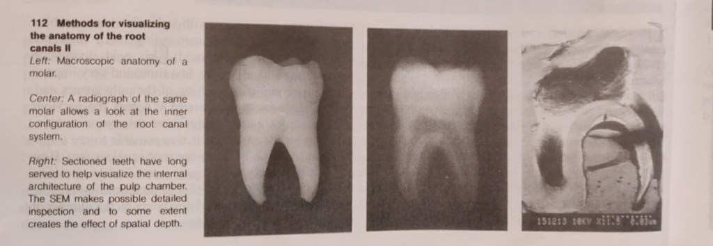
METHODS OF REPRODUCING ROOT CANAL ANATOMY

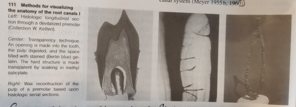
Most techniques require the destruction of the tooth. However, at the beginning of the twentieth century the transparency method was developed (Adolf 1913) in which the integrity of the tooth and the spatial relationships of the root canal and its outer contours were preserved. Various substances (from colored gelatin and paraffin to silicone) were introduced into the pulp space through an access opening, and the tooth was then made transparent by means of oil of cedar, benzol, salicylic acid compounds.

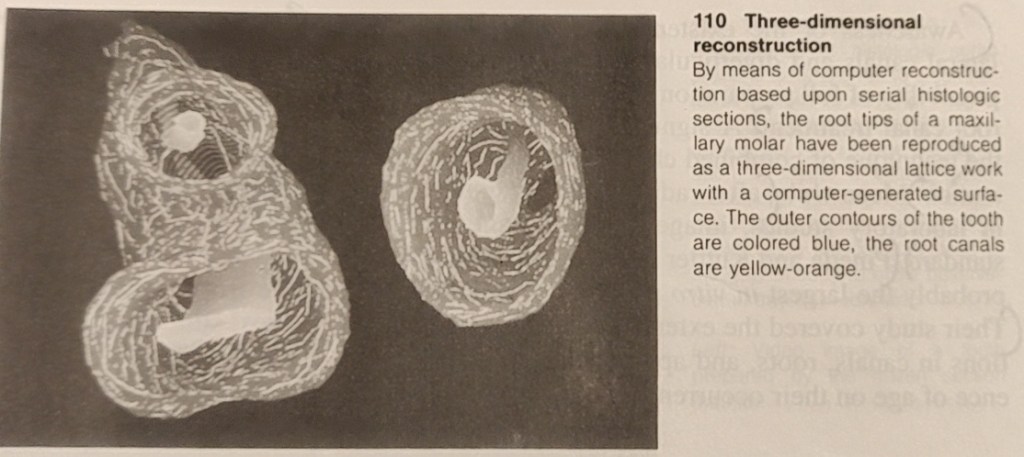
While histologic sections have long provided information on the structure of the root canal and the pulp tissue, Meyer (1955-1970) set new standards. From special sections of all 16 types of permannet teeth he made 50x scale models of the apical canals (the last 6 mm of each) of 800 teeth by projecting the circumference of the canals and building wax models layer by layer. This study further clarified the complexity of the pulp space, from then on called the root canal system (Meyer 1955 b, 1960).

Awareness of the existence of large numbers of lateral canals and diverticula renders obvious the impossibility of full preparation of all the branches during root canal treatment. Asignificant outcome of this is the technique of combined chemical-mechanical preparation. After radiographs began to be used in laboratory studies, images in two planes became standard. Pineda and Kutler (1972) performed what is probably the largest in vitro study on over 40000 teeth. Their study covered the extent of branching and variations in canals, roots, and apical deltas, and the influence of age on their occurence.

Hession (1977a-d) showed the shape of the root canal system radiographically before and after in vitro treatment. The abundant range of research tools is complemented by in vivo radiographs, microradiographs, scanning electron microscopy (SEM), computer reconstructions, monographs of individual cases, and many other aids (Baumann 1995). Subsequently, an immense body of facts has been accumulated and these are presented in excellent didactic style in books,videos, slide series, reports, seminars, and demonstration. This new information should be offered in further education courses (Baumann 1994, 1955).

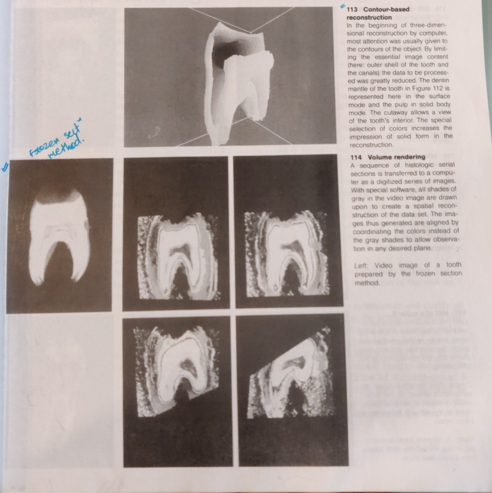
THREE – DIMENSIONAL COMPUTER RECONSTRUCTION

From a historical perspective we see a long tradition of striving to better describe the anatomy of the teeth. Preparations of 20 micrometer thick frozen sections were continuously recorded on videotape, producing data to serve as the basis for computerized three-dimensional reconstructions. In a contour-based reconstruction only the surface outlines of the tooth and the canals are used for input (Baumann et al. 1993d, 1994b). From this emerges a contour line, surface, or solid body model that can be viewed from any desired angle.

Faster computers permit the use of all shades of gray in a video image to create a volume-based reconstruction (volume rendering). Through ray-tracing, isotropic voxels (points in space) are created in which the raw unaltered data is drawn upon for three-dimensional reconstruction (Baumann 195, Baumann et al. 1993d).

Images are created that can be viewed, sectioned, colored, zoomed, or rotated in any desired plane. This makes possible views into the endodontic space that were prevoiusly unknown.

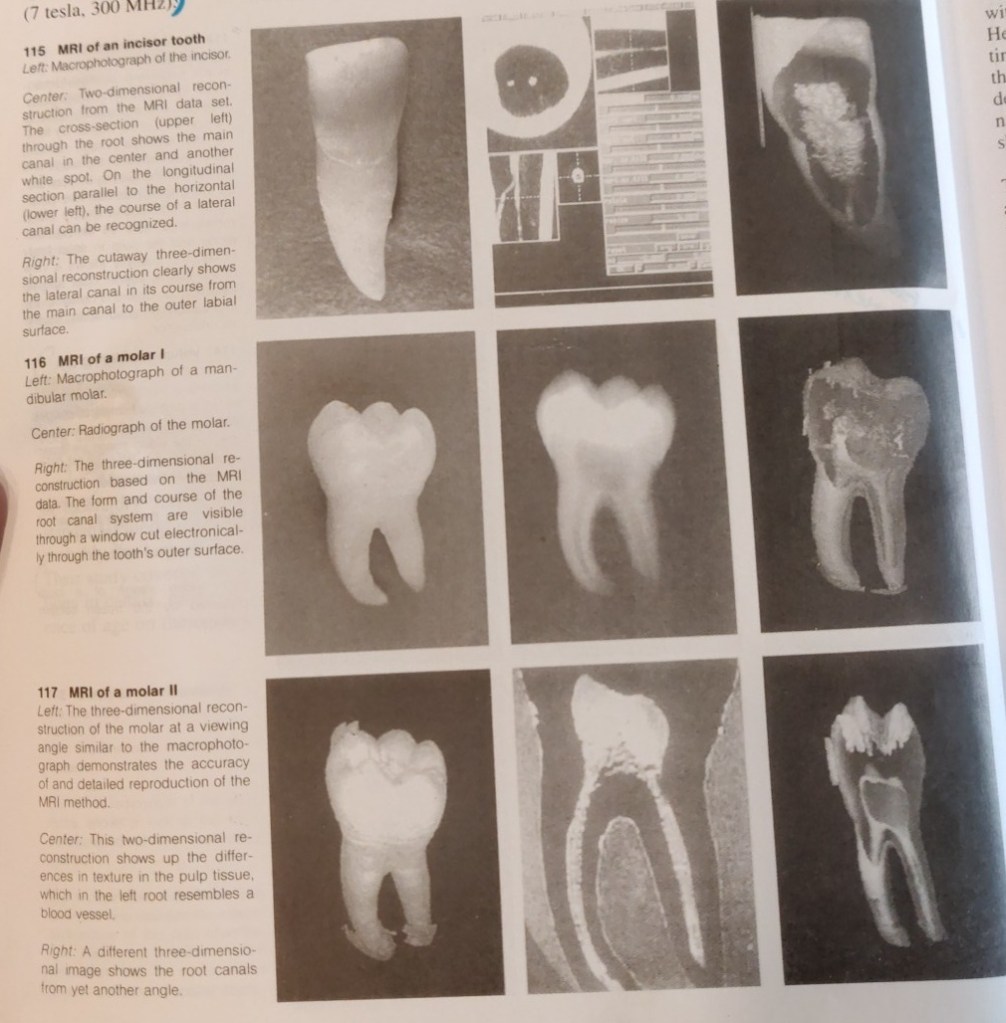
MAGNETIC RESONANCE IMAGING (MRI)

Normally, only vague images of bone and tooth can be obtained by magnetic resonance tomography (MRT). Baumann (1995; Baumann et al. 1993 a-d) was the first to succeed in producing a visual representation of the H+ protons of dental hard structures by using measurement sequences from solid body spectroscopy and especially strong magnetic fields. The soft pulpal tissue is elusive becuase of the small scale of the MRI. The first magnetic resonance images have now been realized with BRUKER SPECTOMETER AMX 300 WB (7 tesla, 300MHz).

Computer processing of data from the MRI permits creation of two and three-dimensional reconstructions that can be rotated and sectioned (Bauman 1995; Baumann and Doll, in press). Now for the first time we have a nondestructive method that does not use ionizing radiation. Two-dimensional sections of molars give rise to the hope that it will be possible to depict differences in tissue texture, which would be a great aid in the diagnosis of pulpitis. The spatial reconstruction of an individual canal configuration would be a great enlightment for endodontic treatment.

Reference – Color Atlas of Dental Medicine

ENDODONTOLOGY

Rudolf Beer, Michael A. Baumann, and Syngcuk Kim

Kriti Keshav Sherigar: Bridging the Gap in Cleft Lip and Palate Treatment

In the bustling world of Navi Mumbai, where careers are cultivated and passions pursued, Kriti Keshav Sherigar shines as a dental intern at MGM Dental College and Hospital. Committed to transforming lives one smile at a time, Kriti is not only a dedicated dental professional but also a former national-level athlete with a penchant for dancing and sports.

1) Could you provide a brief overview of your research project?
My research project had the intriguing title: “Parent’s responses to early presurgical nasoalveolar molding (PNAM) treatment in infants with cleft lip and palate: A qualitative analysis.” Our primary aim was to delve into the responses, coping mechanisms, and psychosocial adjustments of parents whose infants were undergoing PNAM therapy. We conducted face-to-face interviews with parents, discussing various aspects related to PNAM treatment. These interviews were comprehensive and open-ended, allowing us to understand the impact of PNAM on parents and their experiences. We then analyzed the data using inductive thematic analysis.

2) What motivated you to choose this particular research topic?
I was very clear in my head that I wanted pedodontics to be my subject of choice and with the guidance of Dr. Shrirang Sevekar, the Head of the Department of Pedodontics at MGMDCH, we selected this research topic. By reading research papers and articles, we believed that our study could lead to advancements in the treatment and support provided to individuals affected by cleft lip and palate, as well as their families.

3) How did you become interested in the ICMR STS program, and what was your application process like?
I first learned about the ICMR STS program through a lecture at my college that explained the program’s opportunities and benefits. This introduction motivated me to pursue research through the program.

4) What was the main research question or hypothesis you aimed to address in your project?
Our research aimed to address several key questions:

  • How do parents respond to PNAM treatment?
  • What coping strategies do parents employ during PNAM therapy?
  • What patterns of psychosocial adjustments do parents exhibit during PNAM therapy?
  • What are the barriers faced by parents during PNAM therapy?

5) How did you design your research proposal and select your methodology?
Our research proposal was carefully designed in accordance with the guidelines provided by ICMR. Given the nature of our study, which focused on qualitative analysis, we selected our methodology after extensive reading of related articles and papers to ensure its suitability.

6) Can you describe the specific methods and techniques you used to collect and analyze data?
We collected data through face-to-face interviews, and the analysis was conducted using inductive thematic analysis. The process involved familiarizing ourselves with the collected data, generating initial codes, collating codes into themes, and then further categorizing themes into subthemes. Ultimately, we created a thematic map based on these themes and subthemes.

7) Were there any surprising or noteworthy discoveries during your research?
Yes, our research yielded some interesting findings. We discovered that various coping strategies, such as social support from family, parents of infants with similar conditions, and the healthcare team, positively benefited parents. Successfully completing NAM therapy resulted in positive outcomes, including increased empowerment through successful feedings and effective management of daily challenges during therapy. Interestingly, our study also concluded that the financial burden may not be the critical factor influencing parents’ rejection of NAM treatment for infants with cleft lip and palate.

8) Were there any unexpected challenges or obstacles that you encountered during your research process? How did you overcome them?
I encountered a few unexpected challenges during the initial phases of my research journey. This was an entirely new experience for me, so understanding the entire research process posed an initial challenge. Additionally, as I was nearing the end of my third year and had university exams looming just a month or two away, juggling my studies, clinical postings, and meeting the ICMR deadlines proved to be quite a task. However, what made all the difference was the unwavering support and guidance of my mentor, Dr. Shrirang Sevekar. His expertise and encouragement were instrumental in helping me navigate these challenges. His guidance motivated me to work harder and approach the research with determination. Ultimately, our research project was selected by ICMR, and I also achieved a good score in my third-year university exams. The feeling of accomplishment and success was truly incredible and made all the effort worthwhile.

8) Are there any specific tips or insights you would offer to future STS applicants to increase their chances of success?
I’d like to offer a piece of advice to future STS applicants: Give it your best effort. The hard work and effort you put into your project are truly worth it. The sense of satisfaction and joy upon completing your project is incomparable. At the start, it might seem confusing, but as you progress, you’ll gain a deeper understanding of the process. The journey is both memorable and highly educational, so work diligently and never give up!

9) Is there anyone you would like to acknowledge or express gratitude to for their support during your research project?
I want to express my heartfelt gratitude to my mentor, Dr. Shrirang Sevekar. He went above and beyond, dedicating extra hours after college to assist me with my doubts and ensure we completed our project on time. His knowledge, patience, and hard work were not only motivating but also truly inspiring. I’d also like to thank my parents and my brother for their unwavering encouragement, which continually pushes me to take on new challenges and supports me along the way.

Kriti Keshav Sherigar’s dedication to oral health and her commitment to enhancing the lives of individuals with cleft lip and palate reflect the transformative power of research, compassion, and the pursuit of one’s passions. As she continues to dance through life, Kriti’s contributions to both the field of dentistry and her artistic endeavors are bound to leave an enduring mark.

Pavithra B Nair: Illuminating Rural Oral Hygiene Practices through her Research

In the vibrant city of Mangalore, where diverse talents and passions converge, Pavithra B Nair, a dental intern at A B Shetty Memorial Institute of Dental Sciences, stands out not only for her commitment to oral health but also for her passion for classical dance. Pavithra believes that dentistry is a canvas where she can explore the artistry of oral health, aligning her profession with her passion.

1) Could you provide a brief overview of your research project?
Certainly, my research project focused on assessing the knowledge, attitude, and practice of night brushing and oral hygiene among the rural population of Dakshinna Karnataka.

2) What motivated you to choose this particular research topic?
My motivation for choosing this research topic stemmed from my experiences at A B Shetty college, where we encounter many patients from rural areas of Karnataka. It became evident that improving the quality of life for these individuals necessitated addressing their knowledge and practices related to night brushing and oral hygiene. This realization led me to conduct a survey to assess their understanding and explore ways to enhance their oral health.

3) How did you become interested in the ICMR STS program, and what was your application process like?
I learned about the ICMR-STS program through my college. Initially, I had reservations about taking on this project, as I was concerned about balancing my studies with research work, especially since it was a new experience for me. However, my college provided invaluable support and encouragement, urging me to give it a try. The application process was straightforward, involving submission through the official ICMR-STS website, where all the necessary details were provided.

4) What was the main research question or hypothesis you aimed to address in your project?
As dentists, our primary objective is to promote awareness of oral health. While we emphasize the importance of brushing twice a day, the significance of night brushing is often overlooked. Night brushing plays a vital role in maintaining oral hygiene by preventing the accumulation of bacteria in the mouth during the night when saliva flow is reduced. Therefore, my goal was to assess the knowledge and attitude of rural populations regarding night brushing and raise awareness to improve oral hygiene.

5) Were there any unexpected challenges or obstacles that you encountered during your research process? How did you overcome them?
One of the significant challenges was visiting various rural areas in Dakshinna Karnataka and communicating with the local population, as language barriers were a hurdle. Fortunately, I had the support of individuals who helped me navigate this issue. Balancing my semester exams with research work was another challenge, but I received support from my professors and staff to manage the situation.

6) Did you collaborate with any mentors or fellow researchers during the project? How did they support you?
My mentor, Dr. Pushparaj Shetty, a Professor from the Department of Oral Pathology and Oral Microbiology, was a constant source of support throughout the project. His guidance and encouragement were instrumental in helping me achieve my research goals.

7) Are there any specific tips or insights you would offer to future STS applicants to increase their chances of success?
To future STS applicants, I would advise approaching the program with a mindset focused on learning from the project rather than being overly concerned about the results. Despite my initial insecurities, the journey was intriguing and filled with new experiences that have motivated me to continue my research journey.

8) Is there anyone you would like to acknowledge or express gratitude to for their support during your research project?
I’d like to express my gratitude to the almighty for providing me with the strength to see this research project through. My heartfelt thanks go to my mentor, Prof. Dr. Pushparaj, without whom I may not have embarked on this research journey. I’m immensely grateful to my parents, who have always believed in me, even when I doubted myself, and to all my colleagues who lent their assistance in completing this project.

Pavithra B Nair’s dedication to improving oral hygiene and raising awareness among rural populations exemplifies the transformative power of research and the enduring impact that passionate individuals can have on their communities. As she continues her journey, Pavithra is poised to make significant contributions to both the world of dentistry and the realm of classical dance.

Intern Abhinandan Kumar’s Research Unveils Insights into COVID-19 Immune Response

Yenepoya Dental College Intern’s Work on Salivary Antibodies Sheds Light on Pandemic’s Impact

In a commendable display of scientific inquiry, Abhinandan Kumar, a dedicated intern at Yenepoya Dental College, has undertaken pioneering research on the detection of salivary antibodies in both COVID-19 vaccinated and non-vaccinated individuals. His research holds promise in understanding the immune responses to the virus and vaccines, potentially influencing public health measures during the ongoing pandemic.

1) What motivated you to choose this particular research topic?
I was drawn to this research topic due to the unique circumstances presented by the COVID-19 pandemic. The pandemic has posed significant challenges to public health, making it crucial to understand the immune response to the virus and vaccines. This research allows me to contribute to our understanding of COVID-19 and potentially influence public health measures during these unprecedented times.

2) How did you become interested in the ICMR STS program, and what was your application process like?
My interest in the ICMR STS (Indian Council of Medical Research Short-Term Studentship) program stemmed from its esteemed reputation and the opportunity it provides for students like myself to engage in meaningful medical research. The program’s focus on nurturing research skills and promoting scientific inquiry aligns perfectly with my career aspirations.

3) What was the main research question or hypothesis you aimed to address in your project?
The primary research question and hypothesis in my project revolved around comparing IgG (Immunoglobulin G) antibody levels in two distinct groups: COVID-19 vaccinated individuals and non-vaccinated individuals who had contracted COVID-19. Specifically, we aimed to investigate whether there were significant differences in IgG antibody levels between these two groups, which would provide insights into the effectiveness of vaccination and the durability of immune responses in previously infected individuals.

4) How did you design your research proposal and select your methodology?
In our study, we included 15 vaccinated and 15 non-vaccinated individuals, excluding those with recent fevers. Participants rinsed their mouths with water before providing saliva samples, which were stored at 4°C for up to 6 hours and then at -20°C. IgG levels targeting SARS-CoV-2 RBD were measured using an established ELISA method, with absorbance readings at 450 nm taken within 30 minutes of stopping the reaction.

5) Can you describe the specific methods and techniques you used to collect and analyze data?
We utilized the ELISA (Enzyme-Linked Immunosorbent Assay) method for both data collection and analysis in our study.

6) Were there any unexpected challenges or obstacles that you encountered during your research process?
Yes, we did encounter unexpected challenges during the research process, particularly when collecting samples from non-vaccinated individuals.

7) Did you collaborate with any mentors or fellow researchers during the project? How did they support you?
During the project, I had the privilege of collaborating with two esteemed mentors, Dr. Vishnu Das Prabhu and Prof. Bhandari. Their guidance and support were instrumental in the successful execution of the research.

8) Were there any surprising or noteworthy discoveries during your research?
Yes, during our research, we made a noteworthy discovery. We observed both slight changes and no change in IgG levels among the vaccinated individuals. This finding indicated that the immune response to COVID-19 vaccination can vary among individuals, with some showing slight alterations in IgG levels, while others exhibited no change.

9) Is there anyone you would like to acknowledge or express gratitude to for their support during your research project?
I would like to extend my heartfelt gratitude to several individuals who played instrumental roles in supporting and guiding me throughout my research project. Firstly, I express my sincere appreciation to our principal, Dr. Laxmikanth Chatra, for providing valuable encouragement and resources for this endeavor. I’m also deeply thankful to our dean, Dr. Sham Bhat, for his unwavering support and mentorship. Additionally, I want to acknowledge and thank Prof. Vishnu Prabhu and Prof. Bhandari for their expertise, which significantly enhanced the quality of the research. Last but not least, I express my gratitude to Maji Jos for their invaluable assistance during the project. Their collective support was indispensable in making this research a reality.

Revolutionizing Dental Care: BDS Student Successfully Replicates Enamel-like Structure

In a remarkable achievement, Raahul, a final-year BDS student at Rural Dental College, Pravara Institute of Medical Sciences, Loni, has made significant strides in dental research. His pioneering project, funded by the prestigious Indian Council of Medical Research (ICMR) Short-Term Studentship (STS) program, focuses on replicating enamel-like structures using synthetic materials through biomimetics.

Mr Raahul, a passionate problem-solver with a penchant for non-fiction literature and spreading smiles, this research journey was an opportunity to make a tangible impact in the world of dentistry.

Raahul’s journey into the world of biomimetics began with a strong desire to address dental issues by emulating the structural and functional qualities of natural enamel. His research project aimed to create synthetic materials that closely resembled the composition and properties of enamel, a substance renowned for its hardness and protective role in teeth.

  1. What motivated you to choose this particular research topic?
    Among the various topics I considered, this particular one resonated with me strongly, as I believed that if I could emulate the structural and functional qualities of enamel closely, it could potentially address several issues.
  2. How did you become interested in the ICMR STS program, and what was your application process like?
    I came across the ICMR’s STS program on the Dentowesome page, and though I wasn’t previously aware of it, I decided to explore it further and give it a try.
  3. What was the main research question or hypothesis you aimed to address in your project?
    My primary objective was to successfully mimic the enamel-like structure using synthetic materials, and I can confidently say that I achieved this goal.
  4. How did you design your research proposal and select your methodology?
    My initial reference point was the comprehensive guidelines provided by ICMR, and I also received valuable guidance from my mentor.
  5. Can you describe the specific methods and techniques you used to collect and analyze data?
    The study was conducted in vitro, involving the use of extracted third molars with intact enamel from the OMFS department in our hospital. The procedures were carried out in the biochemistry lab.
  1. Were there any unexpected challenges or obstacles that you encountered during your research process? How did you overcome them?
    I was fairly well-prepared for potential challenges, as I had anticipated many of the issues that might arise during the course of my research.
  2. Did you collaborate with any mentors or fellow researchers during the project? How did they support you?
    Certainly, it was a collaborative effort. The OMFS residents assisted me in obtaining the tooth samples, the biochemistry faculty guided me through the procedures, and, of course, my mentor played a pivotal role.
  3. Were there any surprising or noteworthy discoveries during your research?
    Our research allowed us to comprehend the relationship between a dendrimer called PAMAM and human enamel. This has potential implications for future use as a regenerative material in enamel regeneration.
  4. Are there any specific tips or insights you would offer to future STS applicants to increase their chances of success?
    My advice is to stay true to yourself and give your absolute best. Everything will eventually fall into place, and even when facing difficult moments, don’t give up. Often, things can change for the better if you persist through those challenging times.
  5. Is there anyone you would like to acknowledge or express gratitude to for their support during your research project?
    I’m deeply grateful to my mentor, my seniors, the faculty, and particularly Dr. Anisha. Her valuable interactions and assistance in addressing various challenges have been instrumental. In conclusion, I would like to extend my heartfelt thanks to Dr. Anisha for this opportunity. While I’m uncertain of the extent of its impact, if even one student is inspired to pursue a similar path, I would consider it a success.

Raahul’s achievement stands as a testament to the potential for innovative research within the field of dentistry. His work may one day lead to transformative advancements in dental care, offering brighter smiles and healthier teeth for countless individuals.

ENDODONTOLOGY (PATHOLOGY AND DIAGNOSIS) – Part 1

Intensive microbiologic, immunologic, and morphologic research investigations, especially during the past decade, have shown that colonization of tooth surface by pathogenic bacteria is accompanied by humoral and cellular defense mechanisms of the organism not only during the more advanced stage of infection, but also throughout the initial stages. Penetration of these complex defenses, which is usually of limited duration, disturbs the equilibrium of the system and results in disease. Within the dental pulp, this biologic equilibrium has to do with the balanced calcium and phosphate ion exchange during the continuous demineralization and remineralization of the enamel and exposed dentin. As long as a disease process is reversible, as is incipient caries, the capacity for progression and regression is present. Carious breakdown means that enamel is being demineralized by acidogenic plaque more rapidly than it can be remineralized. In its early stages, the caries has become a chronic destructive process, in which irreversible structural changes will preclude any further remission.

Looking at the dynamics of demineralization and remineralization, and the etiology of caries against the epidemiologic background, and comparing them with the results of therapy, a pattern of active disease spurts alternating with resting phases emerges. During these periods of remission, the chronic destructive process is not reversed, but is only brought to a standstill. This concept of progression and stagnation ( Socransky et al. 1984) is strongly influenced by the defensive capability of the organism.

Progression is defined by invasion of caries into dentin with inflammation and loss of connective tissue. Stagnation means the defenses are increased, there are defensive inflammatory cells in the tissues, and connective tissue is being replaced by secondary dentin or granulation tissue. Histologically, this ever-changing dynamic process in carious teeth is recorded over the years through deposition and destruction of dentin.

For the practitioner, the obvious questions that arise are how to classify the histopathologic condition of the pulp and the apical periodontal tissues, and how to initiate treatment that is appropriate, considering the background of stagnation or progression. Based upon clinical findings, differentiations are made between a clinically sound pulp, reversible pulpitis, irreversible pulpitis, a necrotic pulp, and apical periodontitis. These distinctions are based solely upon clinical observations; generally, a correlation between certain symptoms and a specific pathologic entity cannot be expected. Making the distinction between reversible and irreversible inflammations of the pulpal tissues can be a diagnostic problem, because they can present similar clinical symptoms. Histologically, the diagnosis of acute inflammation is based upon the predominance of neutrophilic granulocytes. However, this diagnostic picture does not always coincide with the appearance of pain symptoms because neutrophilic granulocytes can also be found in cases where there is no pain ( Langeland 1981, Lin and Langeland 1981 b, Lin et al. 1984 ).

DIAGNOSIS OF PROXIMAL CARIES

Caries begins with microscopic demineralization of the affected enamel or cementum surface. As it progresses, the enamel first becomes chalky, then its surface is broken through. In this stage, the caries is easy to detect, but has frequently progressed so far that extensive restorative and endodontic treatment in necessary. More difficult to diagnose, on the other hand, are lesions that are in their early stages and dentinal lesions with macroscopically intact surfaces.

Epidemiologic studies have shown that – coincident with a general decrease in caries prevalence in industrialized countries – the occlusal surfaces of the permanent molars of children and young adults are the surfaces most frequently attacked by caries. In contrast to fissure caries, proximal and smooth surface caries is much less frequent. Radiographically evident incipient lesions in enamel of the proximal surfaces have likewise shown a decline. In adults, the probability that these lesions would penetrate further has increased, and this has caused the proportion of proximal caries to rise again.

During clinical examination using explorer many carious lesions with cavity go undiagnosed, so for their proper diagnosis bitewing radiographs, and fiberoptic transillumination (FOTI) are used. Bitewing radiographs are still the method of choice for the diagnosis of approximal caries, and account for the detection of approximately three-fourths of dentinal carious lesions ( Mileman and van der Weele 1990, Noar and Smith 1990). Studies found that where there is a dentinal lesion, there is a surface that has been broken through, which precludes any chance for remineralization (Marthaler and Germann 1970; Bille and Thylstrup 1982; Mejare and Malmgren 1986). Even though the actual extent of caries is underestimated with the radiograph, it may be concluded that the specificity, that is, the ability to recognize sound teeth as sound, is approximately 95% ( Mileman and van der Weele 1990). As far as caries diagnosis is concerned more sensitive X-ray films seem to be the equal of earlier films as far as caries diagnosis is concerned but as they produce same degree of contrast with significantly less radiation, their use is now highly recommended. Preventive measures can impede further penetration and even promote remineralization, provided that the enamel surface has not yet been disrupted.

The progression of caries can be monitored with periodic radiographs. Their interval depends, among other things, upon the individual’s susceptibility to caries. Patients at high risk of caries should be radiographed every year while those at very low risk need only be radiographed every 2-4 years. The time in which it takes caries to penetrate the enamel of a mature permanent molar in a patient with good oral hygiene can exceed 5 years. This offers the opportunity to post-pone invasive restorative treatment and to observe whether the caries progresses or regresses. The rate at which penetration progresses can be estimated by comparing radiographs produced at different times by a standardized technique. Recently erupted teeth, on the other hand, demonstrate a markedly reduced penetration time (Marthaler and Wiesner 1973, Shwarz et al. 1984).

In order to minimize overlapping of the images of approximating tooth surfaces, a film holder is recommended. A deviation of the horizontal angle of the X-ray tube by only a few degrees will result in a substantial decrease in correct diagnoses. Radiolucency in dentin should be treated as invasive only if there is also an unmistakable radiolucency in the enamel region. The radiograph should be inspected carefully under magnification and away from the influence of any light coming from the sides.

FOTI can be used in addition to bitewing radiographs if there is no interference from adjacent interproximal fillings that are other than tooth colored. More than 70% of dentinal lesions in anterior teeth can be detected by means of FOTI. Dentinal lesions in posterior teeth, however, can be differentiated only with great difficulty ( Pieper and Schurade 1987, Choski et al. 1994 ).

Reference – Color Atlas of Dental Medicine

ENDODONTOLGY

Rudolf Beer, Michael A. Baumann, and Syngcuk Kim

BLOOD SUPPLY OF NOSE

Nose is richly supplied by both the external and internal carotid systems, both on the septum and the lateral walls. NASAL SEPTUM Internal Carotid System – Branches of Ophthalmic Artery External Carotid System LATERAL WALL Internal Carotid System – Branches of Ophthalmic Artery External Carotid System LITTLE’S AREA Situated in the anterior inferior part of […]

Diagnosis Demystified- Case 5/255

A radiolucent lesion was found incidentally on a dental panoramic radiograph in a 30-year-old man. The cyst was located in the lower molar area above the inferior alveolar canal and showed a scalloped outline extending between the roots of the teeth. All teeth in the area were vital and the lamina dura was intact. Clear straw-coloured fluid was aspirated from the lesion

Solitary bone cyst (SBC), also known as simple bone cyst, is a benign bone lesion that most commonly occurs in children and adolescents. While the exact cause of SBC is still unknown, it is believed to be the result of a disturbance in the normal bone remodeling process.

SBC typically presents as a painless swelling or bump in the affected bone, often discovered incidentally on routine imaging. Diagnosis is usually made by imaging studies such as X-rays or MRI, as well as fine needle aspiration to confirm the presence of fluid within the cyst.

In many cases, SBCs will heal on their own without the need for any intervention. However, if the cyst is causing significant pain or functional impairment, or if it is at risk of fracturing or expanding and causing further damage to the bone, treatment may be necessary.

Treatment options for SBC include observation, which involves monitoring the cyst with regular imaging studies to ensure that it is not growing or causing any problems; curettage, which involves surgically removing the cyst and filling the cavity with bone graft material to promote healing; or injection of bone-stimulating agents such as bone morphogenetic protein (BMP) to promote healing and prevent recurrence.

The decision on whether to intervene or not will depend on various factors, including the size and location of the cyst, the age of the patient, and the presence of any associated symptoms or complications.