Diagnosis Demystified – Case 1/255

A 28-year-old woman presented for a check-up. The dentist noticed that the occlusal plane was depressed on the left side. The molar teeth did not make contact with the maxillary teeth on that side, though wear facets were present.

ANSWER

So, your jaw is made up of two parts called the maxilla (which is the upper part) and the mandible (which is the lower part). The mandible has a part called the ascending ramus on each side, which is a bony projection that helps to support the jaw joint.

Sometimes, one side of the ascending ramus can grow more than the other side. This is called condylar hyperplasia. It can cause the jaw to become uneven, with one side being bigger than the other.

When this happens, the teeth on the bigger side can grow more than the teeth on the smaller side. This can cause the teeth on the bigger side to stick out more and make the bite uneven.

Does that make sense to you? Let me know if you have any more questions!

Drill Sergeant: Three Inspiring Dental Case reports of the week #1

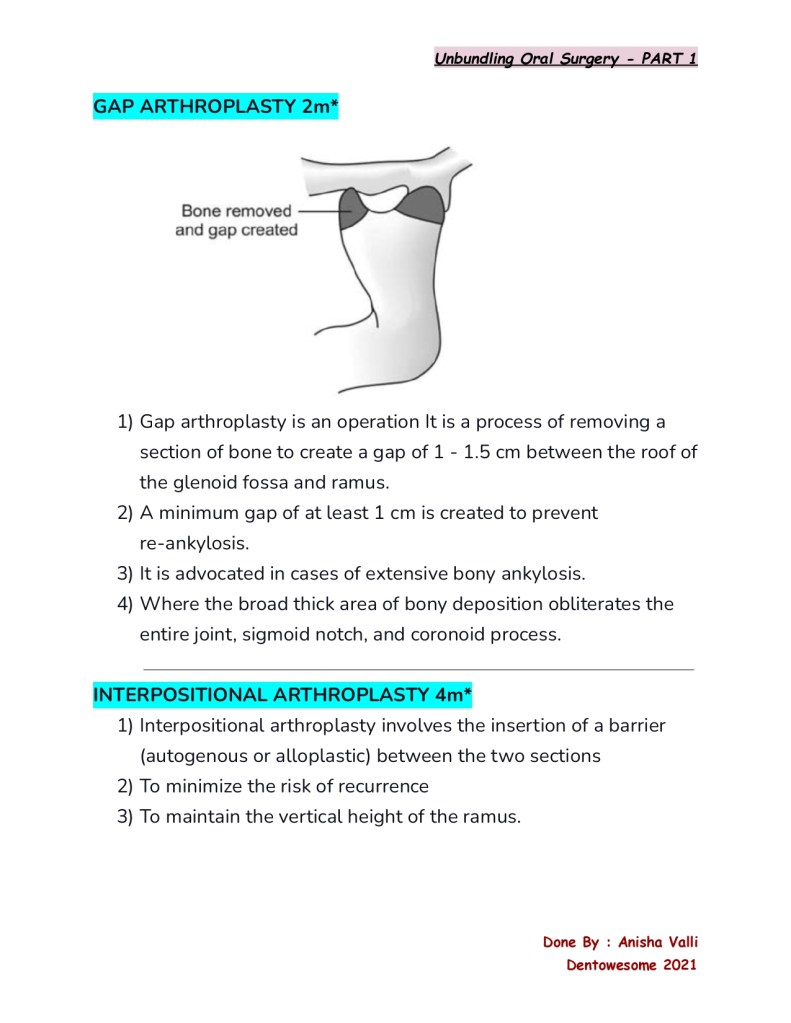
1) Pouch and  tunnel technique in conjunction with connective tissue Graft -a paramount for treating gingival recession

Gingival recession can be a bothersome and unappealing issue for patients. Thankfully, there’s an esthetic correction option available that’s both minimally invasive and promotes fast healing: the Pouch and Tunnel technique with connective tissue grafting (CTG). This approach is an excellent alternative for patients seeking effective recession coverage, and it’s worth considering if they are looking for a solution that’s both friendly to gums and their wallet.

Link – https://doi.org/10.21276/10.21276/ujds.2021.7.1.17

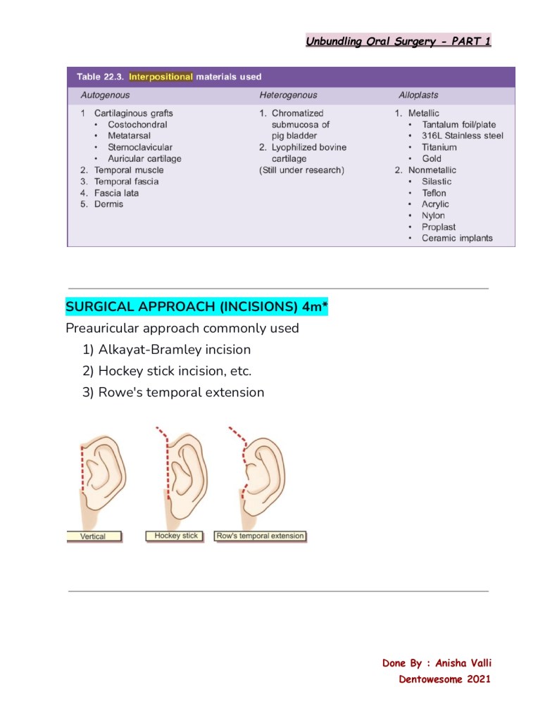
2) The natural tooth pontic and instant idea to retain aesthetics

In cases where a patient experiences sudden tooth loss in the anterior region of their mouth, it can be distressing and affect their confidence. The good news is that there are a range of treatment options available, including removable, tooth-supported, and implant-supported prosthetics.

Regardless of the chosen treatment, it’s important to restore the patient’s smile as quickly as possible while also stabilizing their dental arch. One technique involves using the patient’s own natural tooth as a pontic, which provides an exact match in terms of size, shape, and color, while also preserving the original 3D position of the tooth.

Link : https://www.hindawi.com/journals/crid/2016/8502927/

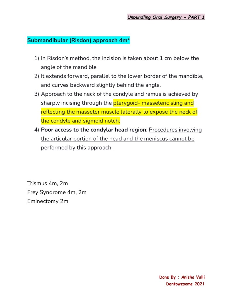
3) Modified roll technique- handy technique to augment the periimplant soft tissue in aesthetic zone

In this randomized controlled trial, researchers are exploring the effectiveness of a modified roll flap (MRF) technique to enhance the appearance of single-tooth implants in the esthetic zone. The MRF is a pedicle flap that utilizes the gingival tissue overlying the covering screw to thicken the labial soft tissue, which can have a significant impact on the overall esthetic outcome.

The study aims to measure the thickness of the labial soft tissue and the implant esthetic score system (IES) to evaluate the success of the MRF technique during stage-two implant surgery. By preserving and utilizing the existing tissue instead of discarding it, the MRF technique could potentially enhance the appearance of the implant site and improve patient satisfaction with the results.

Link: https://www.sciencedirect.com/science/article/pii/S1687857413000231

Ortho Case 4.1

A 12-year-old female presented with a class II division 1 malocclusion on a moderate skeletal class II pattern, with increased vertical dimensions complicated by an increased overjet (12mm), crowding of both dental arches and teasing in relation to her dento-facial appearance.

What is treatment plan?

TREATMENT PLAN

1) Functional appliance like Dynamax or Twin block (more preffered as overjet reduction is more effective) = maxillary retroclination, mandibular incisor proclination, guides the eruption of posterior dentition.

2) Need of Headgear with torqueing spurs = restricts maxillary forward growth and tipping of maxillary incisors.

3) Extraction of four second premolars followed by edge to edge appliances for stability of class II correction.

Picture based diagnosis case 1

Study the two radiographs which are of the same
patient at (a) 19 years and (b) 34 years of age.

1 . What is the most likely periodontal diagnosis at 34
years of age?

Generalised aggressive periodontitis. The severity
of bone resorption and the radiographic absence of
signs of resorption at 19 years preclude a diagnosis of
chronic periodontitis.

2 . What does the initial phase of treatment involve?

Initially conventional cause-related treatment
is instigated: instruction in toothbrushing and
use of adjunctive aids for interproximal and
subgingival cleaning, RSI, prophylaxis. Ultimately,
treatments such as surgery and the adjunctive use of
antimicrobials might be indicated, but conventional
treatment is first in line.

3 . The patient has a sister who is aged 29 years. What
advice might you offer?

Periodontal screening and radiographic examination.
There is evidence that certain subjects are at high risk
from developing aggressive periodontitis and this
risk may be under genetic control. Siblings should
be screened and affected individuals with children
warned that early signs may develop from around
puberty onwards

Case History 4 in Diseases of bone and the maxillary sinus

A 35-year-old man presents with gross loosening of both his lower left premolar teeth. The gingiva around them looks swollen and is purple–brown in colour. A radiograph shows irregular bone destruction to the apices. Incisional biopsy shows multinucleated osteoclast-like giant cells in a haemorrhagic fibrous stroma.

  1. Which investigations should now be performed?
    The serum calcium level should be measured and radiographs reviewed to exclude hyperparathyroidism.
  2. If these prove negative, what treatment should be undertaken?
    The lesion should be treated by local removal with curettage.
  3. Which other lesions in the jaws contain multinucleate giant cells of this type?
    Osteoclast-like giant cells are found in
    1. giant-cell granuloma
    2. brown tumour of hyperparathyroidism
    3. Pagetʼs disease of bone
    4. aneurysmal bone cyst
    5. some fibro-osseous lesions, particularly cherubism. 

INTRODUCTION Over the ages, oral health care has been delivered to the community in different ways. The horseback dentistry of olden days has evolved into the most modern painless dental procedures. In India, about 70% of the population live in rural areas whereas 70% of the dentists practice in urban areas. We seldom find certain dental offices and few government establishments in rural areas, which lack the required infrastructure.WHAT ARE MOBILE DENTALCLINICS? A mobile dental clinic is used primarily when oral health care is be delivered to small pockets of patients that are scattered over a specific geographic area. The mobile clinic generally is parked at a facility such as a school, residential facility or community center.SALIENT FEATURES:- •Useful life is shorter than a fixed facility. •Requirement of water and waste disposal methods. •Dental equipment can be a traditional or a portable one. •Requirement of a generator on board to provide electricity.TARGETED POPULATIONS FOR MOBILE DENTAL SERVICES:- •Low-income individuals or families. •Isolated or very rural rural populations. •Persons in residential care facilities. •People who are “ homebound”, bedridden, very frail or receiving hospital services at home. •Persons with a variety of special health care needs. •Migrant and seasonal workers. •People who are homeless or temporarily displaced.WHY MOBILE CLINICS ARE PREFERRED OVER USUAL ONES? •Moderate start up costs. •It addresses the problem of transportation to the clinics. •It decreases missed appointments when run in conjunction with schools. •Services can be made available at multiple sites. •Services are made available to the needy population.   Even though mobile clinics are preferred over the usual ones, but they do have certain disadvantages. Let’s have a look at certain “disadvantages” of it:- •High maintenance costs may occur. •Difficult to access and store patient record. •Provides limited services and follow up may be difficult. •Requires permission for site use. •Difficult to use during monsoon. ESSENTIALS OF MOBILE DENTAL CLINIC:- The mobile dental clinic should be equipped with 2 dental chairs with all attachments and seating space for 15-20 people. 1.PORTABLE DENTAL UNIT:-•Dental chair should be portable and easy to handle. It should be able to be folded for easy transportation.  All the parts  should be detachable type and well balanced and sturdy. •Mobile suitcase unit: Fitted with aerotar and micromotor hand piece. •Scaler with 3 scaling tips. •Control box with transparent, regulated water tank and foot control.2.OPERATING LIGHT:- Two, intensity fixed with hinge on the top of the van. 3.DENTAL X-RAY UNIT:-  X-ray unit with digital arm timer and day light manual developer. 4.AUTOCLAVE:-  High speed automatic instrument autoclave. 5.METAL CABINETS WITH WASH BASIN 6.WATER TANK: 400 litres capacity. 7.HEALTH EDUCATION MODELSPARTNERSHIPS IN PROVIDING MOBILE DENTAL SERVICES:-Some of the group or  Individuals who could potentially be partners are:- •GOVERNMENT:-State/ Local, Health Department, Department of Social Service. •COMMUNITY:- Local community, Business Leaders, Foundations. •PUBLIC:- Patient Care  Advocate, Organizations that promote health. •POLICY:- Local and Community Policy Makers. •HIGHER/ PROFESSIONAL EDUCATION:- Medical schools ,Dental schools and Allied Health Schools.CONCLUSION A fully-equipped mobile dental clinic is to provide effective dental care at the doorsteps of underprivileged, rural population is the need of the hour. The key to a successful dental practice is a cohesive dental team, which will create an atmosphere of co-operation resulting in the achievement of the goals of oral health in the coming up years.REFERENCE Essentials of public health dentistry-Soben Peter

Case History 1 = Diseases of bone and the maxillary sinus

A 58-year-old woman noticed that her front teeth had become spaced and seeks advice from her dentist. On entering the surgery, the dentist notices that she has difficulty in walking and does not respond to his questions. She has become increasingly deaf and her vision has also deteriorated. On examination, the maxilla and zygoma are enlarged and there is enlargement of the forehead.

1. What diagnosis would you suspect?
Pagetʼs disease of bone results in enlargement of cranial bones and deformation of weight- bearing bones. The cranium is usually expanded in thickness and symptoms may arise from cranial nerve compression.

2. What information might be gained from oral radiographs and blood tests to support this diagnosis?
Radiographs of the jaws may show hypercementosis, cemental masses, abnormal trabeculation and a cotton-wool appearance in the jaws. The alkaline phosphatase level is markedly raised.


3. What are the principal histological features of this disorder?

Disordered bone remodelling is seen; larger osteoclasts are present and the trabeculae show a scalloped outline. Numerous resting and reversal lines, resulting in a mosaic pattern, are seen and the vasculature may be increased. Globular cementum-like masses are seen in the jaws.