A Modification to Enable Controlled Progressive Advancement of the Twin Block Appliance

Welcome to an exciting exploration of one of the most innovative modifications in functional orthodontics! As orthodontic students, mastering the nuances of appliance design and modification is crucial for your future success. Today, we’re diving deep into the groundbreaking Twin Block advancement modification developed by Carmichael, Banks, and Chadwick – a system that has transformed how we approach Class II treatment with enhanced precision and patient comfort.

🎯 Why This Modification Matters for Your Future Practice

The Twin Block appliance, introduced by Clark in 1982, has become one of the most popular functional appliances in the United Kingdom and is arguably the most successful in treating Class II division 1 malocclusions. However, the original design had significant limitations that this modification brilliantly addresses.

The Problem with Traditional Twin Block Reactivation 🚫

  • Inconvenient chairside acrylic additions
  • Unpleasant taste and smell for patients
  • Inaccuracy due to polymerization shrinkage
  • Time-consuming laboratory modifications
  • Limited ability to make small, gradual adjustments

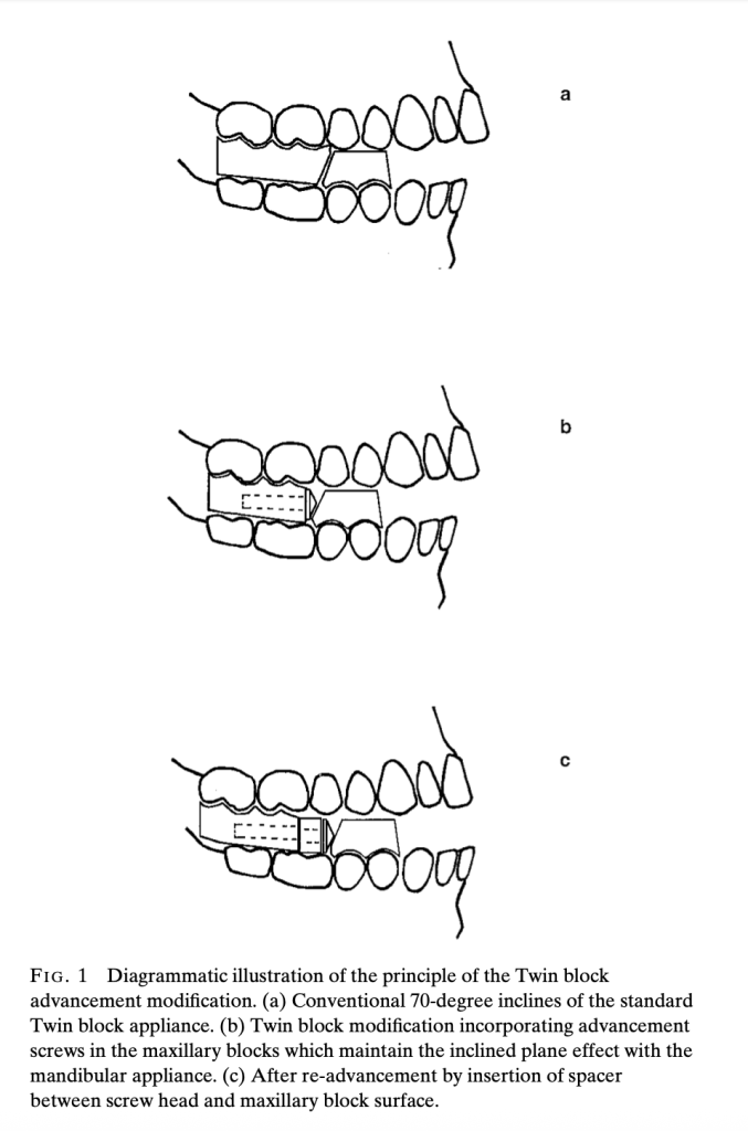
Understanding the Core Principle

The modification incorporates stainless steel screws with conical heads into the upper appliance blocks, maintaining the crucial 70-degree inclined plane effect regardless of screw rotation. This ingenious design allows for controlled, measurable advancement using polyacetal spacers.

Technical Specifications: What You Need to Know

ComponentSpecificationClinical Purpose
Stainless Steel Screws3mm diameter, 18/8 M3 gradeProvide structural strength and stability
Screw140° included angle (70° working angle)Maintain 70° inclined plane regardless of rotation
Screw Lengths12mm and 16mm (longer for >5mm advancement)Accommodate various advancement needs
Spacers MaterialPolyacetal co-polymer resinEnable precise, measurable advancement
Spacer Lengths1mm, 2mm, 3mm, 4mm, 5mmAllow stepwise progression (2-3mm typical)
Spacer Diameter6mm diameterEnsure proper fit and function
Thread HousingInjection-molded acetal resin with lateral tagsPrevent fractures and ensure consistent fit

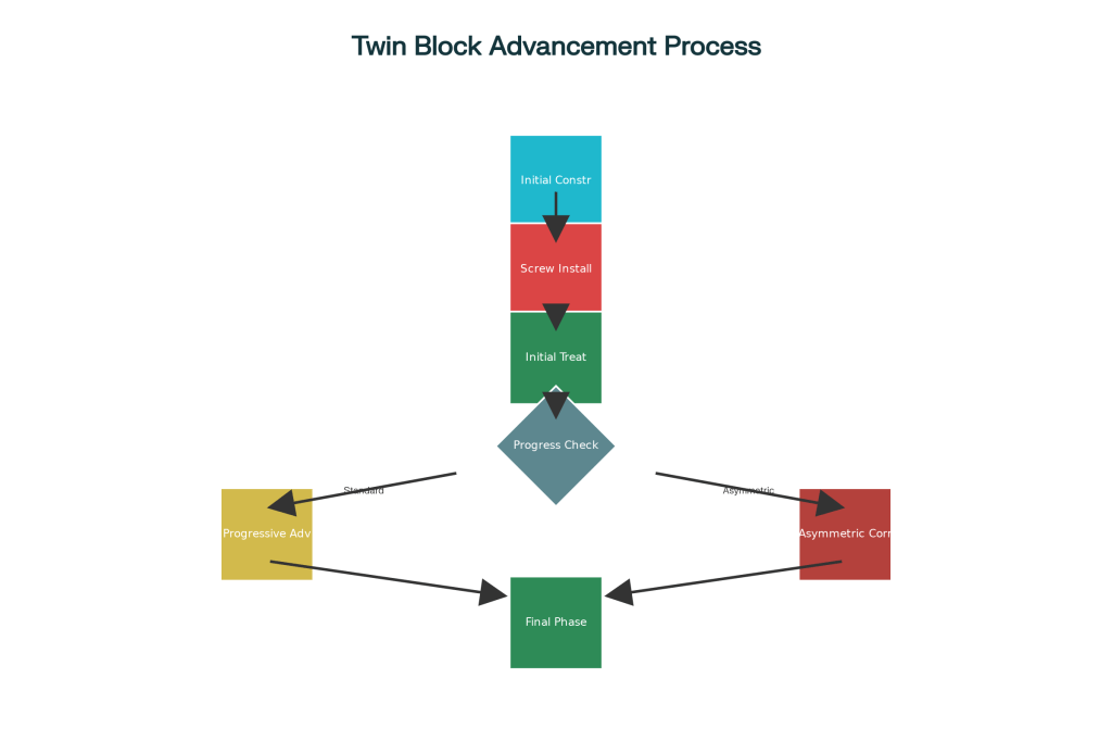
The treatment process follows a logical, patient-friendly progression that maximizes compliance and comfort while achieving optimal results.

Phase 1: Initial Construction and Setup

  1. Bite Registration: Take protrusive wax bite with comfortable advancement (may be as little as 2-3mm in some patients)
  2. Screw Installation: Insert 3mm diameter stainless steel screws with 140° conical heads into upper blocks
  3. Initial Delivery: Begin treatment with screws inserted without any spacers

Phase 2: Progressive Advancement

  • Monitoring: Assess overjet reduction at each visit
  • Advancement: Add 1-5mm polyacetal spacers between screw heads and blocks
  • Typical Increments: 2-3mm per advancement visit
  • Maximum Advancement: Up to 9mm using longer 16mm screws

🎭 Clinical Scenarios: Real-World Applications

Scenario 1: The Dolichofacial Challenge 😰

Patient: 12-year-old female with long face pattern

  • Challenge: Weak craniomandibular musculature, poor tolerance for large protrusions
  • Traditional Problem: Patient bites blocks together instead of maintaining protrusive position
  • Modified Solution: Start with minimal 2mm advancement, progress gradually with 1-2mm spacers
  • Outcome: Improved compliance and comfort, successful Class II correction

Scenario 2: The Large Overjet Case 📏

Patient: 13-year-old male with 12mm overjet

  • Challenge: Requires significant mandibular advancement but limited initial tolerance
  • Traditional Problem: Would require multiple appliance remakes or uncomfortable large advances
  • Modified Solution: Begin with comfortable 3mm advancement, systematically add spacers over 6 months
  • Outcome: Achieved 9mm total advancement with excellent patient acceptance

Scenario 3: The Asymmetric Correction 🎯

Patient: 11-year-old with Class II and dental centerline deviation

  • Challenge: Need for different advancement amounts on each side
  • Traditional Problem: Difficult to achieve asymmetric correction with conventional methods
  • Modified Solution: Use different spacer lengths – 3mm right side, 5mm left side
  • Outcome: Successful centerline correction along with Class II improvement

Scenario 4: The Class III Application 🔄

Patient: 10-year-old with developing Class III malocclusion

  • Challenge: Requires gradual reactivation for optimal growth modification
  • Modified Solution: Incorporate screws into maxillary appliance for controlled reactivation
  • Advantage: Small increments reduce patient discomfort and improve compliance

🎨 Material Science: Understanding Polyacetal Resin

Why Polyacetal is Perfect for This Application:

  • Strength: 10 times stronger than conventional acrylic resin
  • Safety: Non-toxic and non-allergenic properties
  • Durability: High resistance to surface wear and low water absorption
  • Workability: Can be trimmed and polished with standard dental instruments
  • Biocompatibility: Proven safe for intraoral use over extended periods

🎯 Facial Pattern Considerations: Tailored Treatment Approaches

Facial PatternCharacteristicsTreatment ConsiderationsRecommended Approach
Dolichofacial (Long Face)Weak craniomandibular musculatureLess tolerance for large protrusions, gradual advancement essentialStart with 2-3mm advancement, progress gradually
Brachyfacial (Short Face)Deep overbites presentOverbite reduction more problematic due to reduced block trimmingUse Phase 1 appliance or plan fixed appliances to follow
Mesofacial (Average)Balanced growth patternStandard advancement protocol works wellStandard 2-3mm increments per visit
Class III CasesRequires gradual reactivationSmall increments of reactivation necessaryUtilize modification for controlled gradual advancement

💡 Clinical Tips for Success

For Dolichofacial Patients 📐

  • Start conservatively with minimal advancement
  • Monitor for tendency to bite blocks together
  • Consider Phase 1 appliance for overbite reduction
  • Emphasize proper appliance positioning during sleep

For Brachyfacial Patients 🔽

  • Plan for overbite management strategies:
    • Option 1: Use initial upper removable appliance (Phase 1)
    • Option 2: Gradual Twin Block wear reduction during retention
    • Option 3: Upper removable retainer with anterior inclined bite plane

General Clinical Guidelines 📋

  • Advancement Frequency: Every 3-4 weeks based on patient adaptation
  • Typical Increments: 2-3mm spacers for most patients
  • Maximum Achievement: Up to 9mm total advancement reported
  • Block Height Requirement: Minimum 6mm between second premolars

⚠️ Troubleshooting Common Issues

Problem: Block Cracking After Advancement 🔧

Cause: Inadequate block height or retrospective screw insertion
Prevention: Ensure adequate 6mm block height, incorporate screws during initial construction
Solution: Use screw thread housing system for reinforcement

Problem: Difficulty Removing Screws 🔄

Cause: Direct screw insertion into acrylic creating tight fit
Solution: Use screw thread housing to facilitate easy removal and adjustment

Problem: Screw Alignment Issues 📏

Cause: Manual positioning without proper guides
Solution: Use alignment rods during construction for precise positioning

SPOTIFY LINK: https://open.spotify.com/episode/3Nrv4Z2HB1AWzmvTphGnb5?si=BvSquCggS2CPKQggskdNrQ

Recalibrating Dental Education: Passion for Profession and Compassion for Patients – An Interview with Prof (Dr.) Ghanta Sunil

In this exclusive interview, we sat down with Prof (Dr.) Ghanta Sunil — a passionate academician and curriculum reform advocate — to talk about the urgent need to upgrade the dental curriculum. With decades of experience, an eye on the future, and feet firmly grounded in educational values, Dr. Ghanta Sunil breaks down what’s missing, what must change, and how the next generation of dentists can be better prepared for a complex and compassionate future.

Q1: Why do you think there should be a upgradation of curriculum in the field of dentistry?

The contemporary curriculum is a synergetic contribution of many teachers and thinkers through their unwavering commitment and radiant receptivity towards dentistry. We are grateful for the intuitive insights and inspirational wisdom that is evident through their incisive, instructive and informative teaching that will be respected, remembered and revered for days to come and years to go.

However, it is important that we accept, analyze and acknowledge the compounding pace of changing trends in the field of science, technology, research, development, innovation and entrepreneurship, along with the professional paradigm shift in the areas of patient expectation, parent aspirations, public perceptions, pupils transformation, human and moral values, ethical consideration and legal implications in the field of medical and allied sciences.

Considering the above it is important and inevitable that we should involve, evolve, adapt and integrate new methods and methodologies, newer modes and modalities, latest techniques and technologies, thus recalibrating dentistry as an enduring classic with a rarified stature.

Q2: What do you mean by pupils transformation in your list of paradigm shift and can you explain its relevance in your recalibration concept?

The transgenerational transformative transition driven by the man-machine complex has transcended from biologic and organic evolution (biceps to neurons) to mechanical and inorganic revolution (hardware-software) leading to Transhumanisation. This mechanical and inorganic revolution is going to be a million times faster than its predecessor for which we need to plan and prepare our students for a complex interconnected future while nurturing their holistic growth.

Q3: What are the guides and constructs that you think that the comprehensive standardized syllabus should be based on?

The constructs of the course and curriculum should be both descriptive in its content and prescriptive in application within the analytical and dialectical framework of the regulatory body. It should be patient centered, and student mentored in spheres of personal, personality and professional development. The comprehensive standardized syllabus should be guided by a holistic integrated set of principles that are priceless and techniques that are tested and timeless. It is important to balance the magical dialect of preserving the core principles, but at the same time stimulating progress by enriching faculty teaching skills and enhancing student learning cognitive abilities that are patient centered. The importance of human touch, humility, empathy and patience should be inculcated as an interwoven fabric while designing, developing, creating and curating the course and curriculum which makes it less materialistic and more humanistic/alluristic.  By weaving these constructs we can create a robust, adaptable and compassionate educational framework that prepares students for complexities of modern practices.

Q4: Who do you think should be involved in the curriculum development to bring out a comprehensive standardized syllabus?

A curriculum is a culmination of subject content, educational strategies and environment, learning outcomes and opportunities along with assessments. Hence to ensure its effectiveness and relevance it is essential to involve stakeholders to contribute their insights and inputs at different levels of the system based on their areas of experience and expertise. The collaborative approach should take into account the future needs of both community and the profession. The stakeholders are:

  • Policy makers – Government, University, Institution
  • Professors
  • Pupils
  • Parents
  • Private practitioner
  • Public innovators and entrepreneurs

Q5: What are the core areas that should be addressed in developing a comprehensive standardized syllabus?

The core of the curriculum design should be conceptualized on “entrustable professional activity” which is a culmination of several competences that the student/clinician should achieve to transcend this therapeutical proficiency (preclinical) into clinical procedural proficiency, transforming them from a novice into an expert.

  • Establishing gap analysis in the existing system.
  • Deciphering and Deconstructing the gap analysis.
  • Curriculum redesign based on the analyzed and assimilated gap analysis.
  • Implementation of training protocols based on the designed curriculum.
  • Inculcating multi model elements (Faculty Development Program, Continuing Dental Education) in order to increase the familiarity between the trainee- trainer-technique-technology-method-methodology-mode and modality complex interface, thus helping to translate the true therapeutic proficiency of the student/clinician into procedural performance (preclinical to clinical skills).
  • Assessment methods and Validation tools.
  • Feedback and sustainability.
  • The curriculum can effectively bridge the gap between theoretical knowledge and practical skills, fostering the development of competent healthcare professions.

Q6. How do you envisage the final success of a new comprehensive standardized syllabus for the dental profession?

  • Patient centric
  • Student centric
  • Teacher centric

Patient centric: The patient centric success of the new curriculum can be reflected in improvised evaluation and outcomes in patient care and enhanced safety due to

  • Precision in the procedural planning
  • Perfection in execution of professional procedures
  • Accuracy and predictability in treatment outcomes
  • Reduced treatment time
  • Reduced scope of procedural errors
  • Safer and faster post-operative recovery

Student centric: The student centric parameter to assess the success of the comprehensive standardized syllabus should be based on the evaluation of their Intellect, attitude and skills in different spheres of overall student development. Bringing an insight into students: –

  • Personal development
  • Personality development
  • Professional development

Giving an insight to the students that it is “better to make mistakes than fake perfection” thus making them revered doctors, responsible citizens and respectable humans.

Teacher centric:

  • Professional enrichment through Faculty development programs.
  • To demarcate the role of the teachers, responsibilities of the parents and duties of the students.
  • To make the students themselves involved in the internal self-assessment process through professional assessment and validation tools.
  • To enhance and create a platform to promote Implementation Research, Innovation and Entrepreneurship abilities through multiple incentivized opportunities making them role models for their peer group and the students alike.

Q7: Do you think the present system is not good?

Although remembering, respecting and revering our teachers for their incisive, instructive, informative and memorable teachings, we need to accept, analyze and acknowledge the changing trends and times making it inevitable and important to let the conventional methods take guiding roles.

Any curriculum should have its basics very strong for which we need to preserve the core and stimulate progress keeping in pace with the advances in science, technology, research, development, innovation.

While welcoming the transgenerational transformation transition involving interface of that will help the man-machine complex bring about innovative, productive and sustainable solutions in the area.

Thank you for reading our interview with Dr. Ghanta Sunil. We’re excited to continue the conversation live soon, where we’ll delve even deeper into the topics discussed and share fresh insights. Be sure to stay tuned for the upcoming installment — you won’t want to miss what’s next.

Hemimandibular hyperplasia (H.H.) and Hemimandibular elongation (H.E.)

🔍 1. Distinguishing H.H. vs H.E. — Clinical & Radiographic

FeatureHemimandibular Hyperplasia (H.H.)Hemimandibular Elongation (H.E.)
Growth DirectionVerticalHorizontal
Chin PositionNot significantly displacedDisplaced to unaffected side
Facial AsymmetryVertical facial height increased on one sideHorizontal deviation of mandible and chin
OcclusionTilted occlusal plane, possible open bite on affected sideCrossbite on unaffected side, straight occlusal plane
Radiographic FindingsEnlarged condyle + condylar neck, thick trabeculae, mandibular canal displaced downwardCondyle often normal, elongated mandibular body, obtuse angle
Symphysis InvolvementEnds exactly at midlineAlso terminates at midline
MaxillaMay follow mandibular downward growthMaxilla usually normal
Midline DeviationMay show mild dental midline deviationMidline shifted to unaffected side

⚙️ 2. Pathophysiological Mechanism of Unilateral Mandibular Overgrowth

  • Growth originates in the fibrocartilaginous layer of the condyle.
  • Two distinct growth regulators hypothesized:
    • One stimulates vertical (bulk) growth → H.H.
    • One stimulates horizontal (length) growth → H.E.
  • Stimulus could be focal or diffuse, explaining pure vs hybrid presentations.
  • Growth usually begins between ages 5–8, often progressing through puberty.

🧬 3. Histological Distinctions & Diagnostic Relevance

Hemimandibular HyperplasiaHemimandibular Elongation
Cartilage LayerDiffuse thickened fibrocartilage across condyleLocalized (cuneiform) hyperplasia centrally
Osteoblast ActivityWidespread bone formation and remodelingFocal ossification within center of condyle
VascularityHigh, with active osteoclastic/osteoblastic zonesLess prominent, but active centrally
InterpretationSuggests global condylar overactivitySuggests directional mandibular displacement

Implication:
Early recognition of histological subtype can guide timing of high condylectomy and prevent secondary maxillary changes.

⚠️ 4. Hybrid & Combined Forms: Diagnostic & Treatment Challenges

  • Hybrid Form: H.H. + H.E. on one side → grotesque asymmetry, both height and length changes, often tilted occlusal plane + midline shift.
  • Combined Form: H.H. on one side + H.E. on the other → complex occlusion, facial rotation, and treatment planning.
  • Diagnostic Pitfall: Unilateral hypoplasia of the opposite side can simulate elongation on the normal side (pseudo-H.E.)

Why It Matters: Treatment plans require asymmetric surgical corrections (e.g., unilateral sagittal split, condylectomy, leveling osteotomies).

⛽ 5. Condyle as a Growth Center — The “Pacemaker” Hypothesis

  • The fibrocartilaginous layer of the condyle has intrinsic growth potential.
  • Condylar resection (high condylectomy) halts H.H. and H.E. — proof of condyle-driven growth.
  • Functional stimuli (mandibular movements) and condylar growth factors complement each other.
  • Growth control can persist even after condylar resection if function is restored (e.g., post-TMJ ankylosis surgery).
  • Thus, condyle = “growth regulator”, influencing not only normal but abnormal skeletal morphology.

📇 Laminated Reference Card: H.H. vs H.E.

Chairside Quick Reference

Clinical CriteriaH.H.H.E.
GrowthVerticalHorizontal
CondyleEnlarged, irregularNormal or slightly enlarged
Condylar NeckThickened, elongatedSlender or normal
Mandibular CanalDisplaced downwardNormal position
Occlusal PlaneTilted, open bite possibleCrossbite on opposite side
Chin DeviationMinimalTo unaffected side
Maxillary CompensationDownward growth on affected sideNone
Radiograph TipLook for vertical ramus elongation, bowed inferior borderLook for extended horizontal body, obtuse angle

🧬 Histology Tip:

  • H.H. = Diffuse hyperplasia
  • H.E. = Cuneiform central hyperactivity

🩻 Radiographic Sign:

  • H.H. = Rounded angle, mandibular canal displacement, thick trabeculae
  • H.E. = Oblique angle, elongated body, normal trabeculae

SPOTIFY PODCAST LINK: https://open.spotify.com/episode/5DYWP1mioPvtgt2NQ6ccl3?si=ojHcZmrgSCKGLFvK734ffg

PDF link: Check the link below!

Facial soft tissue response to anterior segmental osteotomies: A systematic review

🔍 Overview

  • Procedure: ASO corrects bimaxillary dentoalveolar protrusion, primarily in Asian populations.
  • Goal: Predict soft tissue (ST) changes from hard tissue (HT) movements.
  • Method: Systematic review of 11 studies (199 patients; lateral cephalometry used in all).

📈 Common Soft Tissue Changes

RegionChange
Upper lip (Ls)Retrusion: −0.9 to −7.25 mm
Vertical change: −2.4 mm to +1.2 mm
Lower lip (Li)Retrusion: −1.1 to −8.36 mm
Vertical change: +0.92 to +2.6 mm
Nasolabial angleIncreased by +8.9° to +18.8° (except mandibular-only ASO = slight decrease)
Interlabial gapReduced (improved lip competence)
Nasal tip (Pn)Minimal or variable changes (−0.5 mm to +0.4 mm)
Philtrum lengthIncreased by ~3% (PARK et al.)
Lip widthDecreased by ~6% (PARK et al.)

🔄 Soft Tissue:Hard Tissue (ST:HT) Ratios

LandmarkRatio
Upper lip retraction33–67% of maxillary incisor setback
Lower lip retraction67–89% of mandibular incisor setback
A’ to A (soft vs hard tissue A point)~63%
B’ to B~81% (LEW et al.)

Clinical Considerations

  • Greater effect on labial prominence than nasal or chin structures.
  • Nasolabial angle mostly affected by upper lip retraction—not nasal tip.
  • Genial and nasal landmarks remain relatively stable.
  • Lip competence improves (reduced interlabial gap).
  • Be cautious with patients with obtuse nasolabial angle—ASO may exaggerate nasal tip prominence.

🔵 MCQ 1: Predictive Analysis

A 24-year-old female patient with bimaxillary dentoalveolar protrusion is scheduled for bimaxillary anterior segmental osteotomy (ASO). If the maxillary incisor segment is planned for a 6 mm posterior movement, what is the most likely range of upper lip retraction based on systematic review evidence?

A. 1–2 mm
B. 3–4 mm
C. 4–6 mm
D. 5–7 mm

✅ Answer: C. 4–6 mm
Explanation: The upper lip typically retracts 33–67% of the hard tissue incisor movement. For a 6 mm setback, soft tissue movement would be approximately 2–4 mm (though some cases may show more).

🔵 MCQ 2: Clinical Decision-Making

A patient undergoing ASO shows an obtuse nasolabial angle preoperatively. What is the most appropriate surgical consideration to prevent worsening facial esthetics?

A. Proceed with ASO alone
B. Perform rhinoplasty simultaneously
C. Opt for mandibular setback only
D. Combine ASO with subnasal augmentation

✅ Answer: B. Perform rhinoplasty simultaneously
Explanation: ASO increases the nasolabial angle. In a patient with an already obtuse nasolabial angle, this can make the nose appear more prominent. Rhinoplasty may help balance facial esthetics.

🔵 MCQ 3: Application in Treatment Planning

Which of the following ST landmarks consistently showed minimal movement following ASO, making them less predictable targets for esthetic changes?

A. Labrale superius (Ls)
B. Subnasale (Sn)
C. Pronasale (Pn)
D. Labrale inferius (Li)

✅ Answer: C. Pronasale (Pn)
Explanation: Multiple studies showed minimal to no horizontal or vertical movement of the nasal tip (pronasale), suggesting limited nasal ST change from ASO alone.

T-LOOP POSITIONING QUICK REFERENCE CARD

ScenarioT-Loop PositionResulting Effect
Standard retraction with equal controlCenteredBalanced α and β moments; negligible vertical force
Need to anchor molars (prevent mesial drift)Posterior↑ Beta moment, molars stabilize; anteriors retract + intrude
Need strong anterior retraction with minimal molar effectAnterior↑ Alpha moment, anteriors retract efficiently, but risk of extrusion
Patient with deep bitePosteriorHelps intrude anteriors
Open bite or no vertical concernAnterior or CenteredUse depending on anchorage needs

SPOTIFY LINK: https://open.spotify.com/episode/4Apa24ASMddoT0tybm0d0L?si=QN7tQyAASgyZ0eY121503w

Functional genioplasty in growing patients

🎯 You’re an orthodontic student wondering: “When should a genioplasty be done? What’s the deal with remodeling? Does age really matter?”
Here’s your answer – all decoded from the Angle Orthodontist (2015) paper by Chamberland, Proffit, and Chamberland — in a crisp, clinical, and structured format. 💡📐

🦴 Wait… What’s This Fancy “Functional Genioplasty”?

Back in 1957, two legends—Trauner and Obwegeser—decided the chin needed a glow-up and introduced the inferior border osteotomy of the mandible. 💥 Boom! Chin augmentation was born—not just to make selfies better but to actually help patients functionally. That’s what we call a win-win. 🙌

🪛 More Than Just A Pretty Face: Why Move the Chin?

Let’s break it down:

  • Got a patient with a horizontal deficiency (aka retruded chin)?
  • Or maybe some vertical excess (think long lower face)?

With functional genioplasty, you can move that chin forward and upward—like giving it a motivational speech. 📈😎

And guess what? It’s not just cosmetic. Precious and Delaire (yes, they sound like a law firm, but they’re ortho legends) coined this combo the “functional genioplasty” because it:

  • 💋 Improves lip function
  • 😌 Helps achieve lip competence at rest
  • 💪 Reduces lip pressure on lower incisors (bye-bye proclination problems!)

🔍 Study Recap:

  • 54 patients underwent forward-upward genioplasty.
  • Divided into 3 age groups (<15, 15–19, >19 years).
  • Followed over 2 years to assess bone remodelingsymphysis changes, and post-surgical stability.
  • Compared to a control group that refused surgery.

📊 What This Study Wanted to Figure Out (And Why You Should Care)

This particular study wasn’t just chin-wagging for fun—it had serious ortho goals:

  1. Understand how the chin bone remodels after genioplasty (Does it behave or act out? 🧐)
  2. Track post-surgical stability in both growing and nongrowing patients (Spoiler: not all chins like to stay put! 👀)
🔬 Parameter👶 <15 yrs (Group 1)🧑 15–19 yrs (Group 2)🧔 >19 yrs (Group 3)🧍 Control Group💡 Clinical Significance
Bone Remodeling✅ Most remodeling⚠️ Moderate❌ Least❌ NoneYounger = better regenerative potential
Inferior Border Notch↓ 1.2 mm(Sig.)↓ 0.6 mm (Sig.)↓ 0.3 mm (NS)No changeEarly surgery improves contour smoothing
Apposition at B Point0.7–1.0 mmSameSame-0.4 mm (Resorption)Positive changes across all surgical groups
Symphysis Thickness↑ Significantly↑ Moderate↑ Slight↓ Thin over timeChin strengthens structurally post-surgery
Facial Alveolar Bone Support🆙 Enhanced⚠️ Moderate⚠️ Moderate❌ DeterioratesImproves incisor stability in younger patients
Lingual Bone Apposition✅ Prominent⚠️ Moderate⚠️ Slight❌ AbsentLong-term gain in chin bulk = aesthetic & functional support
Mandibular Growth↔ Not affected↔ Not affected↔ Not affectedNatural progressionNo hindrance to growth post-genioplasty
Relapse (Pg Position)❌ Minimal❌ Minimal❌ MinimalGenioplasty remains highly stable, even in growing patients
Surgical Limitations✅ Canines erupted✅ Canines erupted✅ Canines eruptedNADon’t operate before mandibular canines erupt (~12–13 yrs)

🧑‍⚕️ Scenario 1: Meet Aarav, Age 13 — Class II with a Retruded Chin

You’re finishing Aarav’s orthodontic treatment. He has:

  • retruded chin
  • Lip incompetence at rest
  • Mild lower incisor proclination (thanks to elastics and arch expansion)

Your options:

  1. Retract lower incisors? Risk: bone dehiscence, relapse.
  2. Advance the chin (Functional Genioplasty)? Potential benefits:
    • 🦴 More bone formation (especially at the inferior border)
    • 💪 Improved lip competence
    • 🎯 Enhanced incisor stability

🔬 What the study shows:

  • Aarav’s age (<15) puts him in Group 1 — the best bone response!
  • Greater remodeling = smoother chin contours, stronger symphysis
  • Plus, no negative effect on mandibular growth was found.

🧓 Scenario 2: Nikhil, Age 23 — Same Malocclusion, Same Chin Deficiency

Nikhil finishes treatment with a similar skeletal profile as Aarav. You suggest genioplasty.

🧬 What the data shows:

  • Adults (Group 3) had less remodeling.
  • That notch at the osteotomy cut? Barely remodels in adults.
  • Symphysis thickness improves less (only ~1 mm vs. 3+ mm in younger patients)
  • No evidence of harm, but less biological benefit.

Clinical Insight: Functional genioplasty is safe at any age, but biologically more rewarding when done before age 15.


🦷 Scenario 3: Reena, Age 15, Refuses Surgery

She has:

  • Facial convexity
  • Lip strain
  • Thin symphysis
  • Minor chin deficiency

She opts out of genioplasty. You compare her 2-year follow-up with someone who had surgery.

📊 Study Control Group Data:

  • No bone gain. In fact, symphysis got thinner.
  • Bone resorption at B point occurred naturally.
  • Lip incompetence persisted.
  • Lower incisors still looked proclined.

🧠 Conclusion: Without genioplasty, facial convexity and esthetic imbalance remain. Growth alone won’t fix chin deficiency.


🦴 Remodeling Magic: What’s Happening to the Bone?

Functional genioplasty in adolescents causes:

  • Bone apposition at B point (above the chin) – smoothing out facial profile
  • New alveolar bone formation facial to lower incisors – supports tooth roots, reduces relapse risk
  • Lingual bone formation – adds symphysis thickness = stronger chin structure

And all this happens without any bone grafts (unlike some other studies).


❓ Skeletal vs. Chronologic Age?

Good question!

The study used chronologic age instead of skeletal age (like cervical vertebral maturation) because:

  • It’s simplerradiation-free, and surprisingly more accurate in predicting pubertal growth spurts.
  • It also aligned with prior landmark studies (e.g., Martinez).

🚫 What About Growth Inhibition?

Fear: Early surgery could mess with mandibular growth.

📉 Study results: NO negative effect seen.

  • Growth at the chin remained normal.
  • Mandibular plane angle changes were the same in surgical and control groups.
  • Vertical growth of the lower face continued normally in younger patients.

🧪 Verdict: Genioplasty doesn’t stunt mandibular growth—you’re good to go if permanent teeth have erupted (especially canines around 12–13 yrs).


🔧 Fixation Type: Wire vs Screws?

91% of patients in this study had wire fixation—and it worked beautifully. 💪

  • Stable results.
  • Minimal relapse.
  • Cost-effective!

So don’t feel pressured to use fancy plates or bone screws unless you’re combining with other osteotomies.


📉 Relapse? Myth Busted.

📍 Previous studies said young patients may relapse more.
📍 This study says: Nope!

  • Pg (pogonion) changes were maintained.
  • No significant relapse.
  • Functional genioplasty = super stable (one of the most stable orthognathic procedures out there).

✨ Real-Life Application:

As an ortho student or resident, when you see a patient with:

  • Class II profile
  • Lip incompetence
  • Proclined lower incisors
  • Thin symphysis
  • Low self-esteem due to facial esthetics…

Think beyond elastics and IPR. Functional genioplasty could be the missing piece for long-term stability, function, and confidence.

🦷👨‍⚕️ Remember: You’re not just aligning teeth—you’re shaping faces and futures.

Next time the chin looks shy, help it step up—literally! 😄

Piggyback archwires

Hey ortho enthusiasts! 👋
You’ve probably heard the legend: nickel-titanium (NiTi) archwires are the magic wands of orthodontics. Pop them in, tie up those wild teeth, and—voilà!—straight smiles for everyone. But is it really that simple? Let’s dig deeper.

The Superpowers of NiTi Archwires

Nickel-titanium wires are like the superheroes of the archwire world:

  • Super Flexible: They can be bent out of shape and still bounce back.
  • Shape Memory: They “remember” their original shape and gently coax teeth into alignment.

They also got two personalities:

  • Martensitic phase (soft, bendy 🤸‍♀️) — activated in cold 🍦
  • Austenitic phase (strong, springy 💪) — activated in heat ☕
    So, every time your patient eats an ice cream and sips a hot coffee, the wire is having an identity crisis. 😅

This thermo-active property gives them the ability to keep applying light continuous forces over a range of tooth movements — and that’s a blessing for alignment! 🙌

So, what’s the catch? 🤔

Imagine you’re almost done with alignment, but there’s that one stubborn tooth (or maybe two) still out of place. The rest are lined up like a well-behaved marching band, but this one’s doing its own thing. 🕺

1. Losing Space You Worked Hard to Gain

  • Result? Space closes up again—like your hard work just vanished! 😱
  • You’ve created space for the rebel tooth using stiffer wires and maybe some springs.
  • If you switch back to a super-flexible NiTi wire to pull in that last tooth, the wire might not hold the space.

2. Vertical Problems: Intrusion and Spreading

  • Trying to engage a partially erupted tooth? The wire might push down (intrude) or spread the neighboring teeth.
  • If your patient has a normal or shallow overbite, this can mess up the bite and cause occlusal issues.
  • (Deep overbite? You might get away with it—but don’t push your luck! 😅)

3. Arch Form Distortion

  • Flexible wires are great, but if you force them to pick up a tooth way out of line, they can distort the whole arch.
  • Imagine pulling a bungee cord from the middle — the arch becomes a mess!

So, What’s the Solution? 🛠️

Don’t just rely on flexible NiTi wires for those last tough teeth!
Instead, use a combination approach:

  1. Start smart with round NiTi – Great for general alignment.
  2. Progress to rectangular NiTi → rectangular SS – This gives control over torque and arch form.
  3. Use auxiliaries smartly – Compressed coil springs, lacebacks, etc., to gain space for stubborn teeth.
  4. DO NOT go back to floppy NiTi wires 😵 if you’ve already moved up to SS wires. That’s like going from a steel sword to a rubber noodle in battle ⚔️🍝.
    Step/ComponentDescriptionWhy?
    Base Archwire0.018 high-tensile stainless steel wire formed to the desired arch form.Provides rigidity to maintain arch form and prevent distortion in horizontal & vertical planes.
    Space Creation (Optional)Compressed NiTi push coil can be placed on the base wire to create space for misaligned teeth.Allows controlled space gain without losing arch form stability.
    Piggyback Archwire0.014 NiTi wire cut to length, including two teeth on either side of the displaced tooth.Flexible and elastic, used specifically to align the displaced tooth without affecting the whole arch.
    Partial Ligation (Localising Modules)Piggyback wire is ligated only on one wing of brackets adjacent to displaced tooth initially.Keeps wire in place but allows sliding movement for gradual alignment.
    Full EngagementOnce positioned, piggyback wire is fully ligated on all four wings of the displaced tooth’s bracket.Ensures the tooth is fully engaged for effective alignment.
    Base Archwire PlacementBase wire placed on top of piggyback wire; ligated on all teeth except those with localising modules.Maintains arch form while piggyback wire does its job underneath.
    Removing Localising ModulesLocalising modules removed after base wire is slightly lifted; replaced with full ligation modules.Frees piggyback wire to slide smoothly while keeping everything stable.
    Final Alignment & Wire RemovalAfter alignment, piggyback wire is removed; displaced tooth fully ligated to base wire.Simplifies final stages and allows progression to regular archwires.
    Alternative MethodUse full-sized rectangular wire instead of base + space coil wire.More rigidity and no need to bend wire; but requires displaced tooth to be very close for engagement.

    SPOTIFY PODCAST LINK: https://open.spotify.com/episode/0sUI6FVwql0HnCjgeie8pM?si=zZCtSBq0Qd6GasGiaWFlDg