Elastic Limit – Concept Explained

Sure! Let’s imagine you have a spring. When you push or pull on it, it stretches or compresses, right? The elastic limit in dentistry is like the maximum amount that a material, such as a dental appliance or orthodontic wire, can stretch or bend and still go back to its original shape.

Think of it like this: you have a spring that you’re stretching. At first, it stretches easily and returns to its original shape when you let go. But if you keep pulling harder and harder, there will come a point where the spring doesn’t go back to its original shape anymore. It gets permanently stretched or bent. That’s the elastic limit.

In dentistry, materials like orthodontic wires or dental retainers need to be able to flex or bend without permanently changing their shape. The elastic limit tells us how much bending or stretching a material can handle before it gets permanently deformed.

So, the elastic limit is like a limit or a point where a material can’t stretch or bend anymore without getting permanently changed. In dentistry, it’s important to know the elastic limit of materials to make sure they can withstand the forces they’ll encounter without getting permanently deformed or damaged.

Proportional Limit – Concept Explained

Sure! Let’s imagine you have a rubber band. When you stretch it, it gets longer, right? But if you stretch it too much, it might break! The proportional limit in dentistry is like the maximum stretchiness a material can handle without breaking or permanently changing its shape.

Think of it like this: you have a rubber band that you’re stretching. At first, it stretches easily and returns to its original size when you let go. But if you keep pulling harder and harder, there will come a point where the rubber band won’t stretch anymore and it starts to get permanently longer. That’s the proportional limit.

In dentistry, materials like braces wires or orthodontic appliances need to be able to handle forces without getting permanently deformed or breaking. The proportional limit tells us how much force or pressure a material can handle before it changes shape or breaks.

So, the proportional limit is like a limit or a point where a material can’t stretch anymore without getting damaged. In dentistry, it’s important to know the proportional limit of materials to make sure they can withstand the forces they’ll encounter without breaking or getting permanently deformed.

Elastic Modulus – Concept Explained

Certainly! In dentistry, elastic modulus is a property that helps us understand how strong or flexible materials used for dental treatments are. Let me explain it using an example.

Think of a rubber band again, but this time, let’s imagine it’s a special rubber band that dentists use for braces. When you wear braces, the dentist puts these little brackets on your teeth and connects them with a rubber band.

Now, different people have different teeth, right? Some people’s teeth might be very close together, while others might have more space between their teeth. Dentists use different types of rubber bands depending on how much space there is between the teeth.

If your teeth have a lot of space between them, the dentist will use a rubber band with a low elastic modulus. This means the rubber band is stretchier and more flexible. It can be easily stretched to connect the brackets and close the gaps between your teeth.

But if your teeth are very close together, the dentist will use a rubber band with a high elastic modulus. This rubber band is stronger and less stretchy. It will help your teeth stay in place without stretching too much.

So, in dentistry, the elastic modulus helps dentists choose the right materials, like rubber bands, that are strong enough to move your teeth or hold them in place but still flexible enough to avoid hurting your mouth.

I hope that helps you understand what elastic modulus means in dentistry!

Prosthodontic Strategies for Bruxism Management and Dental Protection

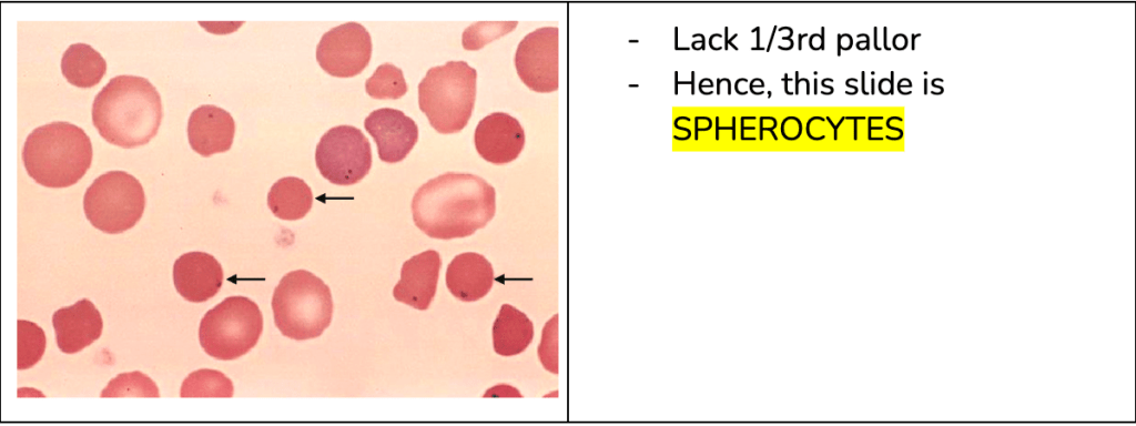
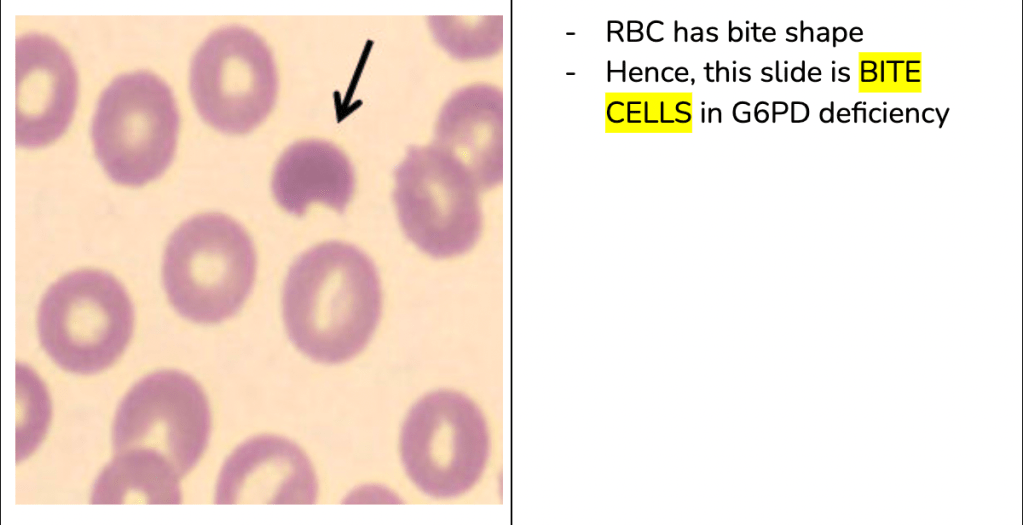
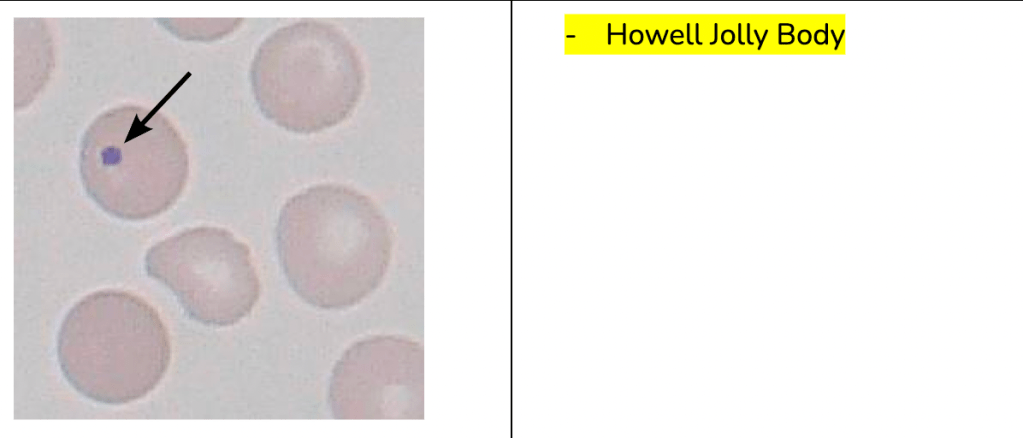
Bruxism, which is characterized by the repetitive clenching or grinding of teeth, is a common phenomenon that can have negative consequences on oral health and overall well-being (Yap & Chua, 2016). It is important to manage bruxism to prevent dental problems such as tooth wear, fractures of dental restorations, and pain in the oro-facial region (Koyano et al., 2008). The management strategies for bruxism mainly focus on reducing the potential negative consequences and controlling the symptoms associated with bruxism (Gouw et al., 2018).

One approach to managing bruxism is through the use of occlusal splints or oral appliances. Occlusal splints are commonly used for the diagnosis and treatment of bruxism, and they work by providing a protective barrier between the upper and lower teeth, reducing the impact of grinding and clenching (Ali et al., 2023). These splints can be effective in preventing tooth wear and reducing muscle pain and headaches associated with bruxism (Raby et al., 2018). However, it is important to note that occlusal splints do not eliminate bruxism, but rather serve as a means of managing its consequences (Raby et al., 2018).

Another management strategy for bruxism is the use of botulinum toxin injections into the masseter muscles. This treatment temporarily reduces the frequency of bruxism events and can provide relief from symptoms such as muscle pain and headaches (Serrera-Figallo et al., 2020). However, it is important to note that the current treatment modalities for bruxism are not effective and feasible for most patients with sleep bruxism (Gouw et al., 2018). Therefore, a multimodal approach that combines different treatment modalities may be recommended for managing bruxism (Gouw et al., 2018).

In addition to these treatment modalities, it is important to consider the underlying causes and contributing factors of bruxism. Bruxism is believed to be regulated centrally, with pathophysiological and psychosocial factors playing a role in its development (Yap & Chua, 2016). Stress sensitivity and anxious personality traits have been identified as potential factors that may contribute to bruxism activities and temporomandibular pain (Manfredini et al., 2017). Therefore, addressing these factors through stress management techniques, relaxation training, and behavioral therapy may be beneficial in managing bruxism (Kumar et al., 2022).

Furthermore, the management of bruxism should also take into consideration the potential impact on dental restorations and implants. Bruxism is considered a contraindication for dental implants, as it may cause overload and failure of the implants (Lobbezoo et al., 2006). Therefore, careful consideration should be given to the use of dental implants in patients with bruxism, and protective measures such as occlusal guards may be recommended to minimize the risk of implant failure (Yang et al., 2022).

It is worth noting that the management of bruxism should be tailored to the individual patient, taking into account their specific needs and circumstances. The use of observational and non-interventional management strategies may be appropriate for younger children, as the majority of bruxist children do not continue to brux during adolescence and adulthood (Manfredini et al., 2013). On the other hand, adults with bruxism may require more comprehensive management strategies to address the consequences of bruxism and alleviate symptoms (Manfredini et al., 2019).

In conclusion, the management of bruxism involves a combination of strategies aimed at reducing the negative consequences of bruxism and controlling its symptoms. These strategies may include the use of occlusal splints, botulinum toxin injections, stress management techniques, and behavioral therapy. It is important to tailor the management approach to the individual patient and consider the potential impact on dental restorations and implants. Further research is needed to better understand the underlying causes of bruxism and develop more effective treatment modalities.

References:

Ali, F., Alsheri, M., Shami, S., Mohana, A., Abujamilah, E., Alshehri, F. (2023). A Case Report Of Bruxism and Its Management With The Help Of Occlusal Splints.. Int J Life Sci Pharm Res. https://doi.org/10.22376/ijlpr.2023.13.2.l27-l30 Ali, S., Alqutaibi, A., Aboalrejal, A., Elawady, D. (2021). Botulinum Toxin and Occlusal Splints For The Management Of Sleep Bruxism In Individuals With Implant Overdentures: A Randomized Controlled Trial. The Saudi Dental Journal, 8(33), 1004-1011. https://doi.org/10.1016/j.sdentj.2021.07.001 Gouw, S., Wijer, A., Kalaykova, S., Creugers, N. (2018). Masticatory Muscle Stretching For the Management Of Sleep Bruxism: A Randomised Controlled Trial. J Oral Rehabil, 10(45), 770-776. https://doi.org/10.1111/joor.12694 Koyano, K., Tsukiyama, Y., Ichiki, R., T, K. (2008). Assessment Of Bruxism In the Clinic. J Oral Rehabil, 7(35), 495-508. https://doi.org/10.1111/j.1365-2842.2008.01880.x Kumar, A., Nair, A., Faizal, F., S, S., Prasad, M. (2022). Diagnosis and Management Of Sleep Bruxism. JPID. https://doi.org/10.55231/jpid.2022.v05.i02.04 Lobbezoo, F., Brouwers, J., Cune, M., Naeije, M. (2006). Dental Implants In Patients With Bruxing Habits. J Oral Rehabil, 2(33), 152-159. https://doi.org/10.1111/j.1365-2842.2006.01542.x Manfredini, D., Ahlberg, J., Winocur, E., Lobbezoo, F. (2015). Management Of Sleep Bruxism In Adults: a Qualitative Systematic Literature Review. J Oral Rehabil, 11(42), 862-874. https://doi.org/10.1111/joor.12322 Manfredini, D., Colonna, A., Bracci, A., Lobbezoo, F. (2019). Bruxism: a Summary Of Current Knowledge On Aetiology, Assessment And Management. Oral Surg, 4(13), 358-370. https://doi.org/10.1111/ors.12454 Manfredini, D., Restrepo, C., Díaz-Serrano, K., Winocur, E., Lobbezoo, F. (2013). Prevalence Of Sleep Bruxism In Children: a Systematic Review Of The Literature. J Oral Rehabil, 8(40), 631-642. https://doi.org/10.1111/joor.12069 Manfredini, D., Serra-Negra, J., Carboncini, F., Lobbezoo, F. (2017). Current Concepts Of Bruxism. Int J Prosthodont, 5(30), 437-438. https://doi.org/10.11607/ijp.5210 Minervini, G., Fiorillo, L., Russo, D., Lanza, A., D’Amico, C., Cervino, G., … & Francesco, F. (2022). Prosthodontic Treatment In Patients With Temporomandibular Disorders and Orofacial Pain And/or Bruxism: A Review Of The Literature. Prosthesis, 2(4), 253-262. https://doi.org/10.3390/prosthesis4020025 Raby, I., Quiroz, D., Galleguillos, P. (2018). Freely Available or Over-the-counter Occlusal Splints Obtainable In Commercial Outlets: A Reality Dentists Should Know. J Oral Res, 7(7), 219-226. https://doi.org/10.17126/joralres.2018.063 Serrera-Figallo, M., Ruiz-de-León-Hernández, G., Torres-Lagares, D., Castro-Araya, A., Torres-Ferrerosa, O., Hernández-Pacheco, E., … & Gutiérrez-Pérez, J. (2020). Use Of Botulinum Toxin In Orofacial Clinical Practice. Toxins, 2(12), 112. https://doi.org/10.3390/toxins12020112 Sriharsha, P., Gujjari, A., Dhakshaini, M., Prashant, A. (2018). Comparative Evaluation Of Salivary Cortisol Levels In Bruxism Patients Before and After Using Soft Occlusal Splint: An In Vivo Study. Contemp Clin Dent, 2(9), 182. https://doi.org/10.4103/ccd.ccd_756_17 Yang, J., Siow, L., Zhang, X., Wang, Y., Wang, H., Wang, B. (2022). Dental Reimplantation Treatment and Clinical Care For Patients With Previous Implant Failure—a Retrospective Study. IJERPH, 23(19), 15939. https://doi.org/10.3390/ijerph192315939 Yap, A., Chua, A. (2016). Sleep Bruxism: Current Knowledge and Contemporary Management. J Conserv Dent, 5(19), 383. https://doi.org/10.4103/0972-0707.190007

Diagnosis Demystified – Case 19

A 62-year-old woman gives a history of a sharp pain lasting for a few seconds whenever she touches the skin over her lower jaw. It is making it difficult for her to carry out daily activities like washing her face or eating. The pain is confined to the left-hand side. Once an episode of pain is complete she can be pain free for about an hour, even if she touches her face in the area affected by the pain. She has been taking paracetamol at regular intervals but this has made no difference to the pain.

The patient gives a classic description including the distribution of pain, trigger zone and a refractory period after stimulation. While Trigeminal neuralgia (classical trigeminal neuralgia; CTN) is highly likely, MRI is obligatory to exclude STN and assess the possibility of DREZ compression.

Trigeminal neuralgia is a disorder characterized by paroxysms of high-intensity facial pain in the distribution of the fifth cranial nerve (Katusic et al., 1990). The treatment options for trigeminal neuralgia include both medical and surgical approaches. Carbamazepine and oxcarbazepine are the first-line pharmacological treatments for trigeminal neuralgia (Szok et al., 2019; Stefano et al., 2021). However, if these drugs fail to provide sufficient pain relief or are poorly tolerated, other medications such as lamotrigine, baclofen, gabapentin, pregabalin, and botulinum toxin type A may be considered (Stefano et al., 2021). Surgical options include microvascular decompression (MVD), which involves relieving vascular compression of the trigeminal nerve (Broggi et al., 2000). Stereotactic radiosurgery (SRS) can also be used for tumor-related trigeminal neuralgia (Kano et al., 2010). In cases where medical therapies and surgical procedures are ineffective, neuromodulation techniques may be considered (Chung & Huh, 2022). It is important to note that the choice of treatment depends on the individual patient’s condition and response to previous treatments (Perszke et al., 2022).

References:

Broggi, G., Ferroli, P., Franzini, A., Servello, D., Dones, I. (2000). Microvascular Decompression For Trigeminal Neuralgia: Comments On a Series Of 250 Cases, Including 10 Patients With Multiple Sclerosis. Journal of Neurology Neurosurgery & Psychiatry, 1(68), 59-64. https://doi.org/10.1136/jnnp.68.1.59 Chung, M., Huh, R. (2022). Neuromodulation For Trigeminal Neuralgia. J Korean Neurosurg Soc, 5(65), 640-651. https://doi.org/10.3340/jkns.2022.0004 Kano, H., Awan, N., Flannery, T., Iyer, A., Flickinger, J., Lunsford, L., … & Kondziolka, D. (2010). Stereotactic Radiosurgery For Patients With Trigeminal Neuralgia Associated With Petroclival Meningiomas. Stereotact Funct Neurosurg, 1(89), 17-24. https://doi.org/10.1159/000321187 Katusic, S., Beard, C., Bergstralh, E., Kurland, L. (1990). Incidence and Clinical Features Of Trigeminal Neuralgia, Rochester, Minnesota, 1945-1984. Ann Neurol., 1(27), 89-95. https://doi.org/10.1002/ana.410270114 Perszke, M., Egierska, D., Martynowicz, P. (2022). Trigeminal Neuralgia – Where Are We Today?. J Educ Health Sport, 9(12), 113-127. https://doi.org/10.12775/jehs.2022.12.09.015 Stefano, G., Nurmikko, T., Zakrzewska, J. (2021). Treatment Of Trigeminal Neuralgia: Pharmacological., 89-104. https://doi.org/10.1093/med/9780198871606.003.0009 Szok, D., Tajti, J., Nyári, A., Vécsei, L. (2019). Therapeutic Approaches For Peripheral and Central Neuropathic Pain. BN, (2019), 1-13. https://doi.org/10.1155/2019/8685954

    Injectable Platelet Rich Fibrin in Periodontal Regeneration

    Revolutionizing Periodontal Regeneration: The Power of Platelet-Rich Fibrin (PRF)”

    Platelet-rich fibrin (PRF) is an autologous platelet concentrate that has been studied for its potential in improving the effect of periodontal regeneration (Chen et al., 2021). PRF is a type of platelet concentrate that is easy to obtain and cost-effective (Chen et al., 2021). It has been validated as an entirely autologous, injectable cell delivery system that overcomes histocompatibility issues related to synthetic scaffolds (Chen & Liu, 2016). PRF is non-cytotoxic, biocompatible, and non-immunogenic, making it suitable for use in tissue engineering (Chen & Liu, 2016). It has been characterized morphologically, in terms of cell content and protein composition, to better understand its clinical effects and improve clinical guidelines for various medical applications (Varela et al., 2018).

    PRF has been investigated for its antimicrobial efficacy in the treatment of periodontal soft- and hard-tissue regeneration (Kour et al., 2018). It has been compared to other platelet concentrates, such as platelet-rich plasma (PRP) and plasma rich in growth factors (PRGF), and has shown similar effectiveness in periodontal bone regeneration (Lei et al., 2019). The use of PRF in combination with other agents, such as 1% alendronate, has been proposed to enhance bone formation and reduce bone resorption in regenerative periodontal treatment (Li et al., 2019). However, there is a lack of evidence-based studies to determine the superiority of this concurrent application (Li et al., 2019).

    PRF has been shown to promote craniofacial bone regeneration through the activation of the Runx2 pathway (Li et al., 2014). It is a second-generation platelet concentrate that is prepared from centrifuged blood and is strictly autologous (Li et al., 2014). PRF has been investigated as a wound healing promoter in various clinical applications, including periodontal regeneration (Tavelli et al., 2022). It is one of the autologous platelet concentrates that are generated after the centrifugation of the patient’s blood to obtain fractions containing a supraphysiologic concentration of platelets and growth factors (Tavelli et al., 2022).

    Injectable platelet-rich fibrin (I-PRF) is a liquid autologous platelet concentrate that has been introduced as a low-cost alternative to PRF (Alshoiby et al., 2023). It has been used in combination with demineralized freeze-dried bone allograft (DFDBA) in the treatment of intrabony defects in patients with stage-III periodontitis (Alshoiby et al., 2023). I-PRF contains growth/differentiation factors, including bone morphogenetic proteins (BMPs), which promote periodontal repair and regeneration (Alshoiby et al., 2023).

    PRF has been studied in the context of dental tissue engineering, where it has shown potential for use in tooth rejuvenation and the enhancement of osteogenic differentiation (Zhao & Gao, 2023). It has been injected into the root canal of necrotic teeth and has been found to promote dentinal wall thickening, root extension, reduction of periapical lesions, and apical closure (Zhao & Gao, 2023). Additionally, PRF has been used in combination with collagen chitosan hydrogel to promote alkaline phosphatase activity and calcium deposition in osteoblast-like cell lines (Sidharta et al., 2023).

    The efficacy of PRF in periodontal regeneration has been evaluated in various clinical trials and systematic reviews. A systematic review and meta-analysis assessed the use of PRF in the treatment of periodontal intrabony defects and found positive effects on clinical and radiological outcomes (Chen et al., 2021). Another systematic review and meta-analysis evaluated the clinical efficacy of PRF in periodontal regeneration and found significant improvements in probing pocket depth and clinical attachment level (Oza et al., 2023). A randomized controlled clinical trial compared the use of injectable PRF with demineralized freeze-dried bone allograft to demineralized freeze-dried bone allograft alone in intrabony defects and found significant improvements in pocket probing depth, clinical attachment level, and bone fill with the combination treatment (Alshoiby et al., 2023).

    In conclusion, PRF is an autologous platelet concentrate that has been studied for its potential in periodontal regeneration. It has been characterized morphologically, in terms of cell content and protein composition, and has been shown to be non-cytotoxic, biocompatible, and non-immunogenic. PRF has been compared to other platelet concentrates and has shown similar effectiveness in periodontal bone regeneration. It has been used in combination with other agents, such as 1% alendronate and demineralized freeze-dried bone allograft, to enhance bone formation and reduce bone resorption. PRF has been investigated in various clinical trials and systematic reviews, which have demonstrated its positive effects on clinical and radiological outcomes in periodontal regeneration.

    References:

    Alshoiby, M., El-Sayed, K., Elbattawy, W., Hosny, M. (2023). Injectable Platelet-rich Fibrin With Demineralized Freeze-dried Bone Allograft Compared To Demineralized Freeze-dried Bone Allograft In Intrabony Defects Of Patients With Stage-iii Periodontitis: a Randomized Controlled Clinical Trial. Clin Oral Invest, 7(27), 3457-3467. https://doi.org/10.1007/s00784-023-04954-y Chen, F., Liu, X. (2016). Advancing Biomaterials Of Human Origin For Tissue Engineering. Progress in Polymer Science, (53), 86-168. https://doi.org/10.1016/j.progpolymsci.2015.02.004 Chen, L., Ding, Y., Wang, W., Meng, S. (2021). Use Of Platelet-rich Fibrin In the Treatment Of Periodontal Intrabony Defects: A Systematic Review And Meta-analysis. BioMed Research International, (2021), 1-13. https://doi.org/10.1155/2021/6669168 Kour, P., Pudakalkatti, P., Vas, A., Das, S., Padmanabhan, S. (2018). Comparative Evaluation Of Antimicrobial Efficacy Of Platelet-rich Plasma, Platelet-rich Fibrin, and Injectable Platelet-rich Fibrin On The Standard Strains Of Porphyromonas Gingivalis And Aggregatibacter Actinomycetemcomitans. Contemp Clin Dent, 6(9), 325. https://doi.org/10.4103/ccd.ccd_367_18 Lei, L., Yu, Y., Han, J., Shi, D., Sun, W., Zhang, D., … & Chen, L. (2019). Quantification Of Growth Factors In Advanced Platelet‐rich Fibrin and Concentrated Growth Factors And Their Clinical Efficacy As Adjunctive To The Gtr Procedure In Periodontal Intrabony Defects. J Periodontol, 4(91), 462-472. https://doi.org/10.1002/jper.19-0290 Li, F., Jiang, P., Pan, J., Liu, C., Zheng, L. (2019). Synergistic Application Of Platelet-rich Fibrin and 1% Alendronate In Periodontal Bone Regeneration: A Meta-analysis. BioMed Research International, (2019), 1-12. https://doi.org/10.1155/2019/9148183 Li, Q., Reed, D., Min, L., Gopinathan, G., Li, S., Dangaria, S., … & Diekwisch, T. (2014). Lyophilized Platelet-rich Fibrin (Prf) Promotes Craniofacial Bone Regeneration Through Runx2. IJMS, 5(15), 8509-8525. https://doi.org/10.3390/ijms15058509 Oza, D., Dhadse, D., Bajaj, D., Bhombe, D., Durge, D., Subhadarsanee, D., … & Hassan, D. (2023). Clinical Efficacy Of Titanium Prepared Platelet Rich Fibrin In Periodontal Regeneration: a Systematic Review And Meta-analysis. F1000Res, (12), 393. https://doi.org/10.12688/f1000research.131461.1 Sidharta, K., Suryono, -., Murdiastuti, K., Pritia, M. (2023). Effect Of Collagen Chitosan Hydrogel With Injectable Platelet-rich Fibrin On Alkaline Phosphatase Activity and Calcium Deposition An In Vitro Study On Osteoblast-like Cell Line Mg63.. https://doi.org/10.21203/rs.3.rs-2948824/v1 Tavelli, L., Chen, C., Barootchi, S., Kim, D. (2022). Efficacy Of Biologics For the Treatment Of Periodontal Infrabony Defects: An American Academy Of Periodontology Best Evidence Systematic Review And Network Meta‐analysis. Journal of Periodontology, 12(93), 1803-1826. https://doi.org/10.1002/jper.22-0120 Varela, H., Souza, J., Nascimento, R., Júnior, R., Vasconcelos, R., Cavalcante, R., … & Araújo, A. (2018). Injectable Platelet Rich Fibrin: Cell Content, Morphological, and Protein Characterization. Clin Oral Invest, 3(23), 1309-1318. https://doi.org/10.1007/s00784-018-2555-2 Zhao, Z., Gao, L. (2023). Stem Cells, Scaffolds, and Growth Factors In Dental Tissue Engineering.. https://doi.org/10.1117/12.2673569

    Diagnosis Demystified – Case 18

    A 58-year-old man gave a history of stabbing pain affecting his left temple. Each burst of pain lasts for about 4 minutes and he can experience three of these each waking hour. He is concerned that he may have developed an infection in his left eye, which looks red.

    On first reading, this case has many of the features of trigeminal neuralgia. The 4-minute duration of the pain is possibly longer than one might normally expect for TN and the ophthalmic is the least commonly affected division of the trigeminal nerve. The presence of conjunctival infection shifts the balance in favour of short-lasting unilateral neuralgiform headache attacks with conjunctival injection and tearing but an overlap is recognised between the two conditions. MRI indicated.

    RESEARCH ARTICLES

    Short-lasting unilateral neuralgiform headache attacks (SUNHA) is a rare form of primary headache that falls under the category of trigeminal autonomic cephalalgias (TACs) (Weng et al., 2017). SUNHA is further divided into two subtypes: short-lasting unilateral neuralgiform headache attacks with conjunctival injection and tearing (SUNCT) and short-lasting unilateral neuralgiform headache attacks with cranial autonomic symptoms (SUNA) (Levy & Matharu, 2018). These headache disorders are characterized by severe, short-lasting headaches that occur unilaterally and are accompanied by ipsilateral cranial autonomic symptoms (Goadsby et al., 2007).

    SUNCT is characterized by frequent attacks of unilateral, orbital, supraorbital, or temporal stabbing or pulsating pain that lasts for 5 to 240 seconds (Leone et al., 2005). These attacks are often accompanied by conjunctival injection, tearing, and blocked or runny nostril, all on the same side as the pain (Leone et al., 2005). However, it is important to note that not all patients with SUNCT manifest both conjunctival injection and tearing (Weng et al., 2017).

    SUNA, on the other hand, is characterized by similar short-lasting unilateral headaches, but with additional cranial autonomic symptoms such as ptosis, miosis, eyelid edema, or nasal congestion (Weng et al., 2017). Like SUNCT, SUNA attacks are also frequent and can last from seconds to minutes (Weng et al., 2017).

    The exact cause of SUNHA is not fully understood, but there is evidence suggesting hypothalamic involvement in these conditions (Cohen, 2007). Functional imaging studies have shown activation of the posterior hypothalamus in patients with TACs, including SUNHA (Cohen, 2007). Additionally, deep brain stimulation of the hypothalamus has been used as a treatment option for these headache disorders (Cohen, 2007).

    The diagnosis of SUNHA is based on clinical presentation and the exclusion of other possible causes of headache (Goadsby et al., 2007). The International Classification of Headache Disorders (ICHD) criteria are commonly used for diagnosis (Qaiser et al., 2020). However, diagnosing SUNHA in pediatric patients can be challenging due to limited data and the overlap of symptoms with other headache disorders (Ghosh et al., 2021).

    reatment options for SUNHA include both pharmacotherapy and procedural interventions (Pomeroy & Nahas, 2015). Pharmacological preventive treatments have been tried, but there is no specific medication that is universally effective for all patients (Levy & Matharu, 2018). Some commonly used medications include indomethacin, lamotrigine, topiramate, and gabapentin (Wei & Jensen, 2018). In cases where pharmacotherapy is ineffective, procedural interventions such as occipital nerve stimulation or deep brain stimulation may be considered (Miller et al., 2017).

    In conclusion, short-lasting unilateral neuralgiform headache attacks (SUNHA) is a rare form of primary headache that falls under the category of trigeminal autonomic cephalalgias (TACs). It is characterized by severe, short-lasting headaches that occur unilaterally and are accompanied by ipsilateral cranial autonomic symptoms. The exact cause of SUNHA is not fully understood, but there is evidence suggesting hypothalamic involvement. Diagnosis is based on clinical presentation and the exclusion of other possible causes of headache. Treatment options include pharmacotherapy and procedural interventions, but there is no universally effective medication for all patients. Further research is needed to better understand the pathophysiology and develop more effective treatments for SUNHA.

    References

    Cohen, A. (2007). Short-lasting Unilateral Neuralgiform Headache Attacks With Conjunctival Injection and Tearing. Cephalalgia, 7(27), 824-832. https://doi.org/10.1111/j.1468-2982.2007.01352.x Ghosh, A., Silva, E., Burish, M. (2021). Pediatric-onset Trigeminal Autonomic Cephalalgias: a Systematic Review And Meta-analysis. Cephalalgia, 13(41), 1382-1395. https://doi.org/10.1177/03331024211027560 Goadsby, P., Cohen, A., Matharu, M. (2007). Trigeminal Autonomic Cephalalgias: Diagnosis and Treatment. Curr Neurol Neurosci Rep, 2(7), 117-125. https://doi.org/10.1007/s11910-007-0006-6 Leone, M., Franzini, A., D’Andrea, G., Broggi, G., Casucci, G., Bussone, G. (2005). Deep Brain Stimulation To Relieve Drug-resistant Sunct. Ann Neurol., 6(57), 924-927. https://doi.org/10.1002/ana.20507 Levy, A., Matharu, M. (2018). Short-lasting Unilateral Neuralgiform Headache Attacks. Ann Indian Acad Neurol, 5(21), 31. https://doi.org/10.4103/aian.aian_356_17 Miller, S., Watkins, L., Matharu, M. (2017). Long-term Follow Up Of Intractable Chronic Short Lasting Unilateral Neuralgiform Headache Disorders Treated With Occipital Nerve Stimulation. Cephalalgia, 5(38), 933-942. https://doi.org/10.1177/0333102417721716 Pomeroy, J., Nahas, S. (2015). Sunct/suna: a Review. Curr Pain Headache Rep, 8(19). https://doi.org/10.1007/s11916-015-0511-2 Qaiser, S., Hershey, A., Kacperski, J. (2020). Sunct/suna In Children and Adolescents: Application Of Ichd-3 Criteria And Treatment Response: Case Series Of 13 Sunct/suna Pediatric Cases. Cephalalgia, 1(41), 112-116. https://doi.org/10.1177/0333102420954525 Wei, D., Jensen, R. (2018). Therapeutic Approaches For the Management Of Trigeminal Autonomic Cephalalgias. Neurotherapeutics, 2(15), 346-360. https://doi.org/10.1007/s13311-018-0618-3 Weng, H., Cohen, A., Schankin, C., Goadsby, P. (2017). Phenotypic and Treatment Outcome Data On Sunct And Suna, Including A Randomised Placebo-controlled Trial. Cephalalgia, 9(38), 1554-1563. https://doi.org/10.1177/0333102417739304

      Vitamin D deficiency as an etiological factor in delayed eruption of primary teeth

      Vitamin D deficiency has been implicated as an etiological factor in the delayed eruption of primary teeth. Several studies have shown that vitamin D deficiency can lead to delayed tooth eruption, incomplete calcification of dentin, and unclear lamina dura in primary and permanent teeth (Kim et al., 2018; Swapna & Abdulsalam, 2021). Vitamin D plays a crucial role in tooth and bone mineralization, and its deficiency can result in hypocalcified dentin and enamel hypoplasia (Swapna & Abdulsalam, 2021; Alshukairi, 2019). The formation of primary teeth is normal in individuals with vitamin D deficiency, but the eruption process is delayed (Jensen & Kreiborg, 1990; Kim et al., 2018).

      Delayed eruption of primary teeth can also be caused by other factors such as mechanical obstruction from supernumerary teeth (Pan et al., 2017). Cleidocranial dysplasia (CCD) is a condition characterized by delayed eruption of permanent teeth and the presence of supernumerary teeth (Jensen & Kreiborg, 1990; Pan et al., 2017). In a study of patients with CCD, it was found that all patients except one had supernumerary permanent teeth, which could contribute to the delayed eruption of primary teeth (Jensen & Kreiborg, 1990). However, it is important to note that delayed eruption of primary teeth is relatively rare compared to permanent teeth (Matsuyama et al., 2015).

      In addition to vitamin D deficiency, other systemic factors can also affect the eruption timing of primary teeth. Maternal factors such as smoke exposure during pregnancy, gestational age, and vitamin D levels have been found to possibly affect the eruption timing of the first deciduous tooth (Georgiadou et al., 2021). Malnutrition and growth stunting in children have been associated with delayed eruption of primary teeth (Fadilla et al., 2022; Setiawan et al., 2022). Chronic malnutrition can lead to hypoplasia and delayed eruption of primary teeth (Setiawan et al., 2022). Furthermore, maternal intake of vitamin D during pregnancy has been associated with the risk of childhood wheezing/asthma and the development of early childhood caries (Bener et al., 2011; Schroth et al., 2014).

      It is important to consider the role of vitamin D in tooth development and eruption, as well as the potential impact of other systemic and local factors. Further research is needed to fully understand the mechanisms underlying the relationship between vitamin D deficiency and delayed eruption of primary teeth. However, the available evidence suggests that maintaining adequate vitamin D levels and addressing other potential contributing factors may help prevent or mitigate delayed eruption of primary teeth.

      Diagnosis demystified – Case 17

      A 36-year-old woman gives a 2-week history of a sharp pain lasting for a few seconds which comes on when she touches the left-hand side of her upper lip. One burst of pain can follow immediately after the preceding one. When you are taking her history, she mentions that about a month ago she consulted her GMP about a deterioration in hearing affecting her left ear.

      The clinical features are strongly suggestive of Trigeminal Neuralgia. The patient’s age is a cause for concern as CTN would be extremely unusual for someone of her age. The absence of a refractory period after triggering the pain is also somewhat atypical. Therefore STN should be seriously considered. The presence of ipsilateral hearing loss raises further concerns about the possibility of a central lesion. Even if the patient were in the usual age group for CTN, this would raise the possibility of STN.

      Research papers on – Latest treatment options in trigeminal neuralgia

      Trigeminal neuralgia is a debilitating condition characterized by severe facial pain. The first-line treatment options for trigeminal neuralgia are pharmacological (Stefano et al., 2021). Carbamazepine and oxcarbazepine are the drugs of choice for managing trigeminal neuralgia (Szok et al., 2019; Stefano et al., 2021). These drugs are voltage-gated sodium channel blockers that stabilize hyperexcited neural membranes and inhibit repetitive firing (Stefano et al., 2021). However, they may cause sedative and cognitive side effects (Stefano et al., 2021). If the first-line drugs are ineffective or poorly tolerated, other medications such as lamotrigine, baclofen, gabapentin, pregabalin, and botulinum toxin type A can be considered (Stefano et al., 2021). Surgical interventions, such as microvascular decompression, gamma knife radiosurgery, and percutaneous rhizotomies, are available for patients who do not respond to medications or cannot tolerate them (Al-Quliti, 2015). Radiofrequency ablation is a minimally invasive procedure that has shown promising results in managing trigeminal neuralgia pain (Usman & Herbiyanto, 2022). It is important to note that treatment decisions should be based on individual patient characteristics and preferences (Stefano et al., 2021).

      References:

      Al-Quliti, K. (2015). Update On Neuropathic Pain Treatment For Trigeminal Neuralgia. NSJ, 2(20), 107-114. https://doi.org/10.17712/nsj.2015.2.20140501 Stefano, G., Nurmikko, T., Zakrzewska, J. (2021). Treatment Of Trigeminal Neuralgia: Pharmacological., 89-104. https://doi.org/10.1093/med/9780198871606.003.0009 Szok, D., Tajti, J., Nyári, A., Vécsei, L. (2019). Therapeutic Approaches For Peripheral and Central Neuropathic Pain. BN, (2019), 1-13. https://doi.org/10.1155/2019/8685954 Usman, N., Herbiyanto, J. (2022). Radiofrequency Ablation For Trigeminal Neuralgia Patient: a Case Report. JACR, 2(3), 342-348. https://doi.org/10.37275/jacr.v3i2.256