Diagnosis Demystified – Case 18

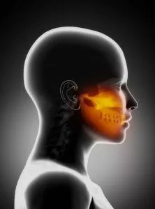
A 58-year-old man gave a history of stabbing pain affecting his left temple. Each burst of pain lasts for about 4 minutes and he can experience three of these each waking hour. He is concerned that he may have developed an infection in his left eye, which looks red.

On first reading, this case has many of the features of trigeminal neuralgia. The 4-minute duration of the pain is possibly longer than one might normally expect for TN and the ophthalmic is the least commonly affected division of the trigeminal nerve. The presence of conjunctival infection shifts the balance in favour of short-lasting unilateral neuralgiform headache attacks with conjunctival injection and tearing but an overlap is recognised between the two conditions. MRI indicated.

RESEARCH ARTICLES

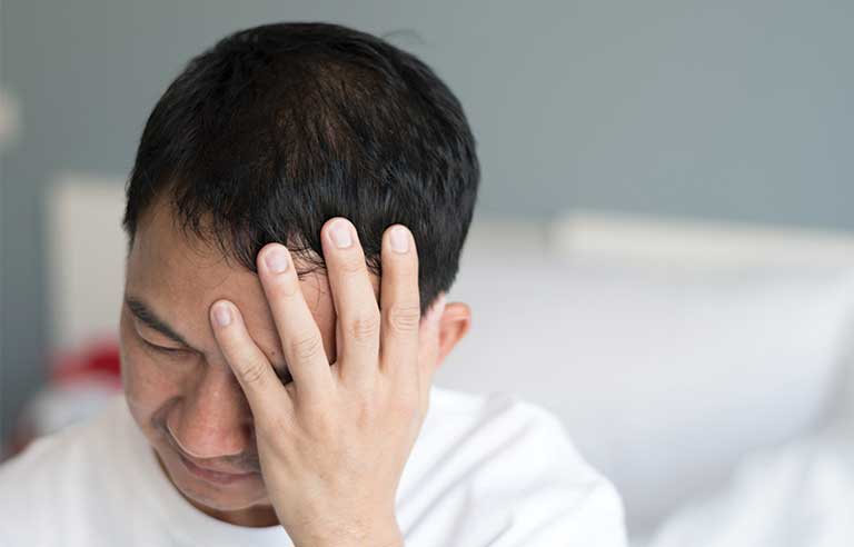
Short-lasting unilateral neuralgiform headache attacks (SUNHA) is a rare form of primary headache that falls under the category of trigeminal autonomic cephalalgias (TACs) (Weng et al., 2017). SUNHA is further divided into two subtypes: short-lasting unilateral neuralgiform headache attacks with conjunctival injection and tearing (SUNCT) and short-lasting unilateral neuralgiform headache attacks with cranial autonomic symptoms (SUNA) (Levy & Matharu, 2018). These headache disorders are characterized by severe, short-lasting headaches that occur unilaterally and are accompanied by ipsilateral cranial autonomic symptoms (Goadsby et al., 2007).

SUNCT is characterized by frequent attacks of unilateral, orbital, supraorbital, or temporal stabbing or pulsating pain that lasts for 5 to 240 seconds (Leone et al., 2005). These attacks are often accompanied by conjunctival injection, tearing, and blocked or runny nostril, all on the same side as the pain (Leone et al., 2005). However, it is important to note that not all patients with SUNCT manifest both conjunctival injection and tearing (Weng et al., 2017).

SUNA, on the other hand, is characterized by similar short-lasting unilateral headaches, but with additional cranial autonomic symptoms such as ptosis, miosis, eyelid edema, or nasal congestion (Weng et al., 2017). Like SUNCT, SUNA attacks are also frequent and can last from seconds to minutes (Weng et al., 2017).

The exact cause of SUNHA is not fully understood, but there is evidence suggesting hypothalamic involvement in these conditions (Cohen, 2007). Functional imaging studies have shown activation of the posterior hypothalamus in patients with TACs, including SUNHA (Cohen, 2007). Additionally, deep brain stimulation of the hypothalamus has been used as a treatment option for these headache disorders (Cohen, 2007).

The diagnosis of SUNHA is based on clinical presentation and the exclusion of other possible causes of headache (Goadsby et al., 2007). The International Classification of Headache Disorders (ICHD) criteria are commonly used for diagnosis (Qaiser et al., 2020). However, diagnosing SUNHA in pediatric patients can be challenging due to limited data and the overlap of symptoms with other headache disorders (Ghosh et al., 2021).

reatment options for SUNHA include both pharmacotherapy and procedural interventions (Pomeroy & Nahas, 2015). Pharmacological preventive treatments have been tried, but there is no specific medication that is universally effective for all patients (Levy & Matharu, 2018). Some commonly used medications include indomethacin, lamotrigine, topiramate, and gabapentin (Wei & Jensen, 2018). In cases where pharmacotherapy is ineffective, procedural interventions such as occipital nerve stimulation or deep brain stimulation may be considered (Miller et al., 2017).

In conclusion, short-lasting unilateral neuralgiform headache attacks (SUNHA) is a rare form of primary headache that falls under the category of trigeminal autonomic cephalalgias (TACs). It is characterized by severe, short-lasting headaches that occur unilaterally and are accompanied by ipsilateral cranial autonomic symptoms. The exact cause of SUNHA is not fully understood, but there is evidence suggesting hypothalamic involvement. Diagnosis is based on clinical presentation and the exclusion of other possible causes of headache. Treatment options include pharmacotherapy and procedural interventions, but there is no universally effective medication for all patients. Further research is needed to better understand the pathophysiology and develop more effective treatments for SUNHA.

References

Cohen, A. (2007). Short-lasting Unilateral Neuralgiform Headache Attacks With Conjunctival Injection and Tearing. Cephalalgia, 7(27), 824-832. https://doi.org/10.1111/j.1468-2982.2007.01352.x Ghosh, A., Silva, E., Burish, M. (2021). Pediatric-onset Trigeminal Autonomic Cephalalgias: a Systematic Review And Meta-analysis. Cephalalgia, 13(41), 1382-1395. https://doi.org/10.1177/03331024211027560 Goadsby, P., Cohen, A., Matharu, M. (2007). Trigeminal Autonomic Cephalalgias: Diagnosis and Treatment. Curr Neurol Neurosci Rep, 2(7), 117-125. https://doi.org/10.1007/s11910-007-0006-6 Leone, M., Franzini, A., D’Andrea, G., Broggi, G., Casucci, G., Bussone, G. (2005). Deep Brain Stimulation To Relieve Drug-resistant Sunct. Ann Neurol., 6(57), 924-927. https://doi.org/10.1002/ana.20507 Levy, A., Matharu, M. (2018). Short-lasting Unilateral Neuralgiform Headache Attacks. Ann Indian Acad Neurol, 5(21), 31. https://doi.org/10.4103/aian.aian_356_17 Miller, S., Watkins, L., Matharu, M. (2017). Long-term Follow Up Of Intractable Chronic Short Lasting Unilateral Neuralgiform Headache Disorders Treated With Occipital Nerve Stimulation. Cephalalgia, 5(38), 933-942. https://doi.org/10.1177/0333102417721716 Pomeroy, J., Nahas, S. (2015). Sunct/suna: a Review. Curr Pain Headache Rep, 8(19). https://doi.org/10.1007/s11916-015-0511-2 Qaiser, S., Hershey, A., Kacperski, J. (2020). Sunct/suna In Children and Adolescents: Application Of Ichd-3 Criteria And Treatment Response: Case Series Of 13 Sunct/suna Pediatric Cases. Cephalalgia, 1(41), 112-116. https://doi.org/10.1177/0333102420954525 Wei, D., Jensen, R. (2018). Therapeutic Approaches For the Management Of Trigeminal Autonomic Cephalalgias. Neurotherapeutics, 2(15), 346-360. https://doi.org/10.1007/s13311-018-0618-3 Weng, H., Cohen, A., Schankin, C., Goadsby, P. (2017). Phenotypic and Treatment Outcome Data On Sunct And Suna, Including A Randomised Placebo-controlled Trial. Cephalalgia, 9(38), 1554-1563. https://doi.org/10.1177/0333102417739304

    Vitamin D deficiency as an etiological factor in delayed eruption of primary teeth

    Vitamin D deficiency has been implicated as an etiological factor in the delayed eruption of primary teeth. Several studies have shown that vitamin D deficiency can lead to delayed tooth eruption, incomplete calcification of dentin, and unclear lamina dura in primary and permanent teeth (Kim et al., 2018; Swapna & Abdulsalam, 2021). Vitamin D plays a crucial role in tooth and bone mineralization, and its deficiency can result in hypocalcified dentin and enamel hypoplasia (Swapna & Abdulsalam, 2021; Alshukairi, 2019). The formation of primary teeth is normal in individuals with vitamin D deficiency, but the eruption process is delayed (Jensen & Kreiborg, 1990; Kim et al., 2018).

    Delayed eruption of primary teeth can also be caused by other factors such as mechanical obstruction from supernumerary teeth (Pan et al., 2017). Cleidocranial dysplasia (CCD) is a condition characterized by delayed eruption of permanent teeth and the presence of supernumerary teeth (Jensen & Kreiborg, 1990; Pan et al., 2017). In a study of patients with CCD, it was found that all patients except one had supernumerary permanent teeth, which could contribute to the delayed eruption of primary teeth (Jensen & Kreiborg, 1990). However, it is important to note that delayed eruption of primary teeth is relatively rare compared to permanent teeth (Matsuyama et al., 2015).

    In addition to vitamin D deficiency, other systemic factors can also affect the eruption timing of primary teeth. Maternal factors such as smoke exposure during pregnancy, gestational age, and vitamin D levels have been found to possibly affect the eruption timing of the first deciduous tooth (Georgiadou et al., 2021). Malnutrition and growth stunting in children have been associated with delayed eruption of primary teeth (Fadilla et al., 2022; Setiawan et al., 2022). Chronic malnutrition can lead to hypoplasia and delayed eruption of primary teeth (Setiawan et al., 2022). Furthermore, maternal intake of vitamin D during pregnancy has been associated with the risk of childhood wheezing/asthma and the development of early childhood caries (Bener et al., 2011; Schroth et al., 2014).

    It is important to consider the role of vitamin D in tooth development and eruption, as well as the potential impact of other systemic and local factors. Further research is needed to fully understand the mechanisms underlying the relationship between vitamin D deficiency and delayed eruption of primary teeth. However, the available evidence suggests that maintaining adequate vitamin D levels and addressing other potential contributing factors may help prevent or mitigate delayed eruption of primary teeth.

    Diagnosis demystified – Case 17

    A 36-year-old woman gives a 2-week history of a sharp pain lasting for a few seconds which comes on when she touches the left-hand side of her upper lip. One burst of pain can follow immediately after the preceding one. When you are taking her history, she mentions that about a month ago she consulted her GMP about a deterioration in hearing affecting her left ear.

    The clinical features are strongly suggestive of Trigeminal Neuralgia. The patient’s age is a cause for concern as CTN would be extremely unusual for someone of her age. The absence of a refractory period after triggering the pain is also somewhat atypical. Therefore STN should be seriously considered. The presence of ipsilateral hearing loss raises further concerns about the possibility of a central lesion. Even if the patient were in the usual age group for CTN, this would raise the possibility of STN.

    Research papers on – Latest treatment options in trigeminal neuralgia

    Trigeminal neuralgia is a debilitating condition characterized by severe facial pain. The first-line treatment options for trigeminal neuralgia are pharmacological (Stefano et al., 2021). Carbamazepine and oxcarbazepine are the drugs of choice for managing trigeminal neuralgia (Szok et al., 2019; Stefano et al., 2021). These drugs are voltage-gated sodium channel blockers that stabilize hyperexcited neural membranes and inhibit repetitive firing (Stefano et al., 2021). However, they may cause sedative and cognitive side effects (Stefano et al., 2021). If the first-line drugs are ineffective or poorly tolerated, other medications such as lamotrigine, baclofen, gabapentin, pregabalin, and botulinum toxin type A can be considered (Stefano et al., 2021). Surgical interventions, such as microvascular decompression, gamma knife radiosurgery, and percutaneous rhizotomies, are available for patients who do not respond to medications or cannot tolerate them (Al-Quliti, 2015). Radiofrequency ablation is a minimally invasive procedure that has shown promising results in managing trigeminal neuralgia pain (Usman & Herbiyanto, 2022). It is important to note that treatment decisions should be based on individual patient characteristics and preferences (Stefano et al., 2021).

    References:

    Al-Quliti, K. (2015). Update On Neuropathic Pain Treatment For Trigeminal Neuralgia. NSJ, 2(20), 107-114. https://doi.org/10.17712/nsj.2015.2.20140501 Stefano, G., Nurmikko, T., Zakrzewska, J. (2021). Treatment Of Trigeminal Neuralgia: Pharmacological., 89-104. https://doi.org/10.1093/med/9780198871606.003.0009 Szok, D., Tajti, J., Nyári, A., Vécsei, L. (2019). Therapeutic Approaches For Peripheral and Central Neuropathic Pain. BN, (2019), 1-13. https://doi.org/10.1155/2019/8685954 Usman, N., Herbiyanto, J. (2022). Radiofrequency Ablation For Trigeminal Neuralgia Patient: a Case Report. JACR, 2(3), 342-348. https://doi.org/10.37275/jacr.v3i2.256

    An In-Vitro Evaluation of Occlusal Fissure Morphology in Primary Molars

    The occlusal surfaces of molars, particularly the permanent first molars, are commonly affected by caries (Demirci et al., 2010). This is consistent with the findings of a study that reported over two-thirds of permanent first molars being affected by caries (Demirci et al., 2010). Additionally, the occlusal surfaces of permanent second molars also contribute significantly to caries development (Demirci et al., 2010). The high caries rates in the permanent dentition are primarily limited to the pit and fissure surfaces of molars (Demirci et al., 2010).

    The pattern of caries in primary molars, specifically the maxillary incisors and first molars, is thought to be related to the eruption times of primary teeth and the acquisition of cariogenic bacteria (Xu et al., 2014). Children who develop caries before 2.5 years of age typically exhibit decay on the smooth surfaces of maxillary incisors and occlusal fissures of the first molar teeth (Xu et al., 2014).

    The progression of caries lesions in primary molars is more frequent in second molars compared to first molars (Guedes et al., 2015). This is consistent with previous studies that have reported a higher probability of progression in occlusal surfaces, both in primary and permanent teeth (Guedes et al., 2015).

    Sealants are effective in preventing and arresting pit-and-fissure occlusal caries lesions in primary and permanent molars in children and adolescents (Prabakar et al., 2018). A systematic review conducted by the American Dental Association Council on Scientific Affairs and the American Academy of Pediatric Dentistry recommended the use of pit-and-fissure sealants on the occlusal surfaces of primary and permanent molars (Prabakar et al., 2018).

    The morphology of occlusal fissures in molars plays a significant role in caries prevention (Dindar & Atay, 2022). Understanding the types of fissure morphology is important for preventing caries on the occlusal surfaces of molars and premolars (Dindar & Atay, 2022). The presence of deep occlusal pits and fissures increases the risk of plaque accumulation and subsequent decay (Alqahtani et al., 2022).

    The retention of sealants on primary molars is generally lower compared to permanent molars (AL-Sultani et al., 2020). However, there is conflicting evidence regarding the effectiveness of sealants on primary molars compared to permanent molars (AL-Sultani et al., 2020). Some studies have reported that sealants are more effective in permanent molars, while others have found no significant difference in retention between primary and permanent molars (AL-Sultani et al., 2020).

    The morphology of the pulp chamber in primary molars can be evaluated using spiral computed tomography (SCT) (Selvakumar et al., 2014). SCT can provide information on the distance between the central fissure and furcation, the distance between the central fissure and the floor of the pulp chamber, the height of the pulp chamber, and the distance between the floor of the pulp chamber and the furcation (Selvakumar et al., 2014).

    The use of self-etching self-adhesive flowable composite for fissure sealing in primary molars has been studied (Шхагошева et al., 2021). This material has shown efficiency in sealing fissures and preventing caries in primary molars (Шхагошева et al., 2021).

    The impact of occlusal tooth morphology on the penetration of fissure sealants has been investigated (Petrovic et al., 2006). The study found that the type of occlusal tooth morphology can affect the penetrating abilities of different types of sealants (Petrovic et al., 2006).

    The retention of fissure sealants in primary molars can be influenced by various factors, including etching time (Duggal et al., 2009). Different etching times have been shown to affect the retention of sealants in primary molars (Duggal et al., 2009).

    The efficacy of glass ionomer sealant (GIS) and fluoride varnish (NaFV) in preventing occlusal caries in primary molars has been compared (Lam et al., 2021). Both GIS and NaFV have shown effectiveness in preventing and arresting occlusal caries in primary second molars among preschool children (Lam et al., 2021).

    The microtensile bond strength of bioactive pit and fissure sealants bonded to primary and permanent teeth has been evaluated (Alqahtani et al., 2022). The study found that the bond strength of sealants can vary between primary and permanent teeth (Alqahtani et al., 2022).

    In conclusion, the occlusal surfaces of molars, particularly the permanent first molars, are commonly affected by caries. The pattern of caries in primary molars is related to the eruption times of primary teeth and the acquisition of cariogenic bacteria. The progression of caries lesions is more frequent in second molars compared to first molars. Sealants are effective in preventing and arresting pit-and-fissure occlusal caries lesions in primary and permanent molars. The morphology of occlusal fissures and the retention of sealants can be influenced by various factors. Understanding the occlusal morphology and evaluating the pulp chamber morphology in primary molars can provide valuable information for caries prevention and treatment.

    References:

    AL-Sultani, H., Al-Janabi, W., Hasan, H., Al-Murib, N., Alam, M. (2020). Clinical Evaluation Of Pit and Fissure Sealants Placed By Undergraduate Dental Students In 5-15 Years-old Children In Iraq. Pesqui. Bras. Odontopediatria Clín. Integr., (20). https://doi.org/10.1590/pboci.2020.004 Alqahtani, A., Al-Dlaigan, Y., Almahdy, A. (2022). Microtensile Bond Strength Of Bioactive Pit and Fissure Sealants Bonded To Primary And Permanent Teeth. Materials, 4(15), 1369. https://doi.org/10.3390/ma15041369 Demirci, M., Tuncer, S., Yuceokur, A. (2010). Prevalence Of Caries On Individual Tooth Surfaces and Its Distribution By Age And Gender In University Clinic Patients. Eur J Dent, 03(04), 270-279. https://doi.org/10.1055/s-0039-1697839 Dindar, M., Atay, M. (2022). Microscopic Evaluation Of Fissure Patterns Of Posterior Permanent Teeth: An In Vitro Study. Int Dent Res, 3(12), 107-111. https://doi.org/10.5577/intdentres.2022.vol12.no3.1 Duggal, M., Tahmassebi, J., Toumba, K., Mavromati, C. (2009). The Effect Of Different Etching Times On the Retention Of Fissure Sealants In Second Primary And First Permanent Molars. International Journal of Paediatric Dentistry, 2(7), 81-86. https://doi.org/10.1111/j.1365-263x.1997.tb00283.x Guedes, R., Piovesan, C., Floriano, I., Emmanuelli, B., Braga, M., Ekstrand, K., … & Mendes, F. (2015). Risk Of Initial and Moderate Caries Lesions In Primary Teeth To Progress To Dentine Cavitation: A 2-year Cohort Study. Int J Paediatr Dent, 2(26), 116-124. https://doi.org/10.1111/ipd.12166 Lam, P., Sardana, D., Luo, W., Ekambaram, M., Lee, G., Chu, C., … & Yiu, C. (2021). Glass Ionomer Sealant Versus Fluoride Varnish Application To Prevent Occlusal Caries In Primary Second Molars Among Preschool Children: a Randomized Controlled Trial. Caries Res, 4(55), 322-332. https://doi.org/10.1159/000517390 Petrovic, B., Markovic, D., Blagojevic, D. (2006). The Impact Of Occlusal Morphology On Fissure Sealant Penetration. SERBIAN DENT J, 2(53), 87-94. https://doi.org/10.2298/sgs0602087p Prabakar, J., John, J., Arumugham, I., Kumar, R., Srisakthi, D. (2018). Comparative Evaluation Of Retention, Cariostatic Effect and Discoloration Of Conventional And Hydrophilic Sealants – A Single Blinded Randomized Split Mouth Clinical Trial. Contemp Clin Dent, 6(9), 233. https://doi.org/10.4103/ccd.ccd_132_18 Selvakumar, H., Kavitha, S., Vijayakumar, R., Eapen, T., Bharathan, R. (2014). Study Of Pulp Chamber Morphology Of Primary Mandibular Molars Using Spiral Computed Tomography. The Journal of Contemporary Dental Practice, 6(15), 726-729. https://doi.org/10.5005/jp-journals-10024-1606 Xu, H., Hao, W., Zhou, Q., Wang, W., Xia, Z., Liu, C., … & Chen, F. (2014). Plaque Bacterial Microbiome Diversity In Children Younger Than 30 Months With or Without Caries Prior To Eruption Of Second Primary Molars. PLoS ONE, 2(9), e89269. https://doi.org/10.1371/journal.pone.0089269 Шхагошева, А., Маслак, Е., Фурсик, Д. (2021). The Results Of Self-etching Self-adhesive Flowable Composite Application For Primary Molars Fissure Sealing In Children. jour, 2(21), 113-117. https://doi.org/10.33925/1683-3031-2021-21-2-113-117

    Diagnosis Demystified – Case 16

    A 33-year-old man complains of an intense aching pain that occurs daily and lasts for about an hour. He outlines the affected area which extends upwards from his right cheek to his forehead. The episodes of intense pain have been going on for about a week. His nose feels blocked and runs when the pain is present. He experienced similar symptoms about 3 months ago which lasted for a couple of weeks and resolved spontaneously.

    Pain is unilateral, severe and has occurred previously but age and duration of pain make CTN unlikely, however could be an atypical variant or STN. Associated nasal congestion and rhinorrhea is significant and not usually associated with TN. Therefore, the answer is Cluster headache, but MRI scan indicated in view of suspicion atypical or STN.

    RESEARCH

    There is evidence to suggest an association between cluster headache and dental problems. Patients with cluster headache often report dental or midfacial pain as a primary presentation, leading to misdiagnosis and unnecessary dental treatments (Egloff et al., 2016). In fact, a study found that many patients with cluster headache were initially examined by a dentist before being diagnosed (Peñarrocha-Diago et al., 2012). Additionally, it is not uncommon for patients with cluster headache to consult a dentist and be misdiagnosed (Byun et al., 2014). Furthermore, some individuals with cluster headache may experience cluster headache-like facial pain following dental extraction (Byun et al., 2014). However, it is important to note that the exact mechanism underlying the association between cluster headache and dental problems is still undetermined (Byun et al., 2014). Further research is needed to better understand this relationship.

    REFERENCES

    Byun, J., Jung, J., Choi, J. (2014). Cluster Headache-like Facial Pain Following Dental Extraction: a Case Report. Journal of Oral Medicine and Pain, 3(39), 115-118. https://doi.org/10.14476/jomp.2014.39.3.115 Egloff, C., Camelot, F., Pape, E., Scala-Bertola, J., Yasukawa, K., Clement, C. (2016). Diagnostics: Cluster Headache and Misuse Of Paracetamol. Br Dent J, 8(220), 377-377. https://doi.org/10.1038/sj.bdj.2016.279 Peñarrocha-Diago, M., Boronat, A., Peñarrocha-Oltra, D., Ata-Ali, J., Bagan, J. (2012). Clinical Course Of Patients With Episodic Cluster Headache Treated With Corticosteroids Inproximity To the Sphenopalatine Ganglion: A Preliminary Study Of 23 Patients. Med Oral, e477-e482. https://doi.org/10.4317/medoral.17578

    Indirect Sinus Lift with autogenous graft material reconstituted with Platelets rich plasma

    The indirect sinus lift procedure, combined with autogenous graft material reconstituted with platelet-rich plasma (PRP), has been shown to produce positive outcomes.

    The use of PRF during the procedure has been found to enhance bone regeneration (Choudhary et al., 2022). The present study aimed to evaluate the outcomes of indirect sinus lift with hydraulic pressure and the simultaneous placement of implant using platelet-rich fibrin (PRF)

    Additionally, the combination of autogenous bone and PRP is effective in sinus lift bone grafting (Ogawa et al., 2015). Sinus lift (SL) using cultured autogenous periosteal cells (CAPCs) combined with autogenous bone and platelet‐rich plasma (PRP) was performed to evaluate the effect of cell administration on bone regeneration, by using high‐resolution three‐dimensional computed tomography (CT). Materials and Methods – SL with autogenous bone and PRP plus CAPC [CAPC(+)SL] was performed in 23 patients

    There are various grafting materials used in sinus floor augmentation procedures, including autogenous bone and PRF (Elbalka et al., 2020). Several grafting materials have been used in the sinus floor augmentation procedures including autogenous bone (AB), Xenograft (Bio-Oss), inorganic bovine bone (ABB), platelet-rich fibrin (PRF), plasma-rich fibrin (PRF), hydroxy apatite (HA), calcium sulfate and pegen P15 used AB as a comparator and the other six materials as interventions.

    The use of platelet-rich plasma in sinus lift augmentation has been found to be effective in increasing the height of residual alveolar bone (Riaz et al., 2010). Maxillary sinus lift procedure using a mixture of hydroxyapatite crystals and platelet rich plasma was found to be very effective in increasing the height of residual alveolar bone when compared to the use of autografts alone

    However, there are alternative techniques such as sinus membrane elevation without adding any graft material, which has also shown positive clinical and radiologic results (Lundgren et al., 2004). The aim of the present study was to investigate the clinical and radiologic results of a new surgical technique by which endosseous implants are inserted in a void space created by elevating the sinus membrane without adding any graft material.

    It should also be noted that maxillary sinus lift procedures have been performed with different grafting materials, including autogenous bone grafts and xenografts (Hegde et al., 2016). The direct maxillary sinus lift procedure has been performed with different grafting materials (autogenous bone grafts, alloplasts, allografts, and xenografts) and without grafting material, having new bone formation around the implant

    Overall, a variety of procedures and grafting materials have been utilized to address sinus lift and bone augmentation in the maxillary sinus.

    References

    Choudhary, S., Bali, Y., Kumar, A., Singh, V., Singh, R., Nayan, K. (2022). Outcomes Following Hydraulic Pressure Indirect Sinus Lift In Cases Of Simultaneous Implant Placement With Platelet-rich Fibrin. Cureus. https://doi.org/10.7759/cureus.28087 Elbalka, A., Abdallah, I., El-Ghareeb, T. (2020). A Histomorphometric Meta-analysis Of Sinus Elevation With Various Grafting Materials. Egyptian Dental Journal, 4(66), 2147-2152. https://doi.org/10.21608/edj.2020.39328.1208 Hegde, R., Prasad, K., Shroff, K. (2016). Maxillary Sinus Augmentation Using Sinus Membrane Elevation Without Grafts – a Systematic Review. J Indian Prosthodont Soc, 4(16), 317. https://doi.org/10.4103/0972-4052.191289 Lundgren, S., Anderson, S., Gualini, F., Sennerby, L. (2004). Bone Reformation With Sinus Membrane Elevation: a New Surgical Technique For Maxillary Sinus Floor Augmentation. Clin Implant Dent Rel Res, 3(6), 165-173. https://doi.org/10.1111/j.1708-8208.2004.tb00224.x Ogawa, S., Hoshina, H., Nakata, K., Yamada, K., Uematsu, K., Kawase, T., … & Nagata, M. (2015). High‐resolution Three‐dimensional Computed Tomography Analysis Of the Clinical Efficacy Of Cultured Autogenous Periosteal Cells In Sinus Lift Bone Grafting. Clinical Implant Dentistry and Related Research, 4(18), 707-716. https://doi.org/10.1111/cid.12356 Riaz, R., Ravindran, C. (2010). Efficacy Of Platelet Rich Plasma In Sinus Lift Augmentation. J. Maxillofac. Oral Surg., 3(9), 225-230. https://doi.org/10.1007/s12663-010-0033-8

    Diagnosis Demystified – Case 15

    An 11-year-old patient, following trauma to the jaw as an infant, developed a worsening facial asymmetry and now has extreme limitations in opening. He has no other joint problems in the body.

    The problem is localised to a single joint in the body and has a clear relationship to trauma when an infant, so ankylosis seems a likely diagnosis and juvenile chronic arthritis seems unlikely. Bony ankylosis means that mandibular movement is essentially non-existent, although a few millimetres of movement may be observed through flexing of the bone. Sometimes, however, the ankylosis is due to fibrous union of the joint components, and a little greater movement may be possible. Radiography should be able to differentiate the two types.

    RESEARCH CORNER

    Bony ankylosis in an 11-year-old individual can be caused by trauma. Trauma-induced bony ankylosis has been reported in various studies (Rikhotso & Nkonyane, 2017; Dhupar et al., 2018; Gomes et al., 2017).

    For example, Rikhotso & Nkonyane (2017) reported a case of bony ankylosis in an 11-year-old resulting from untreated zygomatic arch fracture when the patient was 2 years old (Rikhotso & Nkonyane, 2017).

    Similarly, Gomes et al. (2017) stated that trauma is responsible for a significant proportion of cases of ankylosis (Gomes et al., 2017).

    Additionally, young trauma patients are more likely to develop more severe types of ankylosis (Xia et al., 2019).

    Therefore, the occurrence of bony ankylosis in an 11-year-old individual can be attributed to trauma experienced in infancy.

    References

    Dhupar, V., Akkara, F., Khandelwal, P., Louis, A. (2018). Zygomatico-coronoid Ankylosis As Sequel Of Inadequate Treatment. Ann Maxillofac Surg, 1(8), 158. https://doi.org/10.4103/ams.ams_107_15 Gomes, A., Pereira, G., Santos, Í., Matos, J., Franco, J., Santos, M., … & Neto, I. (2017).

    Ankylosis Due Sequel Of Fracture Of the Mandibular Condyle: Case Report. int arch med, (10). https://doi.org/10.3823/2507 Rikhotso, R., Nkonyane, M. (2017).

    Zygomatico-coronoid Ankylosis: a Case Report. OJST, 11(07), 475-480. https://doi.org/10.4236/ojst.2017.711043 Xia, L., An, J., He, Y., Xiao, E., Chen, S., Yan, Y., … & Zhang, Y. (2019).

    Association Between the Clinical Features Of And Types Of Temporomandibular Joint Ankylosis Based On A Modified Classification System. Sci Rep, 1(9). https://doi.org/10.1038/s41598-019-46519-8

    Appliance components for correcting occlusal problems – Case 1

    An 8-year-old boy with both permanent upper central incisors in crossbite; there is an anterior mandibular displacement on closure on 1̲/1̲ and a 5mm overbite on these incisors. 6edc21 are present in each quadrant.

    1. Adams’ clasps 6̲/6̲.
    2. Adams’ clasps d̲/d̲.
    3. Posterior capping.
    4. Z-spring(s).

    An upper removable appliance incorporating these components is required to correct the anterior crossbite.

    Diagnosis Demystified- Case 13

    A 54-year-old woman developed an unsightly cyst on the nape of the neck which oozed cheesy material.

    Epidermal cysts are very common and occur anywhere on the head and neck skin and at other sites. They are lined by stratified squamous epithelium and contain oily keratinous material.

    Epidermal cysts are common skin lesions that can occur in various parts of the body, including the neck, face, and perineal area (Kang et al., 2011; Pawde & Kathale, 2014; Gupta et al., 2020). They are the most common cysts of the skin (Kang et al., 2011). In some cases, epidermal cysts can be associated with genetic syndromes such as Gardner’s syndrome, which is characterized by extraintestinal manifestations including dental abnormalities (Guilmette & Nosé, 2017). Dental abnormalities, including epidermal cysts, can also be seen in other inherited gastrointestinal cancer syndromes (Achatz et al., 2017). The treatment of choice for epidermal cysts is surgical excision, which is considered to be the first-line effective treatment (Suh et al., 2017; Yu et al., 2021). Epidermal cysts are typically benign, but rare cases of squamous cell carcinoma arising from an epidermal cyst have been reported (Al-Zawi et al., 2019).

    References: Achatz, M., Porter, C., Brugières, L., Druker, H., Frebourg, T., Foulkes, W., … & Plon, S. (2017). Cancer Screening Recommendations and Clinical Management Of Inherited Gastrointestinal Cancer Syndromes In Childhood. Clinical Cancer Research, 13(23), e107-e114. https://doi.org/10.1158/1078-0432.ccr-17-0790 Al-Zawi, A., Memon, S., Shah, A., Eldruki, S., Tan, E., Alowami, S. (2019). A Squamous Cell Carcinoma Arising From Scrotal Epidermal Cyst. a Case Report And Review Of 94 Cases From The World Literature. Nowotwory Journal of Oncology, 3-4(69), 150-156. https://doi.org/10.5603/njo.2019.0028 Guilmette, J., Nosé, V. (2017). Hereditary and Familial Thyroid Tumours. Histopathology, 1(72), 70-81. https://doi.org/10.1111/his.13373 Gupta, R., Verma, P., Bansal, N., Semwal, T. (2020). A Case Of Ruptured Perineal Epidermal Cyst. Cureus. https://doi.org/10.7759/cureus.11099 Kang, S., Kim, C., Cho, H., Park, M., Lee, Y., Cho, M. (2011). Two Cases Of Giant Epidermal Cyst Occurring In the Neck. Ann Dermatol, Suppl 1(23), S135. https://doi.org/10.5021/ad.2011.23.s1.s135 Pawde, Y., Kathale, S. (2014). Fine Needle Aspiration Cytology As a Diagnostic Tool In Head And Neck Lesions. jemds, 45(3), 11072-11079. https://doi.org/10.14260/jemds/2014/3445 Suh, K., Kang, D., Park, J., Yang, M., Kim, J., Lee, K., … & Jang, M. (2017). Usefulness Of Dermoscopy In the Differential Diagnosis Of Ruptured And Unruptured Epidermal Cysts. Ann Dermatol, 1(29), 33. https://doi.org/10.5021/ad.2017.29.1.33 Yu, Q., Wang, Y., Shi, Y., Gu, J. (2021). A Rare Case Of Facial Multiple Epidermal Cysts: Successfully Treated By Surgical Excision.. https://doi.org/10.21203/rs.3.rs-892579/v1

    Occluding effects of dentinal tubules

    The occluding effects of dentinal tubules have been extensively studied in the literature. Various materials and techniques have been evaluated for their effectiveness in occluding dentinal tubules and reducing dentin hypersensitivity.

    Chen et al. [1] compared the effectiveness of red propolis extract (RPE), calcium sodium phosphosilicate (Novamin), and arginine-calcium carbonate (ACC) in occluding dentine tubules. They found that RPE demonstrated a higher degree of occlusion following acid challenge, while ACC showed more occlusion following treatment.

    Davies et al [2] evaluated the efficacy of an arginine-containing dentifrice, two strontium-based products, and a fluoride control in occluding dentinal tubules. The study used dentine specimens with patent tubules and found that all the tested pastes were effective in occluding the tubules when subjected to acid challenge.

    Bae et al. [3] investigated the effects of poly(amidoamine) dendrimer-coated mesoporous bioactive glass nanoparticles (PAMAM@MBN) on dentin remineralization. The study demonstrated that PAMAM@MBN had a better occluding effect for dentinal tubules compared to mesoporous bioactive glass nanoparticles (MBN).

    Mahmoodi et al. [4] conducted a quantitative assessment of dentine mineralization and tubule occlusion by NovaMin and stannous fluoride. They compared different in-vitro techniques and found that current techniques used to determine tubule occlusion do not provide the depth of occlusion and are time-consuming and expensive.

    Yu et al. [5] developed epigallocatechin-3-gallate-encapsulated nanohydroxyapatite/mesoporous silica (EGCG@nHAp@MSN) and evaluated its occluding effects on dentinal tubules. The study demonstrated that EGCG@nHAp@MSN effectively occluded dentinal tubules, reduced dentin permeability, and inhibited the formation and growth of S. mutans biofilm on the dentin surface.

    Deus et al. [6] investigated the influence of filling technique on the depth of tubule penetration by root canal sealer. They found that different root-filling techniques influenced the penetration ability of the filling material into the dentinal tubules. Techniques such as the Thermafil system and warm vertical condensation of gutta-percha promoted deeper tubule penetration.

    G et al. [7] mentioned that many studies have used scanning electron microscopy (SEM) to observe the occlusion of open dentinal tubules after treatment. Numerous studies have focused on the decrease of sensitivity following tubule occlusion.

    Berg et al. [8] evaluated the occluding effect and efficacy of amorphous calcium magnesium phosphate (ACMP) particles as an occluding agent. The study demonstrated that ACMP particles incorporated in a gel could penetrate the tubules and occlude exposed tubules.

    Tian et al. [9] studied the effect of mesoporous silica nanoparticles on dentinal tubule occlusion. They found that nanomaterials with superior dispersion, such as mesoporous silica nanoparticles, can easily enter dentinal tubules and be prime candidates for tubule occlusion.

    Ramos et al. [10] investigated the effect of different toothpastes on the permeability and roughness of eroded dentin. They found that fluoride toothpastes occluded dentinal tubules and increased roughness, with sodium fluoride (NaF) toothpaste promoting a greater decrease in dentin permeability.

    In conclusion, various materials and techniques have been studied for their occluding effects on dentinal tubules. These studies have evaluated the effectiveness of different substances, such as red propolis extract, calcium sodium phosphosilicate, arginine-calcium carbonate, poly(amidoamine) dendrimer-coated mesoporous bioactive glass nanoparticles, epigallocatechin-3-gallate-encapsulated nanohydroxyapatite/mesoporous silica, amorphous calcium magnesium phosphate particles, and mesoporous silica nanoparticles. The occlusion of dentinal tubules has been shown to reduce dentin hypersensitivity and improve the stability and permeability of the dentin surface. These findings contribute to the understanding of dentin hypersensitivity and provide insights into potential treatments for this common dental condition.

    References: [1] Chen, C., Parolia, A., Pau, A., Porto, I. (2015). Comparative Evaluation Of the Effectiveness Of Desensitizing Agents In Dentine Tubule Occlusion Using Scanning Electron Microscopy. Aust Dent J, 1(60), 65-72. https://doi.org/10.1111/adj.12275 [2] Davies, M., Paice, E., Jones, S., Leary, S., Curtis, A., West, N. (2011). Efficacy Of Desensitizing Dentifrices To Occlude Dentinal Tubules. European Journal of Oral Sciences, 6(119), 497-503. https://doi.org/10.1111/j.1600-0722.2011.00872.x [3] Bae, J., Son, W., Yoo, K., Yoon, S., Bae, M., Lee, D., … & Kim, Y. (2019). Effects Of Poly(amidoamine) Dendrimer-coated Mesoporous Bioactive Glass Nanoparticles On Dentin Remineralization. Nanomaterials, 4(9), 591. https://doi.org/10.3390/nano9040591 [4] Mahmoodi, B., Goggin, P., Fowler, C., Cook, R. (2020). Quantitative Assessment Of Dentine Mineralization and Tubule Occlusion By Novamin And Stannous Fluoride Using Serial Block Face Scanning Electron Microscopy. J Biomed Mater Res, 5(109), 717-722. https://doi.org/10.1002/jbm.b.34737 [5] Yu, J., Yang, H., Li, K., Lei, J., Huang, C. (2017). Development Of Epigallocatechin-3-gallate-encapsulated Nanohydroxyapatite/mesoporous Silica For Therapeutic Management Of Dentin Surface. ACS Appl. Mater. Interfaces, 31(9), 25796-25807. https://doi.org/10.1021/acsami.7b06597 [6] Deus, G., Gurgel-Filho, E., Maniglia-Ferreira, C., Coulinho‐Filho, T. (2004). The Influence Of Filling Technique On Depth Of Tubule Penetration By Root Canal Sealer: a Study Using Light Microscopy And Digital Image Processing. Australian Endodontic Journal, 1(30), 23-28. https://doi.org/10.1111/j.1747-4477.2004.tb00164.x [7] G, K., A, L., N, A. (2013). An In Vitro Sem Study On the Effect Of Bleaching Gel Enriched With Novamin On Whitening Of Teeth And Dentinal Tubule Occlusion. JCDR. https://doi.org/10.7860/jcdr/2013/5831.3841 [8] Berg, C., Unosson, E., Engqvist, H., Xia, W. (2020). Amorphous Calcium Magnesium Phosphate Particles For Treatment Of Dentin Hypersensitivity: a Mode Of Action Study. ACS Biomater. Sci. Eng., 6(6), 3599-3607. https://doi.org/10.1021/acsbiomaterials.0c00262 [9] Tian, L., Peng, C., Shi, Y., Guo, X., Zhong, B., Qi, J., … & Cui, F. (2014). Effect Of Mesoporous Silica Nanoparticles On Dentinal Tubule Occlusion: An In Vitro Study Using Sem and Image Analysis. Dent. Mater. J., 1(33), 125-132. https://doi.org/10.4012/dmj.2013-215 [10] Ramos, F., Delbem, A., Santos, P., Moda, M., Briso, A., Fagundes, T. (2022). Effect Of Different Toothpastes On Permeability and Roughness Of Eroded Dentin. AOL, 3(35), 229-237. https://doi.org/10.54589/aol.35/3/229