Source – Rangarajan textbook
Source – Rangarajan textbook
Source – Rangarajan textbook
Written by : Dr.Urusa I Inamdar
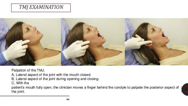
The importance is to determine deviation of jaw from the midline during the opening and closing of the jaws.
The lateral mandibular range of motion or movement is assessed ” Normal 8 to 10 mm ” by having the patient to occlude the teeth and then slide the jaw in both directions. The range of movement from midline and any pain, location and severity is recorded.
Maximum interincisal opening: As a general guide, mobility is considered to be reduced if the subject is unable to open his or her jaw to the width of two fingers ( <30 mm ).
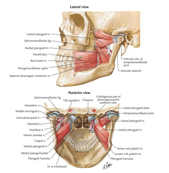
Gravity muscles ( Depression of mandible )
Anti-gravity muscles ( Elevation of mandible )
Protrusion of mandible
Developmental
Traumatic
Inflammatory
Neoplastic
References:
References: TEXTBOOK OF PEDODONTICS SHOBHA TANDON 2ND EDITION
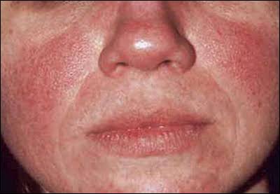
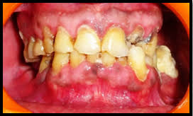
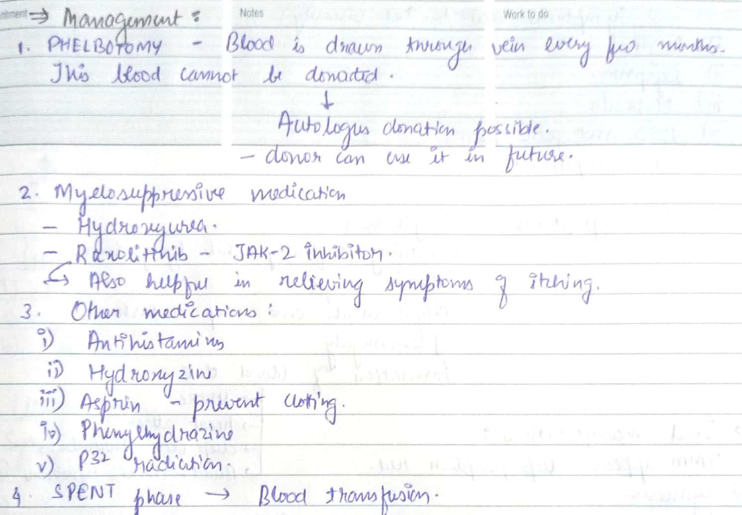
References – Shafer’s textbook of oral pathology , SN Chugh Textbook of clinical medicine for dental students
Image reference : Google images
Plummer Vinson triad :
High risk of “squamous cell carcinoma”
Risk factors :
Pathophysiology :
Signs :
Symptoms :


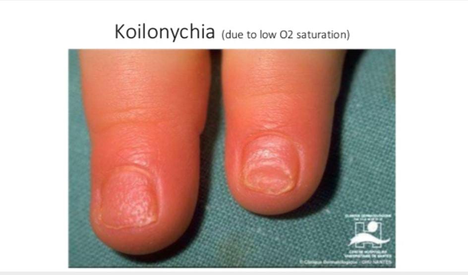
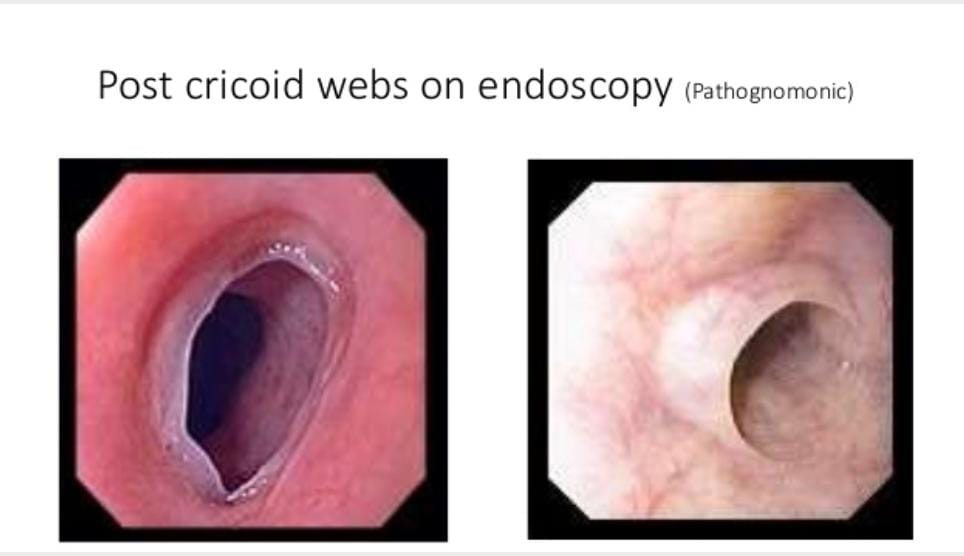
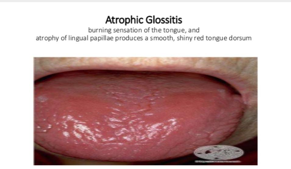
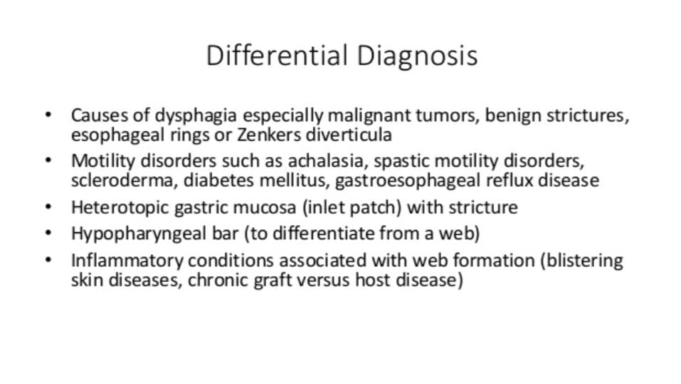


References – Shafer’s textbook of oral pathology , SN Chugh – Textbook of clinical medicine for dental students , slideshare.com
Image reference : Google images.
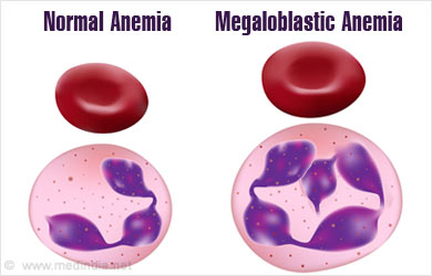
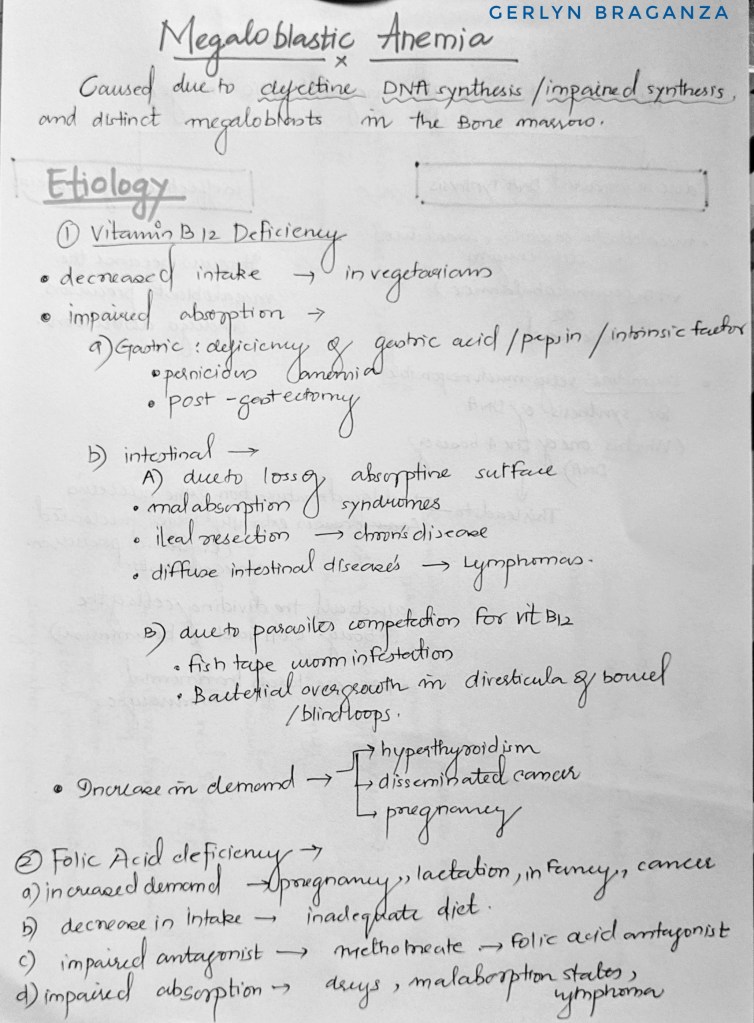
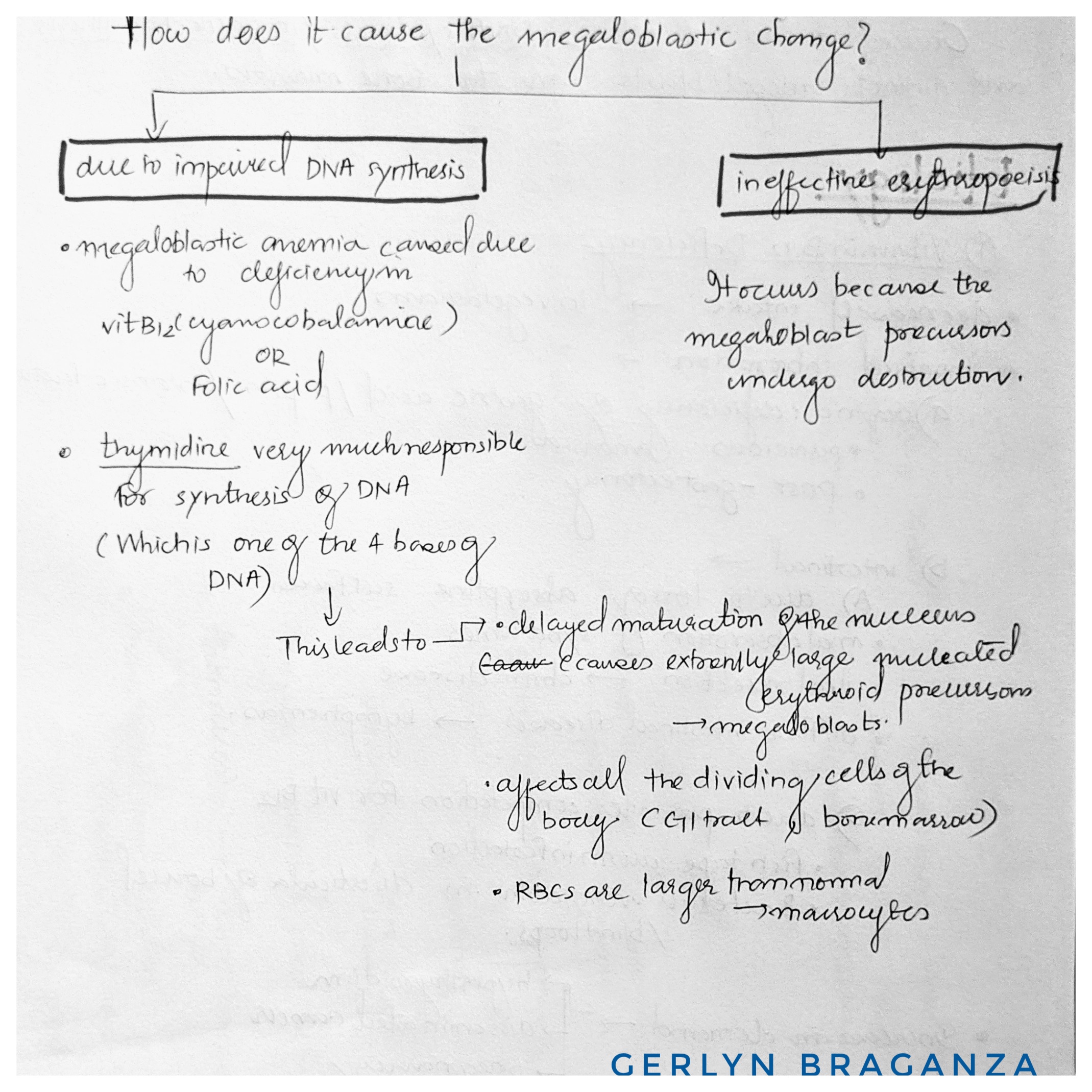
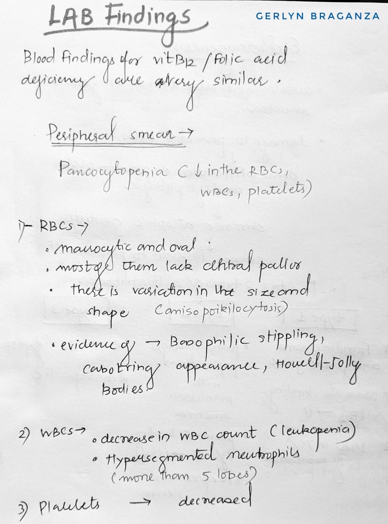

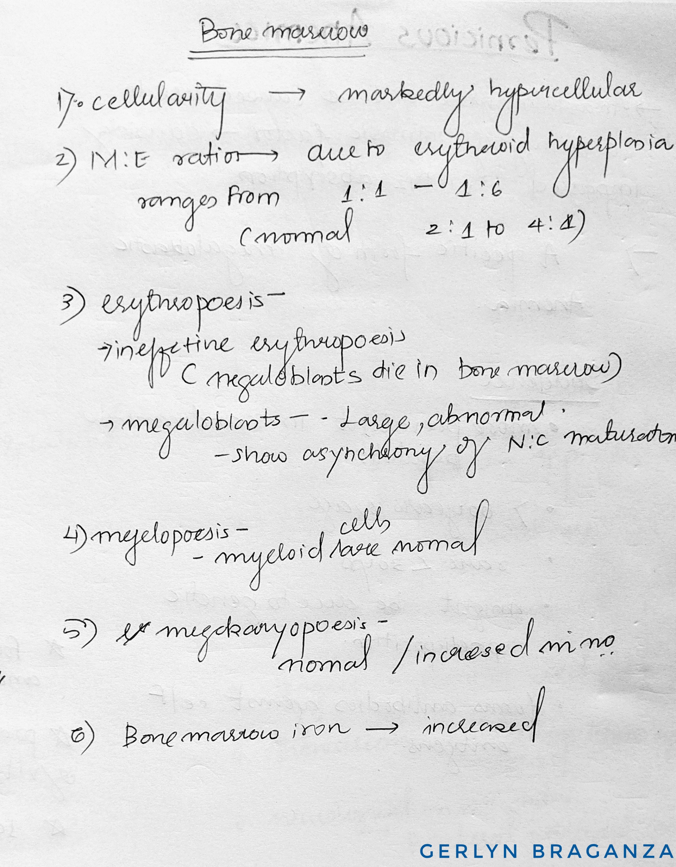
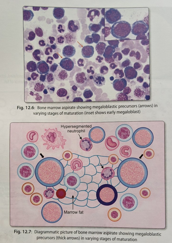
Source – textbook of pathology by ramdas nayak and harsh mohan
Teething is a term limited by common usage to eruption
of primary dentition. Since the time of Hippocrates (460-
377 BC) teething is blamed for ailments such as fever, convulsions, bronchitis, otitis media and diarrhea, for
causing 12% of the deaths in children under four years.
Complete opposite views are also available. It is seen
that these local and systemic factors are associated with
the disease somewhere else also and just as a coinci-
dence appear with teething.
Eruption of primary dentition usually begins in the 4-6th month of a child’s life. The appearance of normal teeth is eagerly awaited by the parents since it represents an important early milestone in development. In most cases eruption of teeth causes no distress to the child or parents, but sometimes the process causes local
irritation, which is usually minor but which may be severe enough to interfere with the child’s sleep.
CLINICAL FEATURES OF TEETHING
LOCAL SIGNS
1. Hyperemia or swelling of the mucosa overlying theerupting teeth.
2. Patches of erythema on the cheeks.
3. Flushing may also occur in the skin of the adjacen cheek.
SYSTEMIC SIGNS
1.General irritability and crying
2.Loss of appetit.
3.Sleeplessness
4.Increased salivation and drooling
5.Insanity
6.Meningitis
7.Increased thirst
8.Circumoral rash
9.Cough
Associated Problems
Systemic
Fever,convsions, diarrhea,vomiting, bronchitis, cholera, tetanus
Local
Eruption hematoma, eruption sequestrum, ectopic eruption, transmigration , transposition
MANAGEMENT
PREVENTIVE MEASURES
1. Maintain child’s oral and general body health.
2.Gums should be wiped after each meal with cotton soaked in a weak antiseptic.
3. Adequate quantities of vitamins, minerals , proteins, are given to the child for increasing body’s resistance .
GENERAL MANAGEMENT
1. Hard , non sweetened rusks
2. Use of toasted bread helps in providing gingival stimulation.
3. Use of hard fruits such as appleor guava .
MEDICAL MANAGEMENT
demulcent and mild antiseptic.
1. Topical application of glycerin: It acts as a protective demulcent and mild antiseptic.
2. Topical application of lignocaine hydrochloride which provide relief in the interval before the analgesic is effective .
3. Topical application of benzyl alcohol can also be effective.
SURGICAL MANAGEMENT
Surgical treatment is sometimes recommended for the relief of pain from an eruption cyst or a hematoma. The technique advocates is to make 2 semilinar incisions over the crowns of the tooth , which meet at their extremities. The intervening portion of the tissue , which lies over the occlusal surface of unerupted tooth is then removed with a pair of tissue foreceps.
REFERENCES:TEXTBOOK OF PEDODONTICS SHOBHA TANDON 2ND EDITION
So what is the pulp – in simple words it’s a tissue that forms the inner tooth structure along with the blood vessels and contains nerves .
Pulp capping is the process of placing a specialized agent in contact with or in a close proximity to the pulp with the intention to encourage formation of new dentin ( secondary dentin) and also promote healing of the pulp.
Even before the discovery pulp capping agents, exposure lead to pulpitis or pulpal infection and ultimately pulp necrosis.
Examples of a very common pulp capping agent used – calcium hydroxide cement.
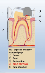
Placement of an agent directly on the exposed pulp is direct pulp capping.
It is done in the following situations-
Note- direct pulp capping is considered oy for immature permanent teeth or for future permanent teeth

Source – Google
Procedure where gross caries are removed from the lesions and cavity is sealed for a time with a biocompatible material.
And secondary dentin formation is induced even when pulp is not exposed but is near exposure.
This induces new dentin formation.
It is done in the following situations-
Note – it promotes formation of reparative dentin, promotes dental sclerosis and remineralizing the carious dentin.
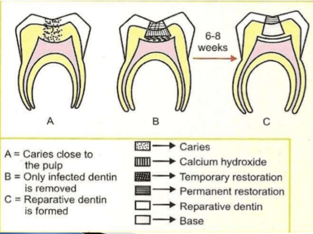
Source-google
•Stimulate reparative dentin formation.
•Preserve pulp vitality
•Release fluoride to prevent secondary caries .
•Bactericidal or bacteriostatic
• Adhere to dentin and restorative material
•Resist forces during mastication .
•Provide bacterial seal.
•Calcium hydroxide
•Zinc-Oxide – Eugenol
• GIC and adhesive resins
• MTA
• Biodentine
•Tricalcium phosphate ceramic
•Polycarboxylate cements
Source – Sturdevant’s art and science of operative dentistry ,slide share and manappallil
Image source – Google and slide share

Source – Rangarajan textbook

