
Source – Rangarajan textbook
Source – Rangarajan textbook

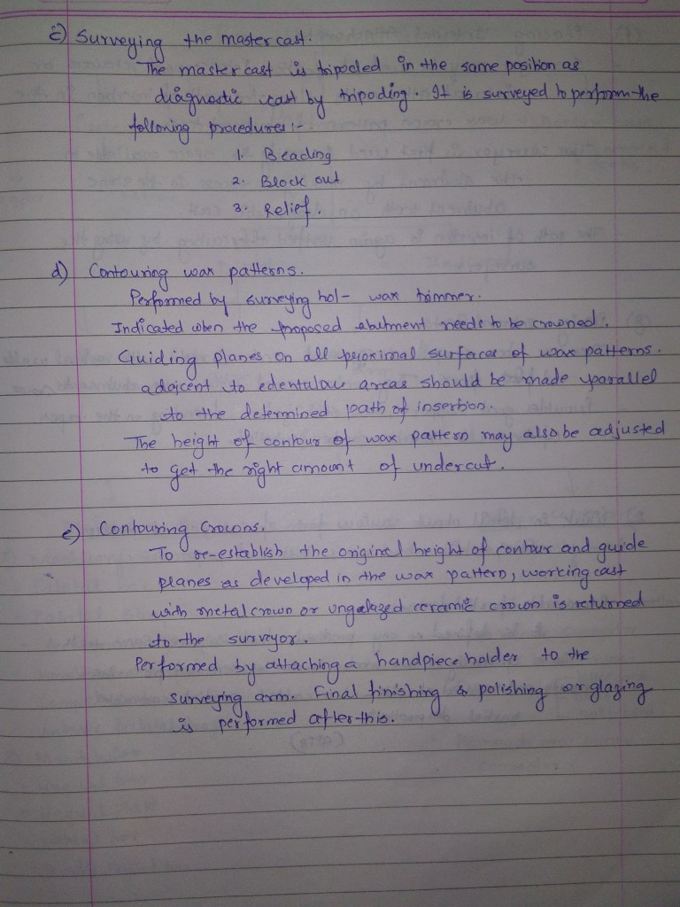
Source – Rangarajan textbook


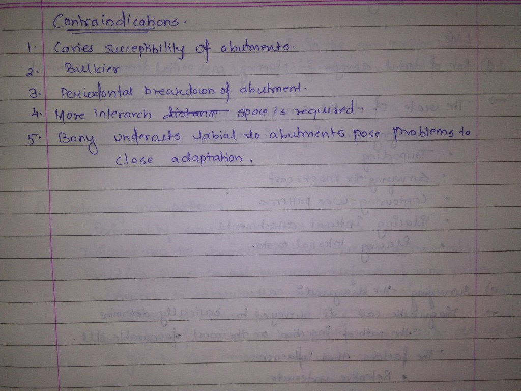
Source – Rangarajan textbook
Examination of neck nodes is important, particularly in head and neck Malignancies and a systematic approach should be followed.Neck nodes are better palpated while standing at the back of the patient. Neck is slightly flexed to achieve relaxation of muscles.
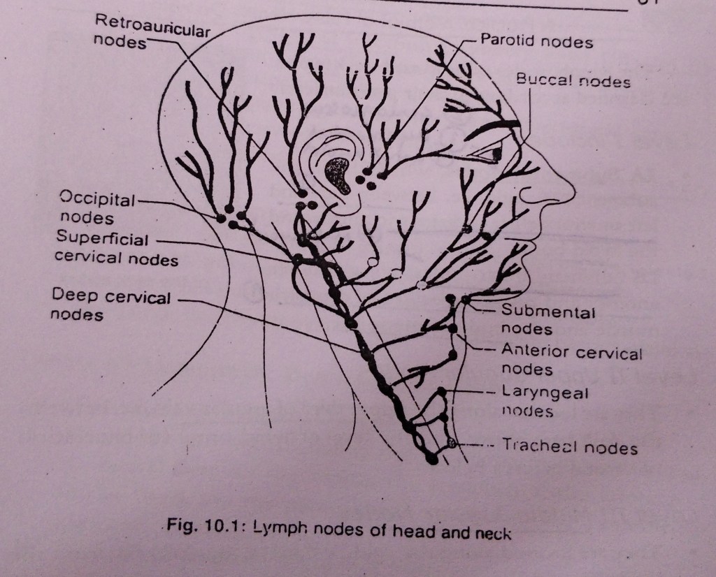
They are located along the upper third of jugular vein, i.e between the skull base above and the level of hyoid bone ( or bifurcation of carotid artery ) below.
They are located along the middle third of jugular vein , from the level of hyoid bone above, to the upper border of cricoid cartilage.
They are located along the lower third of jugular vein , from upper border of cricoid cartilage to the clavicle.
They are located in the posterior triangle ,i.e between posterior border of sternocleidomastoid ( anteriorly ) , anterior border of trapezius ( posteriorly ) , and the clavicle below. They include lymph nodes of spinal accessory chain , transverse cervical nodes and supraclavicular nodes.
They are located between the medial borders of sternocleidomastoid muscles ( or carotid sheath ) on each side, hyoid bone above and suprasternal notch below. They include prelaryngeal, pretracheal , paratracheal nodes .
They are located below the suprasternal notch and include nodes of the upper mediastinum.
A normal lymph node cannot be felt. If a node is palpable, it must be abnormal.
The face has 3 lymphatic territories :
References:
A practical manual of Public Health Dentistry by CM Marya.
Written by - Dr.Urusa I Inamdar
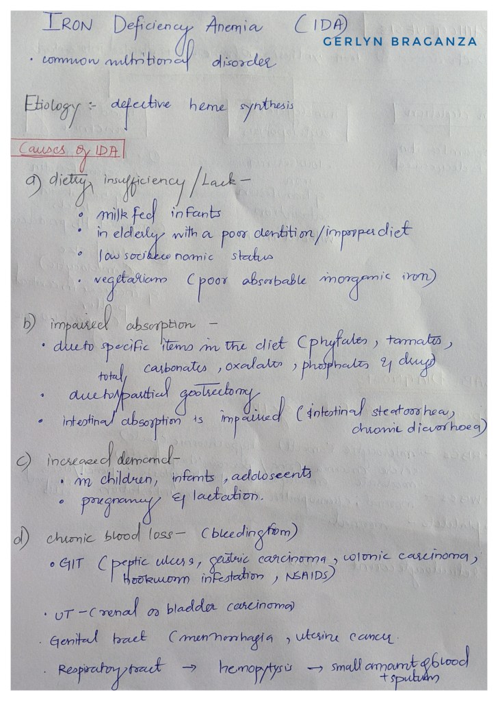


Peripheral smear
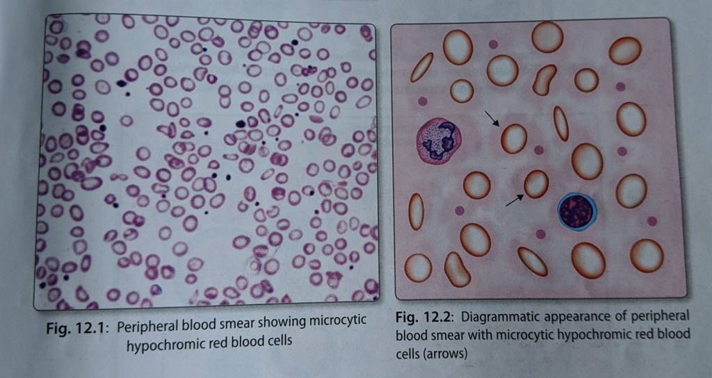
Reference – textbook of pathology for dental students – Ramdas Nayak


source – youtube
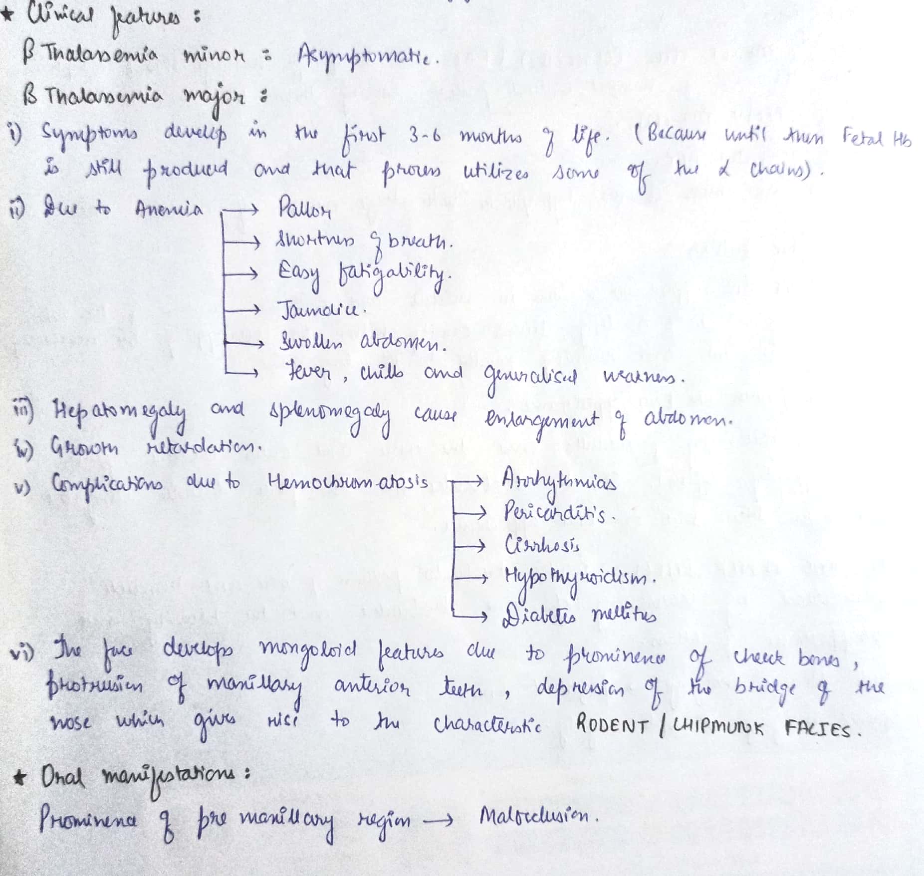

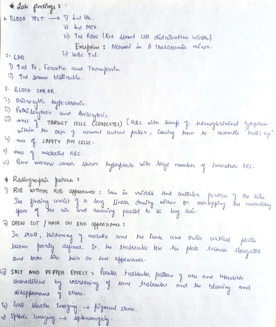

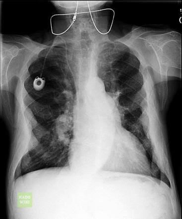
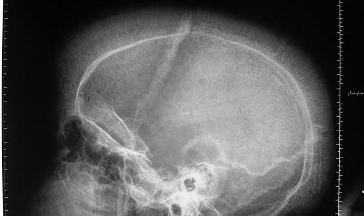

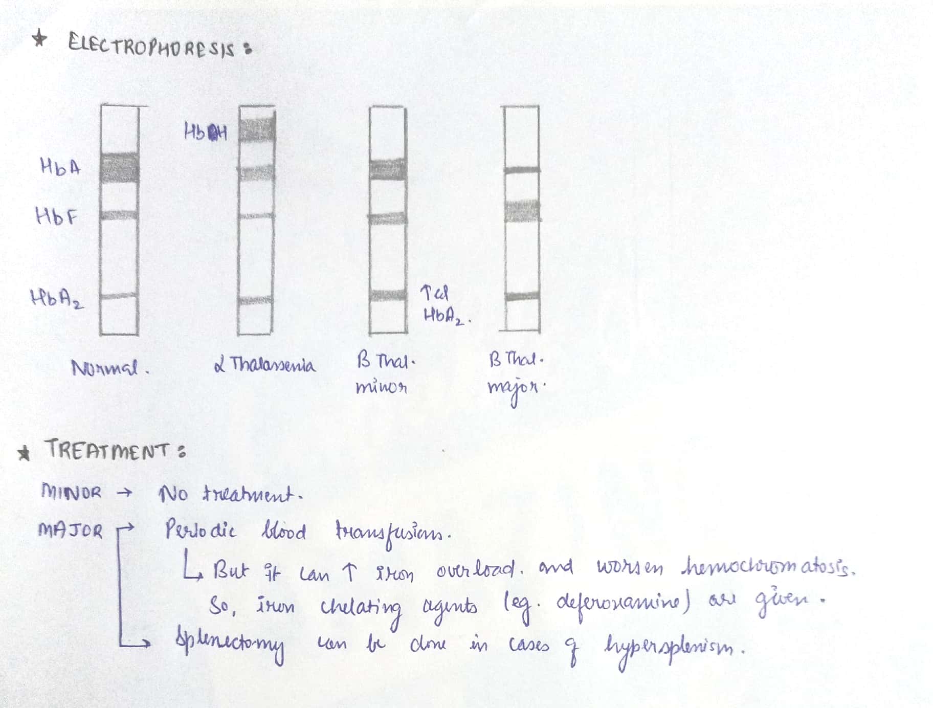
References – Shafer’s Oral pathology , S.N chugh – Textbook of clinical medicine for dental students
Image source – Google images.
Happy reading 🙂
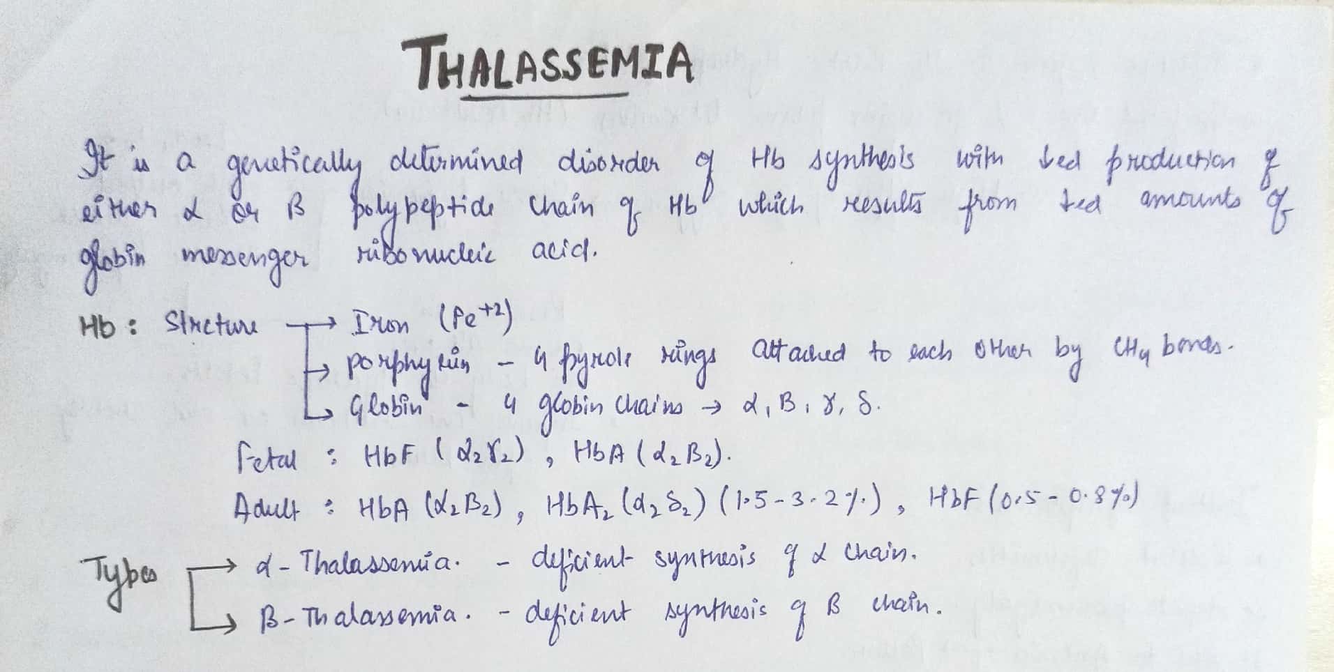
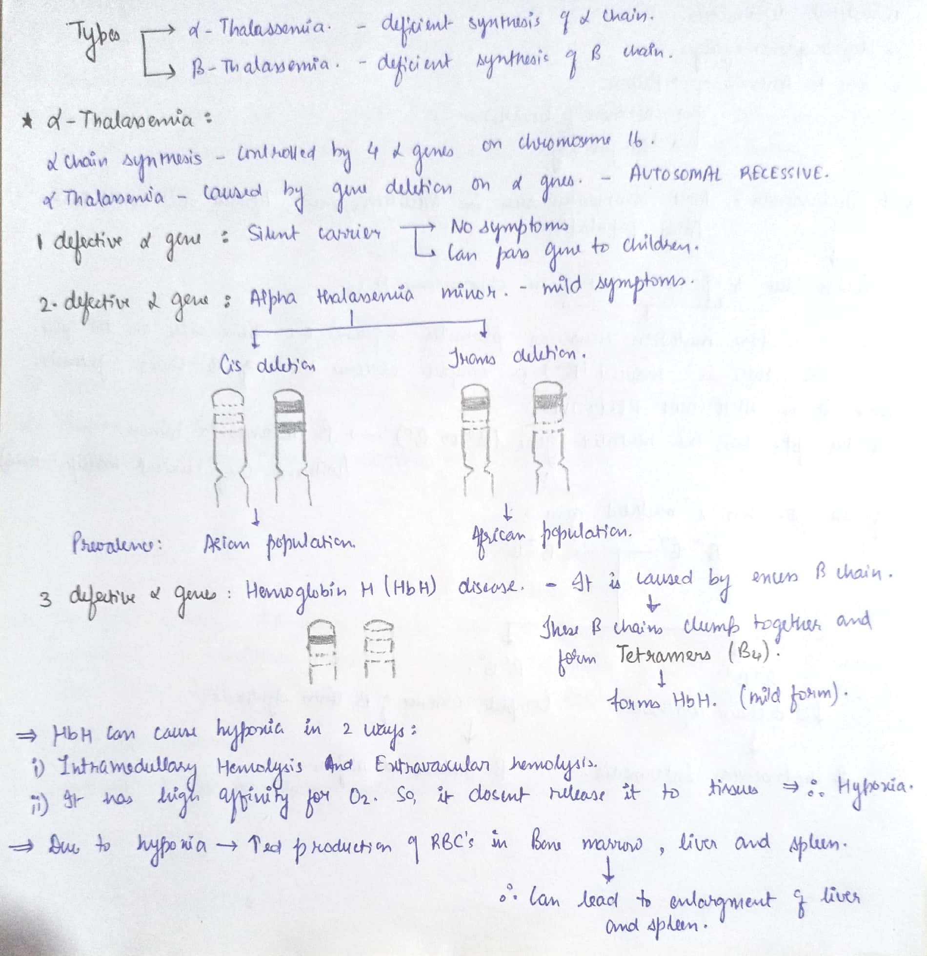


References – Shafer’s oral pathology , SN Chugh – Textbook of clinical medicine for dental students , osmosis.org
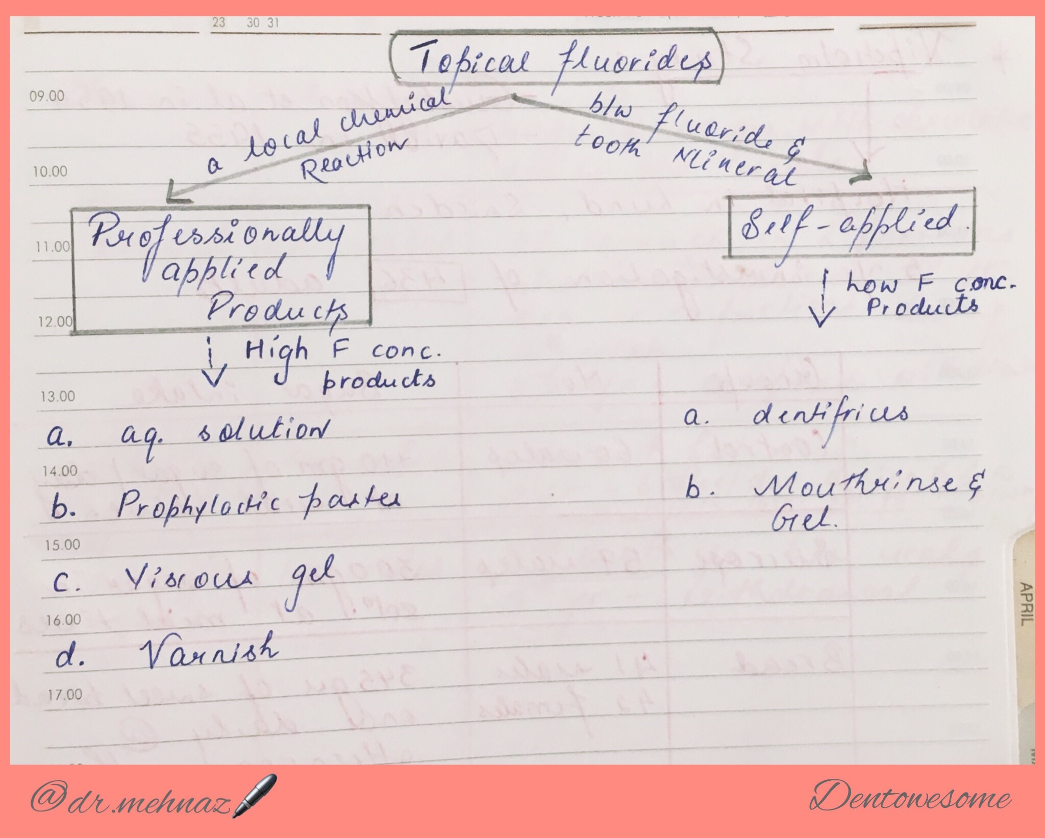

Fluoride Varnish 👇🏻
References: Textbook of Public Health Dentistry-Sober Peter(5th Edition)
Reduction of total circulating red cell mass below normal limits.
Decrease in the oxygen carrying capacity of the blood that leads to tissue hypoxia.
Anemia may be absolute (decreased RBC mass), or relative ( associated with higher plasma volume).
1. Morphological classification
2. Etiological classification – given below in the tables

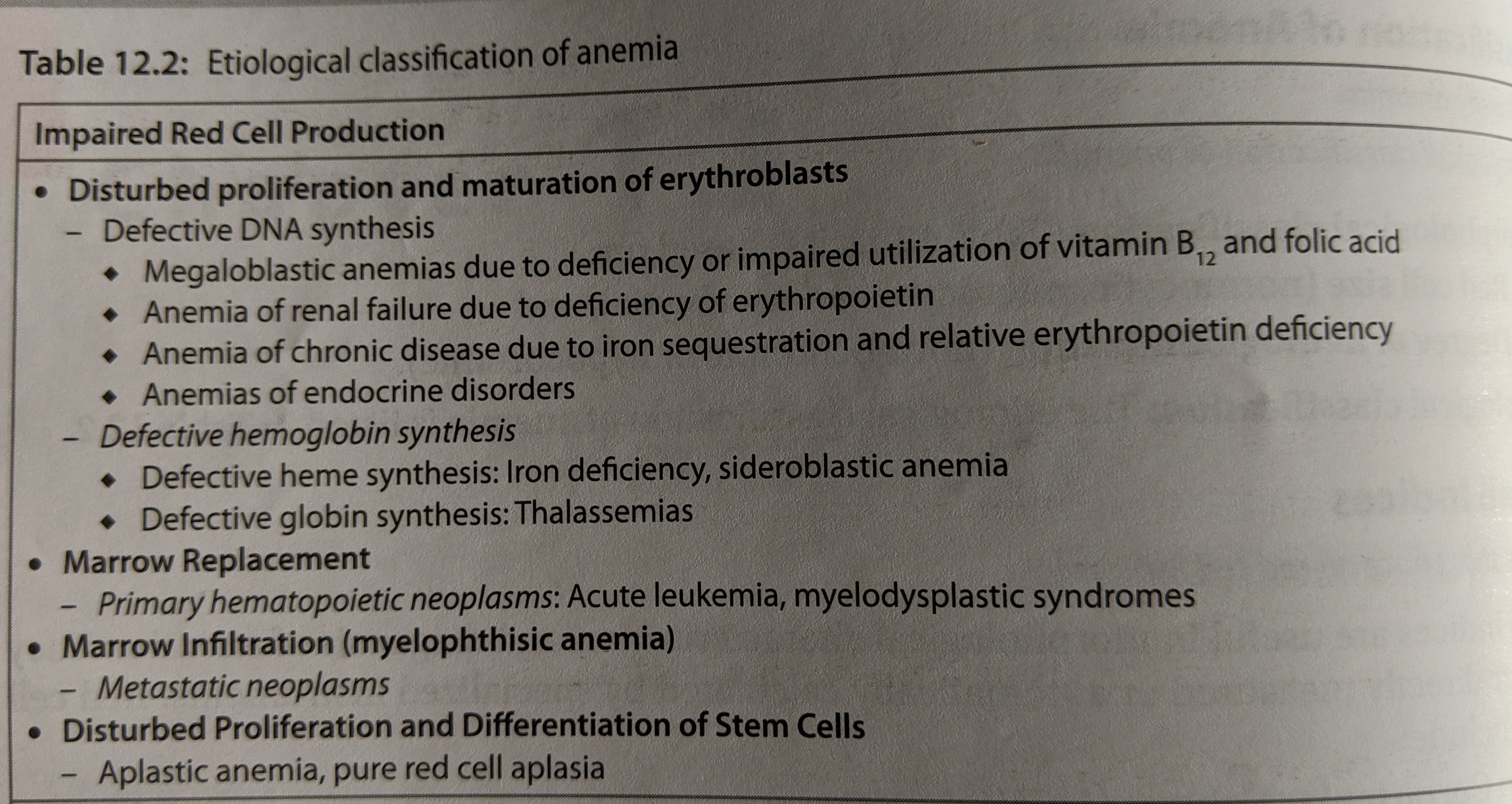

Source – textbook of pathology ( Ramdas Nayak )

