PATHOGENESIS OF POCKET FORMATION

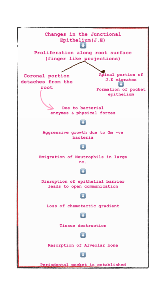
👉🏻 The initial lesion in the development of periodontitis is the inflammation of gingiva following bacterial challenge.

  1. Formation of bacterial colony
  2. Infammatory process
  3. Migration of neutrophils
  4. Tissue destruction
  5. Activation of Neutrophils
  6. Pocket formation

PERIODONTAL POCKET TYPES:

• Bacterial cells in dental plaque

Infammatory changes in the Connective Tissue wall of the gingival sulcus

⬇️

The cellular & fluid inflammatory exudate causes degeneration of Connective tissue & gingival fibres

⬇️

Just apical to Junctional epithelium, collagen fibres are destroyed

⬇️

Infammatory cells + edema

• Mechanisms associated with collagen loss:

  1. Collagenase enzyme secreted by fibroblasts, PMN & macrophages degrade collagen & other matrix molecules into small peptides – MMP’s
  2. Fibroblasts phagocytize collagen fibres by extending cytoplasmic processes to the ligament-cementum interface.

👉🏻 Apical cells of junctional epithelium proliferate along the root, Coronal portion of J.E detaches from the root & is invaded by PMN’s (60%)


Dr. Mehnaz Memon🖊
STAGES OF GINGIVITIS

Gingivitis is defined as an inflammation which is confined to the tissues of the marginal gingiva . It is an observable alteration in tissues associated with changes in vascular permeability and dilation often accompanied with the infiltration of leukocytes into the affected tissues .

The sequence of events cumulating in the clinically apparent gingivitis is categorized into

  • STAGE 1: Initial lesion
  • STAGE 2 : Early lesion
  • STAGE 3 : Established lesion
  • STAGE 4 : Advanced lesion

STAGE 1 : THE INITIAL LESION ( 2- 4days ):

  1. VASCULAR CHANGES : Classic vasculitis sub adjacent to junctional epithelium, dilated capillaries , increased blood flow
  2. MICROSCOPIC CHANGES : Presence of leukocytes, loss of perivascular collagen and presence of serum proteins changes in the coronal most position of junctional epithelium .
  3. CLINICAL CHANGES : Exudation of fluids from the gingival sulcus , sub clinical gingivitis .

STAGE 2 : EARLY LESION (4-7 DAYS ):

  1. VASCULAR CHANGES : Vascular proliferation
  2. MICROSCOPIC CHANGES : Rete pegs formation in junctional epithelium , presence of lymphocytes , loss of collagen is increased , fibroblasts show cytoplasmic alterations .
  3. CLINICAL CHANGES : Bleeding on probing, erythematous gingiva.

STAGE 3 : ESTABLISHES LESION ( 14-21 DAYS ) :

  1. VASCULAR CHANGES : Same as early lesion with blood stasis.
  2. MICROSCOPIC CHANGES: Proliferation , apical migration and lateral extension of junctional epithelium, atrophic areas , plasma cells are predominant , further loss of collagen, increased enzyme levels of acid and alkaline phosphatase.
  3. CLINICAL CHANGES : Changes are seen in color, surface texture and consistency , bluish hue around the reddened gingiva , gingiva appears to be moderately to severely inflamed .

STAGE 4 : THE ADVANCED LESION :

  1. VASCULAR CHANGES: Same as early and established lesions. characterized by the invasion of the lesion to the underlying alveolar bone .
  2. MICROSCOPIC CHANGES: Persistence of features seen in established lesion, extension of inflammation into deeper structures including alveolar bone and periodontal ligament , presence of all types of inflammatory cells , conversion of bone marrow into fibrous tissues.
  3. CLINICAL CHANGES :Formation of periodontal pocket and its associated changes.

REFERENCES: ESSENTIALS OF PERIODONTOLGY SHANTIPRIYA REDDY 5TH EDITION

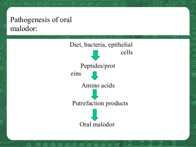
ORAL MALODOR

INTRODUCTION:

Halitosis – or chronic bad breath – is something that mints, mouthwash or a good brushing can’t solve. Unlike “morning breath” or a strong smell that lingers after a tuna sandwich, halitosis remains for an extended amount of time and may be a sign of something more serious. 

CLASSIFICATION :

  1. BASED ON ETIOLOGY :
  • Local factors of pathological origin : poor oral hygiene , extensive caries, periodontal disease , cysts , tumors .
  • Local factors of non pathological origin : stagnation of saliva associated with food debris , dentures, excessive smoking .
  • Systemic factors of pathological origin : diabetes mellitus, liver failure, lung abscess , tuberculosis .
  • Systemic factors of non pathological origin : diet like garlic , onion , meat , excessive alcohol consumption.
  • Xerostomia :conditions like sjogren’s syndrome, radiation therapy.

2. BASED ON PATIENT’S CRITERIA :

  • Genuine halitosis
  • Pseudo halitosis
  • Halitophobia

PATHOGENSIS OF MALODOR :

ETIOLOGY :

  1. Physiological halitosis
  • mouth breathing
  • medications
  • aging and poor dental hygiene
  • fasting / starvation
  • tobacco
  • foods like onion , garlic and alcohol

2. Pathological halitosis:

  • periodontal infection
  • stomatitis
  • xerostomia
  • faulty restorations
  • unclean dentures
  • oral cancers
  • candidiasis
  • nasal infections
  • GERD
  • bronchitis, pneumonia
  • diabetes mellitus
  • renal failure
  • fever

DIAGNOSIS :

  1. Review of medical , dental , personal history
  2. Clinical examination : tongue coating, mouth breathing , xerostomia
  3. Complete periodontal examination
  4. Measurement of malodor : gas chromatography . halimeters, BANA test , chemiluminescence , diamond probe

TREATMENT :

  • Brush your teeth after you eat.
  • Floss at least once a day.
  • Brush your tongue.
  • Clean dentures or dental appliances. .
  • Avoid dry mouth.
  • Adjust your diet. .
  • Regularly get a new toothbrush.
  • Schedule regular dental checkups.
  • Regular use of halita solution which reduces the levels of volatile sulfur compounds .

References: Essentials of clinical periodontology . Shantipirya reddy 5th edition

RUBBER DAM ISOLATION

Written by – Dr.Urusa I Inamdar

In 1864, S.C. Barnum, introduced the rubber dam into dentistry. It is used to define the operating field by isolating one or more teeth from the oral environment. The dam eliminates saliva from the operating site and retracts the soft tissue.

Advantages

  • A dry and clean operating field
  • Improved access and visibility
  • Potentially improved properties of dental materials
  • protection of the patient and operator
  • Operating efficiency

Disadvantages

Time consumption and patient objection are the most frequently quoted disadvantages of the rubber dam.

Certain oral conditions may preclude the use of the rubber dam , these include :

  • Teeth that have not erupted sufficiently to support a retainer
  • Some third molars
  • Extremely malpositioned teeth

In addition, patients suffering from asthma may not tolerate the rubber dam if breathing through the nose is difficult. Latex allergy.

However, latex free rubber dam material is currently available.
Materials

  • Holder
  • Retainer
  • Punch
  • Retainer forceps
  • Napkin
  • Lubricant
  • Modeling compound
  • Anchors

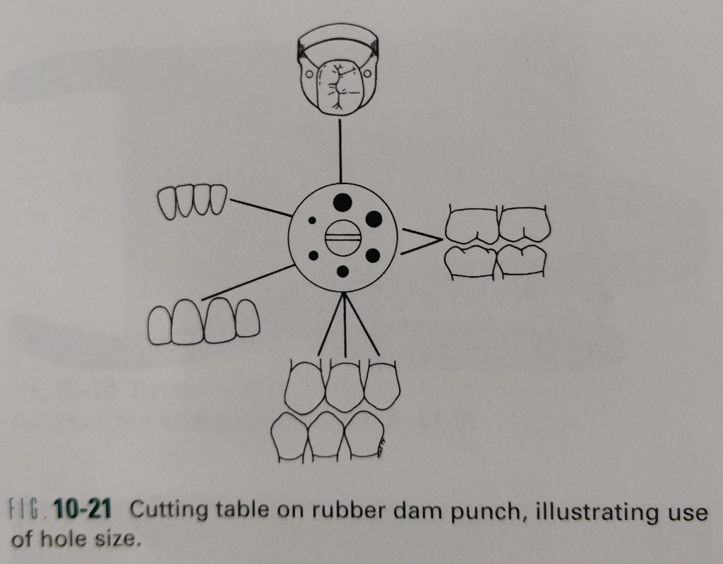
Hole size and position

Placement

  • Testing and lubricating the proximal contacts.
  • Punching the holes.
  • Lubricating the dam.
  • Selecting the retainer.
  • Testing the retainers stability and retention.
  • Positioning the dam over the retainer.
  • Applying the Napkin.
  • Positioning the Napkin.
  • Attaching the frame.
  • Attaching the neck strap ( optional )
  • Passing the dam through posterior contact.
  • Applying low fusing modelling compound ( optional )
  • Applying the anterior anchor ( if needed)
  • Passing the septa through the contacts without tape.
  • Passing the septa through the contacts with tape.
  • Inverting the dam interproximally.
  • Inverting the dam faciolingually.
  • Using a Saliva ejector ( optional )
  • Confirming a properly applied rubber dam .
  • Checking for access and visibility.
  • Inserting the wedges.

Removal

  • Cutting the septa.
  • Removing the retainer.
  • Removing the dam.
  • Wiping the lips.
  • Rinsing the mouth and massaging the tissue.
  • Examining the dam.

Alternate / additional methods and factors

The procedure just detailed describes the method of sequentially placing the retainer and rubber dam on the anchor tooth.

  • Applying the dam and retainer simultaneously.
  • Applying the dam before the retainer.
  • Cervical retainer placement.
  • Fixed bridge isolation.
  • Substitution of a retainer with a matrix.

Errors in application and removal

  • Off center arch form
  • Inappropriate distance between the holes
  • Incorrect arch form of holes
  • Inappropriate retainer
  • Retainer pinched tissue
  • Shredded or torn dam
  • Incorrect location of hole for class v lesions
  • Sharp tips on no. 212 retainer
  • Incorrect technique for cutting septa

Reference:

Sturdevant’s – Art and science of Operative Dentistry (4th edition)