It refers to the cellulitis of the submandibular and submental regions combined with inflammatory oedema.
Causes:
🔍 Streptococcal organisms are responsible for the infection around the submandibular region.
🔍 Anaerobes also play a major role.
Clinical Features:
👩⚕️ Elderly patients who present with a diffuse swelling in the submandibular and submental regions (brawny oedema).
👨⚕️ Oedema of the floor of the mouth is seen which causes elevation of the tongue that in turn results in difficulty in swallowing.
👩⚕️ High grade fever with toxicity.
👨⚕️ Putrid halitosis is also characteristic of Ludwig’s angina.
Treatment:
💊 Rest and hospitalization
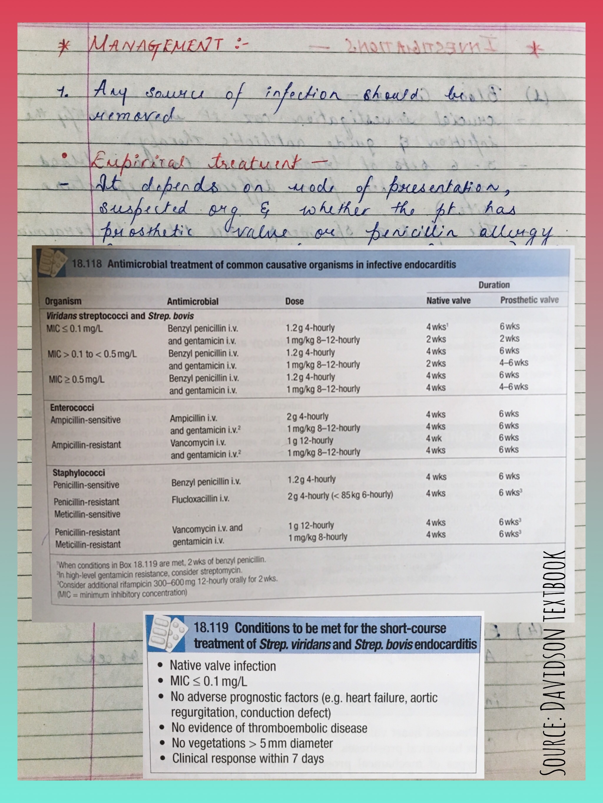
💊 Appropriate antibiotics should be given.
💊 Intravenous fluids to correct dehydration and Ryle’s tube feeding.
💊 If conservative management is not responsive in the patient, then surgery is preferred.
Surgery:
✂️ Under General Anaesthesia, a 5-6 cm curved incision is made below the mandible in the submandibular region and over the most prominent part of the swelling.
✂️ The submandibular gland is mobilised, mylohyoid muscle is divided, and the pus is drained.
✂️ Even if there’s no pus, the oedematous fluid comes out which in itself will improve the patient condition greatly.
✂️ The wound is closed by loose sutures, after cleansing the cavity with antiseptics and a drainage tube is inserted.
Complications:
⚠️ Mediastinitis and Septicaemia.
⚠️ Oedema of the glottis which is caused due to the spread of the cellulitis via a tunnel connecting stylohyoid muscle and submucosa of the glottis.
Crevicular fluid is a serum ultrafiltrate that accumulates in gingival connective tissue resulting from increased vascular permeability. When its production increases, it passes to the gingival crevice.
Gingival crevicular fluid contains products derived from microbial plaque, tissue breakdown, host cells, and host immunity that, in some instances, have been demonstrated to be related to the active phases of periodontal destruction.
Aspartate aminotransferase (AST) is an enzyme normally confined to cell cytoplasm, but is released to the extracellular environment upon damage. AST levels in serum and other body fluids ( cerebrospinal fluid, arthritic joint fluids) have been used for several decades as a laboratory diagnostic aid for assessing tissue destruction (i.e.MI, hepatitis). Furthermore, the amount of AST activity observed generally reflects the extent of cell death and, consequently, the magnitude of tissue destruction. AST activity greatly depends on cellular damage in periodontal tissues. Levels of various inflammatory cytokines can also be determined in GCF.
The periimplant gingival sulci has been shown to be similar to the periodontal crevice with respect to gingival fluid flow. In spite of this observation, to date relatively few studies have focused their attention on the investigation of the periimplant crevicular fluid (PCF) components and their relationship with the peri-implant condition.
Functions of GCF:
• Promotes adhesion of the gingival crevice epithelium to the enamel through the presence of salivary and plasma proteins. • Defense through the presence of inflammatory cells and molecules. • Elimination of micro-organisms and food debris through the washout effect of fluid formation.
Diet can affect teeth: • Pre-eruptively—fluoride is the most important. The effect of calcium,phosphate, vitamins, and sugar is unclear, but is unlikely to be great. • Post-eruptively—again, fluoride is important, as is sugar. Acidic foods or drinks can cause erosion.
Dietary Analysis
Aim: To determine the time for which the teeth are at risk of demineralization and increase the potential remineralization period. Indications:
(i) high caries activity,
(ii) unusual caries pattern,
(iii) suspected dietary erosion.
Dietary advice should be tailored to the individual. This is most easily done after analysing the patient’s present eating pattern.
Method:
A consecutive 3- or 4-day analysis (including at least one weekend day) is the most widely used, with the patient recording the time, content, and quantity of food/drink consumed. In addition, toothbrushing and bed-time should be indicated. When the form is returned the entries should be checked with the patient.
Analysis: • Ring the main meals. If in any doubt,identify those snacks that contain complex carbohydrate. Assess nutritional value of meals. • Underline all sugar intakes in red. • Identify between-meal snacks and note any associations, e.g. following insubstantial meals or at school. • Decide on a maximum of three recommendations.
Dietary advice should include an explanation of the effect of between-meals eating and sugary drinks. It must also be personal, practical,and positive! The suggestion that a child should select crisps when friends are buying sweets is more likely to be followed than total abstinence.
Some helpful hints: • Save sweets to be eaten on a day, e.g. Saturday dinnertime, or to be eaten at the end of a meal. • All-in-one chocolate bars are preferable to packets of individual sweets. • Foods which increase salivary flow (e.g. cheese, sugar-free chewing gum) can help to reverse the pH drop due to sugar if eaten afterwards.
• Treacle, honey, and fruit (especially fruit juice) are cariogenic. • Artificial sweeteners should be avoided in pre-school children. • Fibrous foods, e.g. apples, are preferable to a sucrose snack, but they can still cause decay and there is no evidence that they can clean teeth. Where the nutritional content of meals is inadequate, considerable tact is necessary. It may be possible to suggest that larger meals would reduce the temptation to eat snacks. For children who are ‘picky’ eaters snacks and sweets saved until the end of a meal can act as an encouragement to consume more food at mealtimes.
Remember that while cheese, peanuts, and crisps may constitute a safe snack in dental terms, they are all high in fat, and peanuts can be inhaled by small children. Also, ‘diet’ cola drinks are sugar-free, but can still cause erosion if large quantities are drunk. Therefore, dental dietary advice should be given in the wider context of the general health of the individual, i.e. decrease consumption of sugars and fats, and increase consumption of fibre-rich starchy foods, fresh fruit, and vegetables. Meals provide a better nutritional balance than snacks. Hence good eating/drinking at mealtimes and avoiding between meals snacking is healthy.
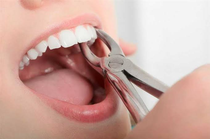
In both these cases, take a radiograph and consider transalveolar method.
3. Fracture of alveolar process – if large piece of bone fractures, replace the bone in position with the periostium intact. Suture the mucosa.
4. Fracture of maxillary tuberosity – check oroantral communication and if present, manage accordingly. If tuberosity has periosteal attachment, reposition it back and suture.
5. Fracture of Jaws – reduction and fixation
6. Mucosal laceration – avoid by holding instruments with proper support. Manage by approximation and suturing.
7. Luxation of adjacent tooth – replace the luxated tooth to its original position and splinting made for a period of 3 – 6 weeks.
8. Oroantral communication – small opening, no treatment, do good suturing of the extraction site. Large opening needs surgical closure and coverage of opening by stent/other materials till healing.
9. Displacement of tooth into the facial spaces – removal of the tooth/root indicated.
10. Nerve injury – patient should be informed and routine follow up done.
11. Hemorrhage – if from soft tissue, (pressure pack/LA pack/sutures/cautery) if from artery, (ligation) if from bone (gel foam/oxidised cellulose/ bone wax)
12. Dislocation of jaw – avoided by supporting the jaw during extraction.
13. Root tip left – leave if deeply buried, give antibiotics and review. However if large pieces of root remains, remove.
Postoperative
Hemorrhage
a) primary – occurs due to inadequate hemostasis at surgery.
b) reactionary – occurs within 48 hours due to rise in BP or slippage of sutures.
C) secondary – 7 days postoperatively usually due to infection
Agents used as packs –
Resorbable mesh (oxidised cellulose)
Transexemic acid
Adrenaline
Epsilon amino capriac acid
Bismuth iodine paraffin paste
Whiteheads varnish
2. Pain and swelling – edema maximum on second day and then it subsides. Administration of anitobiotics and analgesics.
3. Dry socket (Alveolar osteitis) – loss of blood clot and socket appears empty and dry. Irrigation of the site and placement of obtundent dressing.
🔼 Clinical Presentation: Slow growing, non tender, a simple fibroma arising from the periodontal membrane, presents on the gum. May undergo sarcomatous change.
Giant Cell Epulis: (⭐ AKA Myeloid Epulis)
🔼 Consistency: Soft to firm gums with indurated underlying mass due to expansion of bone.
🔼 Clinical Presentation: It is an osteoclastoma arising in the jaw. Presents as hyperaemic vascular, oedematous, may ulcerate and result in haemorrhage. X ray shows pseudo trabeculation.
🔼 Treatment: Small tumours are treated by currettage; Large tumours are treated by radical excision.
Carcinomatous Epulis:
🔼 Consistency: Hard or malignant in consistency.
🔼 Clinical Presentation: This is an epithelioma arising from mucous membrane of the alveolar margin. Presents as a non healing, painless ulcer slowly infiltrating the bone. Hard regional lymph nodes are due to metastasis.
🔼 Treatment: Wide excision which includes removal of segment of the bone.
Broken Bone Fact #1: It’s no secret that smoking is bad for you, but did you realize that it can also have a negative impact on your ability to heal from broken bones?
Broken Bone Fact #2: If you aren’t sure you have a fracture, you can always try treating your injury at home with the “RICE” treatment: Rest, Ice, Compression and Elevation.
Broken Bone Fact #3: Ability to move isn’t a sure indicator of a broken bone, as the only way to confidently identify a fracture is with an X-ray. Infact, broken bones are almost always movable!
Broken Bone Fact #4: Fractures happen more easily as you get older. If you are over 85, your chance of breaking a bone is four times higher than if you are between the ages of 65 and 75.
Broken Bone Fact #5: At the end of the day, it’s better to see a doctor for your concerns. If you take too long to speak to your doctor about your possible broken bone, your bone could begin healing on its own in a crooked, painful and uncomfortable fashion!
STUDY NOTES ⚕️
Fracture:
A fracture is a structural break in the normal continuity of bone. This also includes hairline fractures.
Mechanism of Injury:
Direct violence ➡️ Fracture of tibia or mandible can occur due to a fall.
Indirect violence ➡️ Fracture of clavicle due to fall on outstretched hand.
Traction injuries ➡️ Traction of the ligament attached to the medial malleolus may lead to its fracture.
Compression fracture ➡️ It may occur due to fall from a height. May also be associated with impacted fracture of the vertebral body.
Types of Bones:
🔸Tubular bone
🔸Cancellous bone
Types of Fracture:
• Closed or Simple: There is no communication between the site of Fracture and the exterior.
• Open or Compound: There is a wound leading to the site of Fracture or communication between the Fracture site and the exterior.
Classification:
I. Depending on etiology of Fractures
II. Depending upon the plane of Fracture surface
III. Special Fractures
I. Depending on the etiology of Fractures
1️⃣ Traumatic Fractures
2️⃣ Pathological Fractures:
2a) Tumours 👉 Giant cell tumours, Secondaries in bone, bone cysts.
2b) Infections 👉 Acute osteomyelitis
2c) Metabolic bone diseases 👉 Hyperparathyroidism, Osteoporosis, Paget’s disease, Multiple myeloma, etc.
3️⃣ Stress Fractures:
Commonly seen in metatarsal bones (particularly in the second metatarsal due to prolonged marching).
Hence it is also called as ‘march fracture’.
II. Depending upon the plane of Fracture surface
• Transverse
• Spiral
• Oblique
• Comminuted
• Compression
III. Special Fractures
📍 Depressed Fractures
• Results due to sharp localised blow because of which a cortical bone segment is depressed.
• That needs to be treated with elevation of bone outwards.
• This type of Fracture is commonly seen in the skull.
📍 Fracture-Dislocation
• Fracture-Dislocation gives rise to severe pain and the continues till the dislocation is reduced.
• Classic example: Anterior dislocation of shoulder with Fracture of neck of humerus.
📍 Fracture involving a joint
• These Fractures should be treated with care OTHERWISE joint stiffness and late osteoarthritis can occur.
📍 Complex Fractures
• These Fractures involve major vessels or nerves.
• Example: Fracture of humerus associated with radial nerve injury or fracture of lower femur associated with popliteal artery injury.