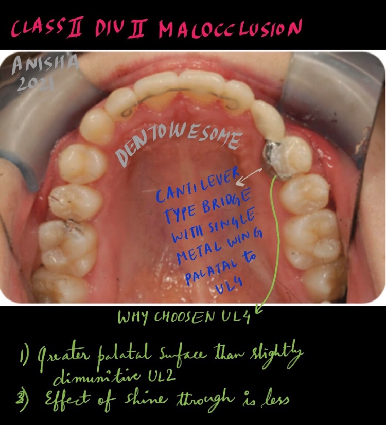
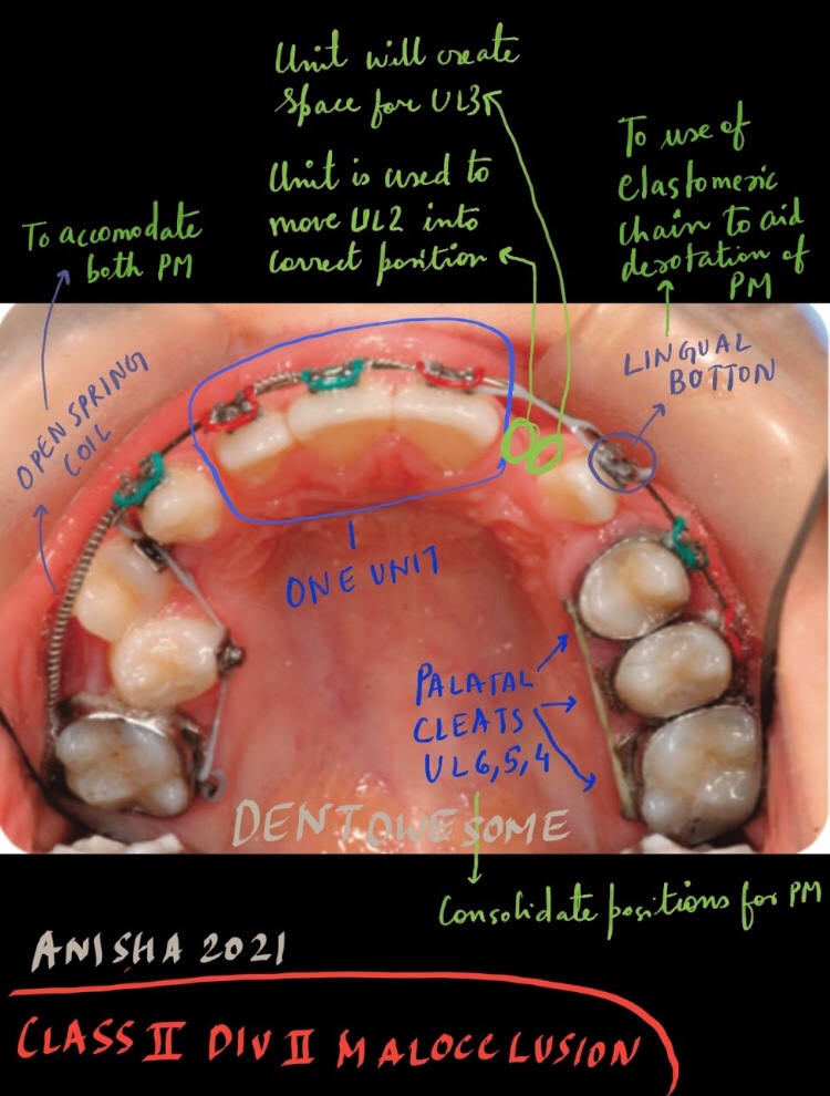
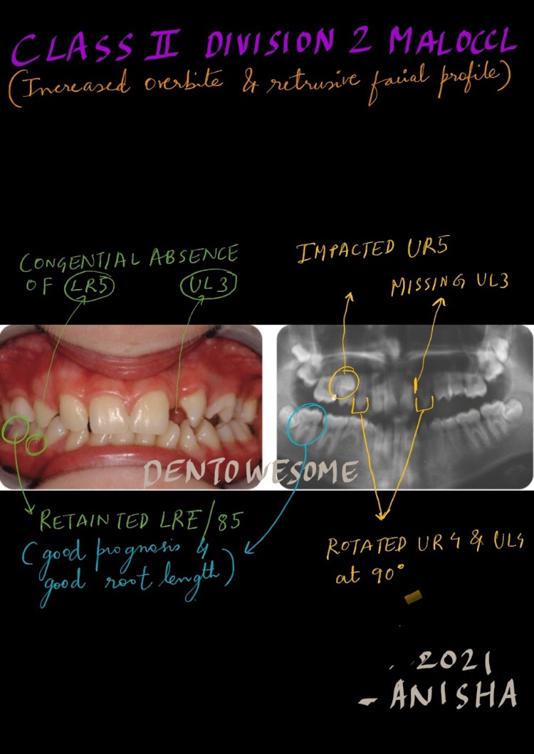
This 12-year-old female presented with a class II division 2 malocclusion on a moderate skeletal class II base with a retrusive profile and agenesis
Link: https://drive.google.com/file/d/1jzdkbvizdmQRovvsN2wBQ8KF42ePM9np/view?usp=drivesdk
This 12-year-old female presented with a class II division 2 malocclusion on a moderate skeletal class II base with a retrusive profile and agenesis
Link: https://drive.google.com/file/d/1jzdkbvizdmQRovvsN2wBQ8KF42ePM9np/view?usp=drivesdk



PA radiolucencies identification made easy! Happy learning..😀📖
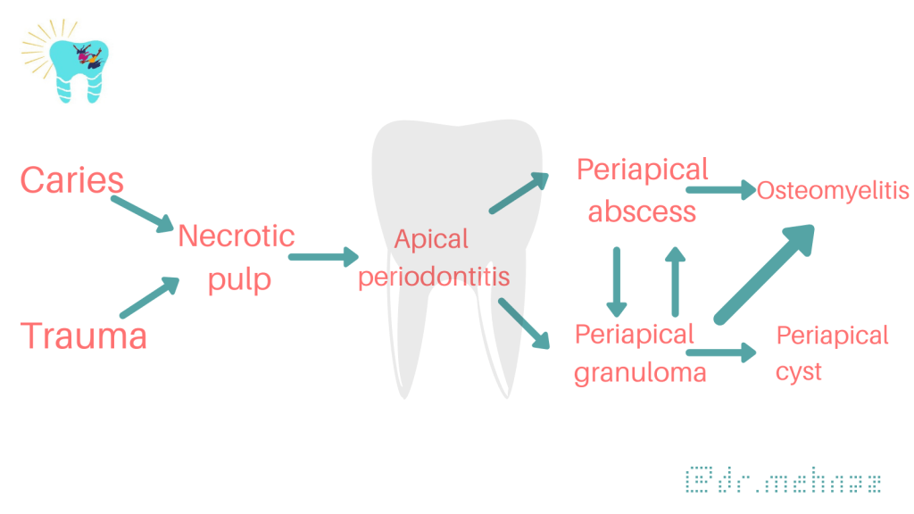
When formulating radiological differential diagnosis, features should be evaluated carefully, such as
• Location: With periapical inflammatory lesions, which are pathological conditions of the pulp, the epicenter typically is located at the apex of a tooth.
• Periphery: Ill defined
• Effects on surrounding structures: Periapical lesions cause widening of PDL space at apical region of the root.

PERIAPICAL RADIOLUCENCIES
Developmental
Lateral periodontal cyst

Inflammatory Lesions
Apical periodontitis, periapical abscess


Cystic Lesions
Periapical (radicular) cyst

Odontogenic keratocyst

Glandular odontogenic cyst
Benign Tumors
Ameloblastoma

Malignant Tumors
Ameloblastic carcinoma


Dentowesome|@drmehnaz🖊
References:
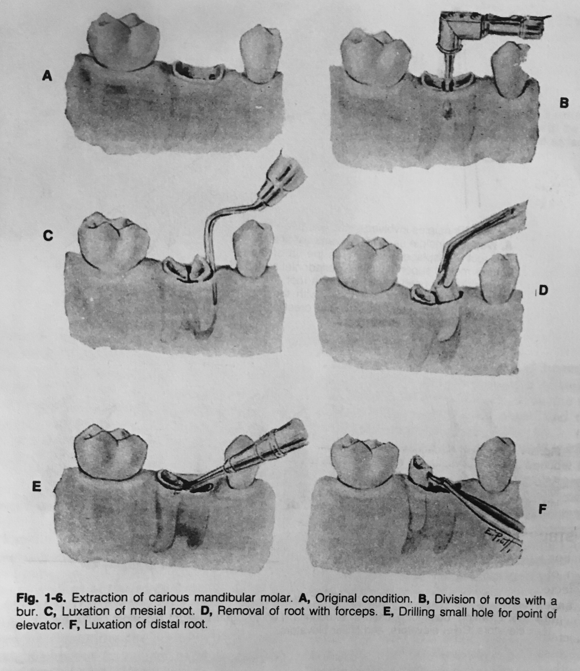
The extraction of a tooth requires the separation of its attachment to the alveolar bone via the crestal and principal fibers of the periodontal ligament. Also involved is a process of expanding the alveolar socket.
After the tooth has been removed, the form of alveolar process is restored by finger pressure. Bleeding is arrested by means of a pressure pack placed between the jaws, and the wound is allowed to fill with a blood clot.
The following techniques may be used for tooth removal:
Odontotomy can be used to facilitate any of these three procedures.

Dentowesome|@drmehnaz🖊
References: Textbook of Oral Surgery – Daniel M Laskin
1) https://pubmed.ncbi.nlm.nih.gov/8832476/
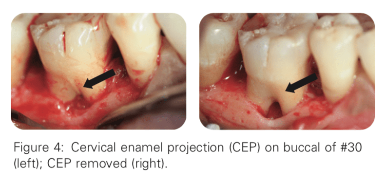
Masters and Hoskins reported that 90% of mandibular furcation invasions have CEPs
2) https://www.ncbi.nlm.nih.gov/pmc/articles/PMC2848791/
Bissada and Abdelmalek reported a 50% correlation between CEPs and furcation invasion
3) https://pubmed.ncbi.nlm.nih.gov/19518034/
The length of root trunk affects attachment loss The longer a given root trunk, the less likely a furcation will be predisposed to attachment loss. Teeth with Taurodontism usually have apically displaced furcation and longer root trunk length
4) https://pubmed.ncbi.nlm.nih.gov/13513891/
Intermediate bifurcation ridges are ridges spanning across the bifurcation of mandibular molars in the mesiodistal direction These ridges are present in 70–77% of the mandibular molars. Just like other anatomic structures, the presence of an intermediate bifurcation ridge may hinder effective plaque control and root preparation by both the patient and dentist
5) https://www.nature.com/articles/bdj.2007.1059
Buccal radicular grooves and palato-gingival grooves are developmental phenomena that affect mainly the maxillary anterior teeth. These grooves run on the roots in the coronal-apical direction Due to their anatomy, the grooves frequently provide a plaque-retentive area that is very difficult to instrument, making teeth with these developmental grooves more prone to attachment loss
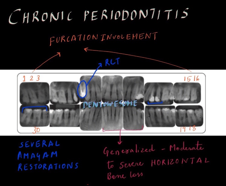
A 47-year-old female presented with a chief complaint of: “My gums around one of the lower right teeth hurts.” The patient reported soreness and discomfort around tooth #30 from time to time, especially on the buccal side. On occasion, the patient experienced bleeding when brushing her teeth
LEARNING GOALS AND OBJECTIVES
To be able to identify local anatomic factors that may contribute to periodontal disease
To understand the anatomy of the furcation and root
To be able to diagnose a furcation invasion using a furcation classification system


LINK: https://drive.google.com/file/d/1R3qBKH78nqyZ-3Fe4YkvMJjsSbcG9EXw/view?usp=sharing
Sinus
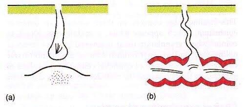
Latin: Hollow (or) a bay
A sinus is a blind tract leading from the surface down to the tissue. There may be a cavity in the tissue which is connected to the surface through a sinus. The sinus is lined by granulation tissue which may be epithelized.
A sinus has one open draining end and the channel ends in a blind ending. An example would be a dental sinus draining from a dental abscess to either the inside of the mouth or the skin.

Fistula
Latin: flute(or) a pipe (or) a tube
It is a communicating tract between 2 epithelial surfaces commonly between hollow viscous & skin or between 2 hollow viscera. The tract is lined by granulation tissue which is subsequently epithelized. A fistula may be an abnormal communication between vessels.
An example would be from the mouth (oral cavity) to the skin surface, usually of the face or neck, and this specific type is called an orocutaneous fistula.


Fistulas and sinuses of the neck and face may be classified by cause.
Fistulas and sinuses due to developmental causes are usually present at birth.

Cysts are lumps in the skin containing fluctuant contents. They may have an opening to the skin surface.
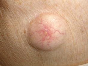
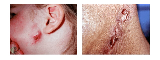
In addition to careful history and examination, one or more of the following tests will usually be required to confirm the diagnosis and determine the cause:
Abscess
Collection of pus
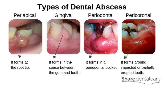
Features of acute inflammation; The four cardinal signs of inflammation are:
Diagnosis of Dental Abscess:
Dentowesome|@drmehnaz🖊
References:
Link: https://drive.google.com/file/d/11de2DeD4_Xsh_PeNEW7sbP2l1J1mA5Ns/view?usp=drivesdk
LEARNING GOALS AND OBJECTIVES
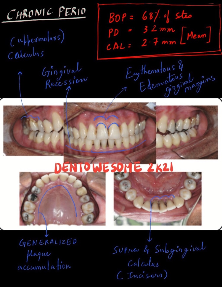
To be able to identify the clinical features and overall characteristics of chronic periodontitis
To be able to list difficulties in the proper diagnosis of early chronic periodontitis
To understand possible overlaps with the
diagnosis of aggressive periodontitis
To know what clinical changes can be anticipated in the response of chronic
periodontitis to anti-infective therapy

A swelling is a value term that denotes only enlargement or protuberance in body due to any cause.
According to the cause a swelling may be congenital, traumatic, inflammatory, Neoplastic or miscellaneous.
Examination of a swelling should be accompanied by a complete history of the patient. Following points should be noted:
• Duration: The clinician may ask ‘when was the swelling first
noticed’? Swellings that are painful and of shorter duration are mostly inflammatory (acute), whereas those with longer duration and without pain are chronic, e.g. a chronic periapical abscess.
• Mode of onset: The clinician may ask ‘how did the swelling start’? The history of any injury or trauma or any inflammation may contribute to the diagnosis and nature of the swelling.
• Progression: The clinician should ask ‘has the lump changed in size since it was first noticed? Benign growths such as bony swellings grow in size very slowly and may remain static for a long period of time. If the swelling decreases in size, this suggests of an inflammatory lesion.
• Site of swelling: The original site where it started must be assessed.
• Other symptoms: Pain, fever, difficulty in swallowing, difficulty in respiration, disfigurement, bleeding or pus discharge are the common symptoms associated with swellings in the orofacial region.
• Recurrence of the swelling: many swellings do recur after removal of the tissue, indicating the presence of precipitating factor, e.g. ranula.

(A) Inspection

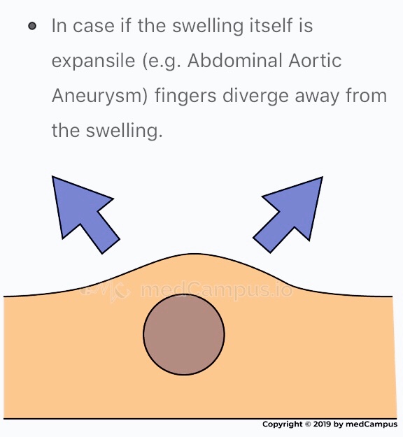

(B) Palpation

Surface: Can be👇🏻
• Smooth (cystic swellings)
• Lobular with smooth lumps (lipoma)
• Nodular (multinodular goitre)
• Matted (lymph nodes)
• Irregular (carcinoma)
Margins: Well defined/indistinct👇🏻
• Malignant growth - irregular margin
• Acute inflammatory swelling - ill defined margin
• Benign tumor - swelling slips & is indistinct
Edge👇🏻
• Well defined & regular-Benign Neoplasms
• Well defined & irregular-Malignant Neoplasms
• Ill defined & diffuse - Inflammatory swellings
Consistency: 👇🏻
• Soft - lipoma
• Cystic - Cyst or chronic abscesses
• Firm - Fibroma
• Hard - Chondroma
• Bony hard - Osteoma
• Stony hard - Carcinoma
• Variable consistency - Malignancy
An ulcer is break in continuity of epithelium, skin or mucous membrane. A proper
history must be taken in case of an ulcer:

• Mode of onset: The clinician may ask ‘how has the ulcer developed’? The patient may provide significant information about the nature and etiology of the ulcer such as any trauma or spontaneously.
• Duration: The clinician may ask ‘how long is the ulcer present here’? It determines the chronicity of the ulcer. For example, traumatic ulcers in oral cavity are acute (occurring for a short period), but if the agent persists; it may become a chronic ulcer.
• Pain: The clinician may ask ‘is the ulcer painful’? Most of the ulcers, being inflammatory in nature, produce pain. Painless ulcers usually suggest nerve diseases (such as peripheral neuritis, syphilis, etc).
• Discharge: Any blood, pus or serum discharge must be noted.
• Associated disease: Any associated generalized systemic problem may be associated with the ulcers of oral cavity (such as
tuberculosis, squamous cell carcinoma, etc).
(A) Inspection
Size & Shape:
• Tuberculous ulcer - oval with irregular border
• Varicose ulcer - vertical & oval in shape
• Carcinomatous ulcer - irregular
Number: Tuberculous, inflammatory ulcer may be more than one in number
Position:
• Arterial ulcer: Tip of the toes, dorsum of the foot
• Varicose ulcer: lower limb
• Perforating ulcers: over the sole at pressure points
• Non-healing ulcers: over the shin
• Rodent ulcer: upper part of face
Edge: An area between margin & floor. In spreading ulcer, edge is inflamed. Undermined edges destroy subcutaneous tissue faster than skin.


Floor: This is the part of the ulcer which is exposed or seen.
• Red granulation tissue - Healing ulcer
• Necrotic tissue, slough - spreading ulcer
• Pale, scanty granulation tissue - tuberculous ulcer
• Wash-leather slough - Gummatous ulcer
Discharge:
• Serous discharge - Healing ulcer
• Purulent discharge - Spreading ulcer
• Bloody discharge - Malignant ulcer
• Discharge with bony spicules - Osteomyelitis
• Greenish diacharge - Pseudomonas infection
(B) Palpation
Tenderness: Characteristic of infected ulcers and arterial ulcers.
Induration: The edge, base and the surrounding area should be examined for induration
• Maximum induration - Squamous cell carcinoma
• Minimal induration - Malignant melanoma
• Brawny induration - Abscess
• Cyanotic induration - Chronic venous congestion as in varicose ulcer
Mobility: Malignant ulcers are usually fixed, benign ulcers are not.
Bleeding: Malignant ulcer is friable like a cauliflower. On gentle palpation, it bleeds. Granulation tissue as in a healing ulcer also causes bleeding.
Surrounding Area:
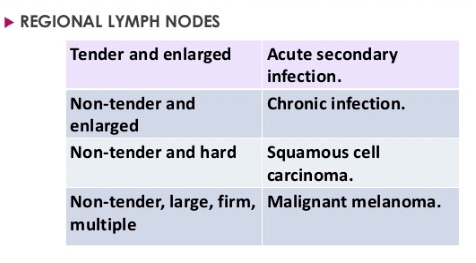
Relevant Clinical Examination:
Dentowesome|@drmehnaz🖊
References:

