Diagnosis Demystified – Case 6/255

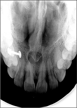
A pear-shaped and well-circumscribed radiolucent lesion with a corticated outline was found on a radiograph related to the root of an upper central incisor. The tooth was not restored and proved vital on testing.

A nasopalatine duct cyst is a specific type of cyst that develops from remnants of the nasopalatine duct. During embryonic development, this duct is involved in the formation of the nasal and oral cavities. Sometimes, remnants of this duct can persist and give rise to a cystic lesion later in life.

The nasopalatine duct extends from a structure called the incisive canal, which is located in the midline of the maxillary bone (the bone that comprises the upper jaw). The cyst can develop anywhere along this tract.

When we examine the lining of a nasopalatine duct cyst under a microscope, we typically observe the presence of both respiratory and squamous epithelium. This finding is significant because it indicates the origin of the cyst from the nasopalatine duct. The respiratory epithelium represents the lining of the nasal portion of the duct, while the squamous epithelium represents the lining of the oral portion.

Furthermore, a nasopalatine duct cyst often contains a neurovascular bundle within its capsule. This bundle consists of nerve fibers and blood vessels that supply the nasopalatine area. Its presence within the cyst is a characteristic feature.

In clinical practice, nasopalatine duct cysts are usually asymptomatic and are often discovered incidentally during routine dental examinations. However, if the cyst becomes large or infected, it can cause pain, swelling, and discomfort in the affected area.

As a dental student, it’s important to be familiar with the clinical presentation, radiographic appearance, and management of nasopalatine duct cysts. Treatment typically involves surgical removal of the cyst to relieve symptoms and prevent potential complications.

Endodontics and Pulp Therapy – NEET and INICET pearls from Dentest and Pulse

  1. Pulp of all primary teeth is active for a period of = 8.3 years/9.6 years
  2. Infected dentin (TRUE STATEMENTS)
    1. Loss of organic network 
    2. Lacking sensation
    3. Irreversible demineralization 
    4. Excavated during cavity preparation 
  3. Affected dentin (TRUE STATEMENTS)
    1. Demineralised but can be mineralized
    2. No loss of organic network 
    3. Sensitive
    4. Should be left out for remineralisation during cavity preparation
  4. The dye used for the detection of dentin caries is = Acid red system
  5. The greatest problem in pulpal diagnosis is estimating the amount of = INFLAMMATION
  6. Radiographs of permanent molar 
    1. Acute apical abscess shows
      1. Early stages – no radiographic changes
      2. Later stages – little change from the normal structure
    2. Acute alveolar abscess = thickened periodontal membrane
    3. Chronic pulpal infection = rarefaction at bony furcation
    4. Carious involvement = Radiolucency in the furcation area 
  7. ACUTE ALVEOLAR ABSCESS (TRUE STATEMENTS)
    1. Primary tooth = more diffuse 
    2. A localized collection of pus at root apex in alveolar bone
    3. There is swelling, mobility and elevation of a tooth from its socket 
  8. Pulse oximetry 
    1. Measures the pulpal circulation directly 
    2. TRUE STATEMENTS
      1. Objective test
      2. Measures and compares amplitudes of ratios of transmitted infrared light with red light 
      3. Capable of evaluating the stature of blood vasculature
      4. Depends on pulsatile blood flow
      5.  
  9. Laser doppler flowmetry
    1. Based on the velocity of RBC cells in capillaries 
    2. A non-invasive method to measure blood flow
    3. GAZELIUS
  10. Indirect Pulp Therapy 
    1. Objective = Eliminate the removal of deep caries
    2. Indicated (imp)
      1. in primary molars when the carious lesion is suspected of producing an exposure of the pulp
      2. when there is no history of spontaneous pain  = Tooth must be asymptomatic 
    3. IPT = It is dependent on the remineralisation of affected dentin and reparative dentin formation  
    4. ​​
    5. The success of IPT = placement of temporary restoration with excellent sealing properties
    6. Most ideal material = Calcium hydroxide
      1. Introduced by FAUCHARD
      2. When applied to residual carious dentin = it remains for 6 – 8 weeks 
      3. Following amputation of the coronal portion of the pulp of an immature permanent first molar = the stump is capped with CaOH
      4. The success of CaOH pulpotomy is determined by = Continuation of root formation and apexogenesis
      5. Dentin bridge is formed = at a level slightly below the amputation
  11. Direct Pulp Therapy 
    1. Amount of repartive/3* dentin formed after 30 days = 1.5 microns/day and then slows down after 48 days 
    2. Contraindicates in primary teeth 
    3. Prognosis is best = mechanically exposed permanent tooth
    4. Indicated in cases of little (Pinpoint exposure = less than 1 mm) or no haemorrhage from the exposure site
    5. The pulp should be vital
    6. Done in traumatic or iatrogenic cases = Reported within 24 hours
    7. High rate of failure = high cellular content of pulp
  12. Pulpotomy
    1. The simplest form of pulp therapy 
    2. Contraindicated = If there is Inflammation of radicular pulp and pain
    3. Mechanical exposure of mesiobuccal pulpal horns in primary maxillary 1st molar with moderate caries lesion on the mesial-distal surface = formocresol PULPOTOMY + stainless steel CROWNS
    4. Pulp preservative material and best   = MTA
    5. The primary function of MTA = Apexification of immature traumatised pulpless tooth
    6. Important criteria of success = Root end completion 
  1. Formocresol Pulpotomy 
    1. SWEET
    2. Composition of Buckley Solution
      1. 1:2 parts F: C
      2. 19% formaldehyde 
      3. 13% cresol
      4. 15% glycerine 
    3. Successful treatment for vital primary second molar with a large carious and pulpal exposure 
    4. The success of the technique depends on vital root pulp 
    5. Cotton pellet applied to the pulpal stumps after formocresol pulpotomy should be = Dampened with formocresol and placed for 5 mins
    6. Amount of pulp to be removed in formocresol technique= entire coronal pulp down to the cervical constriction of each root canal
    7. FERRIC SULPHATE = material with haemostatic effect, used as a substitute for formocresol pulpotomy
    8. EFFECT = Surface fixation of pulpal tissue accompanied by degeneration of odontoblasts 
    9. The first sign of formocresol technique failure = Internal resorption
  2. Glutaraldehyde Pulpotomy 
    1. Superior fixative properties 
    2. Conc as pulpotomy agent = 2 to 5 % concentration 
    3. The cotton pellet is kept on pulp stumps = for 3-4 mins
    4. KOPEL
  3. Ferric Sulphate Pulpotomy 
    1. 2 – 5 seconds 
    2. 15.5% concentration
    3. 100% clinical success rate
  4. CaOH Pulpotomy
    1. contraindicated in primary teeth due to internal resorption – odontoclastic reaction
    2. Immediate reaction = Severe inflammatory reaction
    3. The calcified bridge is evident within = 1 month
    4. Increases in thickness – next 12 months 
    5. Failure of technique = 
  5. Cvek Pulpotomy
    1. Partial technique
    2. Important criteria of success = Root end completion 
  6. Pulpectomy
    1. The entire pulpal tissue is removed
    2. Complete Pulpectomy technique for primary molars is developed by = STARKEY
    3. Pulp extirpation in primary teeth is difficult because = Tortuous anatomy and branching of canals 
    4. The major problem with this technique = is limited knowledge of pulpal anatomy
    5. KRI paste used for obturation in case of pulpectomy is primarily a mixture of = Iodoform and CMCP
    6. MC filling material, traditionally = Zinc Oxide Eugenol 
    7. ROOT CANAL SPREADER = It is not used in the widening of root canals 
  7. Pulp Devitalization 
    1. PARAFORMALDEHYDE = common medicament in all types of devitalization pulpotomy 
    2. Laser = Nd: YAG
  8. Pulp mummification = Indicated in = traumatic exposure of a vital primary tooth 
  9. Apexification
    1. Rx of pulpless/nonvital immature young permanent tooth
    2. A condition that leads to apexification = Pulpectomy of the young permanent tooth with incomplete root formation
    3.  Any excess CaOH periapically = will be removed by multinucleated giant cells 
    4. Increase success rate = Increased blood supply through the wide apex
    5. Best obturated by = OBTURA – thermoplasticized gutta percha system
    6. FRANK technique 
      1. Uses CaOH and CMCP (Camphorated monochlorophenol) 
      2. To stimulate root closure/ apexification in incompletely developed young permanent tooth 
      3. CMCP = 6 months
  10. Rate of resorption in pulpless primary teeth compared to vital teeth = SIMILAR
  11. In the absence of second premolar roots of primary second molar will most likely = resorb more slowly than normal
  12. Ideal Root Canal filling = CaOH
  13. Rx choice in poor prognosis cases, alveolar abscess and necrotic pulp cases = extraction 
  14. Apexogenesis
    1. TRUE STATEMENTS
      1. More of a physiological process
      2. Root development continues
      3. Indicates in pulp tissue with mild inflammation 
    2. Done in the vital and infected tooth
  15. In regenerative endo therapy, Metronidazole is replaced by = Ornidazole 
  16. Reattachment of fractured tooth fragments using the resin bonding technique is called = Fragment Restoration 
  17. STAINLESS STEEL CROWN
    1. Rx of choice for Ankylosis of a primary molar with the absence of a permanent successor
    2. Rx of choice for amelogenesis imperfecta in primary dentition = SSC
    3. HUMPHREY
    4. SSC is a = Semi-permanent restoration 
    5. Contraindicated in Medically compromised patients – Heart problems – VSD, ASD
    6. INDICATIONS 
      1. Restoration of hypoplastic teeth 
      2. Following pulpotomy or pulpectomy 
      3. As an abutment for space maintainers
      4. NOT USED = teeth that are not restorable 
    7. Iron content in 3M SCC = 10%
    8. CHENG CROWNS = pre-veneered crown which is stain resistant and pre-crimped
    9. Countering of stainless steel crown is done at middle 1/3rd of the crown to produce = BELLING EFFECT
    10. MC complication while tooth prep = LEDGE Formation
    11. PREVENTION OF LEDGING = most important for using burs in opening proximal contacts to receive SSC
    12. SCC should extend
      1. intra gingivally or below the gingival crest = 0.5 – 1mm 
      2. Occlusal = 1.5-2mm 
      3. Buccal and lingual = 0.5mm 
      4. Proximal = 1-1.5mm 
    13. Basket Crown technique
      1. Temporary crowns 
      2. Placed in the anterior tooth 
      3. 3/4th crown
      4. The window is prepared on the LABIAL aspect for aesthetic

    14. Finish Line of SSC = FEATHER edge
    15. Difficulty in adopting an SSC to a primary mandibular first molar = Buccal cervical ridge showing constriction
    16. Surface requiring the least amount of reduction = BUCCAL and LINGUAL 
    17. Retention is achieved primarily by = PARALLEL distal and mesial walls
  18. In a con-compliant pulp chamber, average intrapulpal pressure = 10 mm HG and varies with each arterial pulse
  19. PREPOMETER = device to measure the thickness of the dentin layer above the pulp chamber
  20. PULPDENT = most capable of stimulating early dentinal bridge formation
  21. Obturation of deciduous teeth can be done with = Iodoform paste

Diagnosis Demystified- Case 6/255

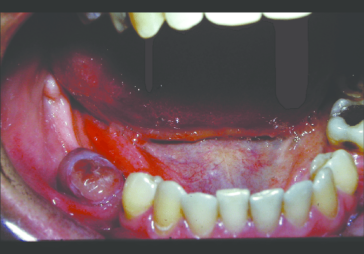
A 26-year-old African patient presented with a rapidly growing lesion that expanded the mandible. There was bone destruction on the radiograph and therefore malignancy was suspected. On biopsy, the lesion was intraosseous and was cavitated. There was profuse bleeding and a small biopsy of the lining was taken. The pathologist reported osteoclast-like giant cells and granulation tissue with blood clots.

An aneurysmal bone cyst is a type of benign bone tumor that can occur in the jawbone. It is called an “aneurysmal” because it contains blood-filled spaces that can resemble the sac-like structure of an aneurysm. These cysts are relatively rare, accounting for less than 1% of all bone tumors.

Clinically, an aneurysmal bone cyst can be dramatic because it can grow rapidly and attain a large size. This can cause symptoms such as pain, swelling, and displacement of nearby teeth. When a biopsy is performed to confirm the diagnosis, bleeding can be profuse due to the presence of fragile blood vessels within the cyst. In some cases, a blood transfusion may be necessary to manage the bleeding.

It is important to note that aneurysmal bone cysts often form around an underlying primary lesion, such as a vascular malformation or bone tumor. Therefore, it is crucial to thoroughly evaluate any aneurysmal bone cyst to determine if there is an underlying primary lesion that needs to be addressed. Treatment options for aneurysmal bone cysts may include surgical excision or minimally invasive procedures such as sclerotherapy or curettage.

As a dental student, it is important to be aware of aneurysmal bone cysts as a potential differential diagnosis when evaluating patients with jaw pain, swelling, or other symptoms. If you suspect a patient may have an aneurysmal bone cyst, referring them to an oral and maxillofacial surgeon for further evaluation and management is important.

Diagnosis Demystified- Case 5/255

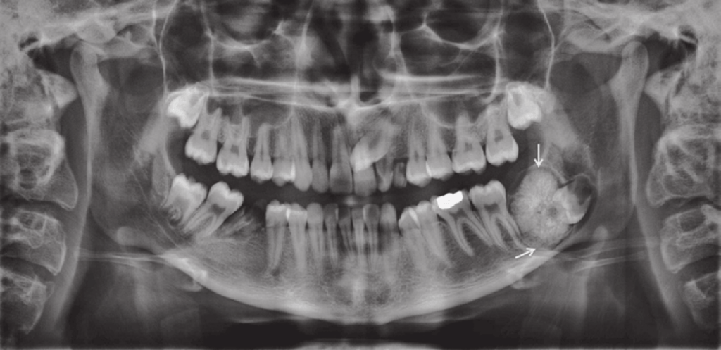
A radiolucent lesion was found incidentally on a dental panoramic radiograph in a 30-year-old man. The cyst was located in the lower molar area above the inferior alveolar canal and showed a scalloped outline extending between the roots of the teeth. All teeth in the area were vital and the lamina dura was intact. Clear straw-coloured fluid was aspirated from the lesion

Solitary bone cyst (SBC), also known as simple bone cyst, is a benign bone lesion that most commonly occurs in children and adolescents. While the exact cause of SBC is still unknown, it is believed to be the result of a disturbance in the normal bone remodeling process.

SBC typically presents as a painless swelling or bump in the affected bone, often discovered incidentally on routine imaging. Diagnosis is usually made by imaging studies such as X-rays or MRI, as well as fine needle aspiration to confirm the presence of fluid within the cyst.

In many cases, SBCs will heal on their own without the need for any intervention. However, if the cyst is causing significant pain or functional impairment, or if it is at risk of fracturing or expanding and causing further damage to the bone, treatment may be necessary.

Treatment options for SBC include observation, which involves monitoring the cyst with regular imaging studies to ensure that it is not growing or causing any problems; curettage, which involves surgically removing the cyst and filling the cavity with bone graft material to promote healing; or injection of bone-stimulating agents such as bone morphogenetic protein (BMP) to promote healing and prevent recurrence.

The decision on whether to intervene or not will depend on various factors, including the size and location of the cyst, the age of the patient, and the presence of any associated symptoms or complications.

Childhood Diseases – NEET Pearls from Dentest and Pulse – 2023

  1. Normal colour of
    1. Primary teeth = China white
    2. Permanent teeth = Ivory white
  2. In primary dentition =
    1. dentine is uniformly calcified 
    2. Enamel and dentin is less mineralized
    3. Enamel and dentin thickness is less
    4. DEJ is less scalloped
  3. Main difference between primary and permanent tooth = Mineral content 
  4. Cleft of secondary palate causes = cleft lip
  5. Ectodermal dysplasia in 3-year-old child with only primary molars and canine and overall appearance of an older person
  6. Treatment of geographic tongue = No treatment
  7. Dentinogenesis Imperfecta 
    1. Grey in colour
    2. Exhibit extensive occlusal and incisal wear
    3. r/g = secondary dentin deposition
    1. Transillumination of soft tissue is useful in detection of = Sialolithiasis 
  8. KOPLIK SPOTS
    1. Small irregular bright red spots with white specks in centre 
    2. Indicate the onset of RUBEOLA/MEASLES
  9. MC cause of acute generalised inflammation 
    1. in pre-school children = Acute herpetic gingivostomatitis/ herpes
      1. Child with fever of 102*C and vesicles in oral cavity
      2. Rx = symptomatic rx for fever and prevent secondary infection and dehydration
    1. In adolescents = ANUG
  10. Apthous ulcer should be treated by = Palliation and Patience
  11. Rx of severe intraoral infection differs from that in adult because = dehydration occurs more rapidly and severely in children
  12. The purulent lesion in the oral vestibule of an 8-year-old child = ODONTOGENIC FISTULA
  13. Bluish dome shaped lesion on the inside of a lip = Mucocele
  14. MC in children = gingivitis
    1. Reaches in its severity at peak of = 11 – 13 years
  15. Gingiva in children = less keratinized, less stippled
  16. NOT seen on attached gingiva = interdental clefts
  17. Gingival stripping in children = due to narrow attached gingiva 
  18. Periodontosis or Juvenile Periodontitis
    1. Vertical pockets around incisors and first molars
    2. Presence of plaque isn’t consistent with inflammatory findings 
  19. NOT related to gingival inflammation = Spirochetal infection
  20. Pulp starts reacting to caries when carious lesion reaches = Dentinoenamel Junction 
  21. Periapical abscess more diffuse in primary teeth because = Alveolar bone surrounding the teeth is less dense
  22. Gingival abscess is common in primary teeth because = More accessory canals are present on buccal surface with porous floor and thin bony surface
  23. MCC of draining sinuses in oral cavity of children = chronic periapical abscess
  24. Hypoplasia of primary enamel that forms before birth is = RARE
  25. Eruptive cysts are best treated by = No treatment, only observation
  26. A disease that only affects formation and eruption of tooth but doesn’t cause hypoplasia = RICKETS
  27. OSTEOGENESIS IMPERFECTA = doesn’t cause delayed eruption
  28. MANDIBULAR second premolar = shows greatest variation relative to the onset of mineralization of crowns
  29. Child with Down syndrome (all are important points)
    1. Affectionate and cooperative
    2. Capable of understanding operative procedures
    3. Fearful of quick movements
    4. Simian crease in hand
    5. Head shape = brachycephalic 
    6. Incidence of caries = less as compared to general population 
    7. High incidence of periodontal disease
    8. Retarded eruption
    9. Delayed exfoliation**
    10. Retained deciduous teeth and microdontia
    11. Typical face = hypoplastic maxilla
    12. IQ = 120 – 139
    13. Syndrome associated with congenital heart lesions
  30. Fluoride is contraindicated in = Chronic renal failure 
  31. DIABETES
    1. Serious complication of juvenile diabetes/ DM – 1 = blindness
    2. MC type of diabetes = DM – type 2
    3. Level of glucose for diabetic ketoacidosis = 300 to 600 mg/dl 
  32. LEUKAEMIA 
    1. Child suffering from acute leukaemia is more susceptible to = Oral infections
    2. Primary cause of death in patients = Infections 
    3. TRUE STATEMENTS 
      1. May be manifested by mucosal pallor
      2. Cause Obvious Purpura 
      3. Lymphoblastic variety 
    4. Line of Rx for periapical abscess = Obtain medical consultation before treatment 
    5. Down Syndrome = 10 – 20 fold increased risk of leukaemia pt  
  33. HEMOPHILIA
    1. Epsilon aminocaproic acid = given to haemophilic child before surgical procedures to control bleeding
    2. Line of Rx for periapical abscess = Obtain blood count before extraction, determine the amount and duration of factor concentrate replacement 
    3. In extraction cases, Minimal level of Factor VIII = 50%
    4. Minimal level needed for adequate hemostasis = over 25%
    5. 5 years old, primary second molar with non vital pulp is treated by = conventional pulpectomy
    6. Mode of anaesthesia contraindicated = INTRAMUSCULAR
    7. MC type = Hemophilia A
  34. CYSTIC FIBROSIS (all points imp)
    1. Maldigestion and malnutrition
    2. Chronic respiratory infection
    3. Thyroid deficiency 
    4. Water and electrolyte imbalance
    5. Staining of teeth = yellow in colour  
  35. Yellowish primary tooth due to trauma indicates = calcific reaction of pulp
  36. Pink primary tooth indicates = Internal resorption
  37. TETRACYCLINE PIGMENTATION
    1. Yellow primary tooth, under UV light – faint overall yellow green autofluorescence = Tetracycline pigmentation
    2. TS occurs in primary tooth during the period of = mineralization of first millimetre of dentin at DEJ
    3. Tetracycline administration causes primary tooth staining = upto 9th month of life
  38. Sensitive period for tetracycline discolouration 
    1. PRIMARY
      1. Max and mand incisors = 4 months in utero to 3 months postpartum 
      2. Max and mand canines = 5 months in utero to 9 months postpartum 
    2. PERMANENT
      1. Max and mand incisors and canines = 3 months postpartum to seventh year of life
  39. SEIZURES
    1. Rx of Petit mal seizure in dental office = Watch until episode passes away
    2. Occurring in children several times in a day with no involvement of aura = Petit Mal 
    3. MC type of epilepsy seen in children = Petit Mal 
  40. Following are associated with slowness of mental retardation in a child
    1. Family history
    2. Metabolic disease
    3. Pregnancy and delivery history 
    4. Phenylketonuria
    5. Galactosaemia 
    6. Non trainable type of mental retardation = IQ level of below 20 
  41. CEREBRAL PALSY
    1. Features seen
      1. Increases Caries
      2. Increased salivation
      3. NOT SEEN = fluorosis 
    2. Most of CP children = SPASTIC
    3. MC type of CP = Spastic and Athetosis
    4. SPASTICITY = characterised by sudden violent involuntary contraction of a muscles
    5. TYPES
      1. Spasticity = muscular contractions which are irregularly spaced and have no purpose 
      2. Athetosis = slow, worm like, constant involuntary uncontrollable purposeless movements 
      3. Ataxia = disturbances of balance and equilibrium
      4. Rigidity = marked resistance to passive motion
    6. MC classifications = physiological and topographic 
    7. Oral Manifestations 
      1. Periodontal diseases
      2. High incidence of caries
      3. Attrition of teeth due to bruxism
    8. Class II Divison 2 = mc malocclusion observed in pt with spastic type 
  42. AUTISM
    1. Delayed milestones, playing with herself, unable to make friends, and difficulty in learning 
    2. It is a severely incapacitating disturbance of mental and emotional development that causes problem in learning, communication and relating to others 
    3. Incapacitance of emotional and mental disturbance
    4. Parrot like repetition speech 
    5. Also known as Kanner’s syndrome
  43. RESTRAINTS
    1. In uncooperative child, last resort is = Physical restraints
    2. TRUE STATEMENTS
      1. Papoose board is for restraining body
      2. Use of restraining device is a passive method
      3. Posey straps are used for restraining extremities
    3. MC restraints in children with neuromuscular diseases = Papoose Board
    4. Mouth props are used as = Restrainers
    5. Bean body is used in dental chair for = Restraining body
  44. BACTERIAL ENDOCARDITIS
    1. Antibiotic prophylaxis = 1 hour before
    2. Prophylactic antibiotic coverage in a child with history of rheumatic fever is precaution against developing = subacute bacterial endocarditis
  45. MOUTH BREATHING
    1. Reliable method for quantifying the extent of mouth breathing = Rhinomanometry
    2. TRUE STATEMENTS
      1. Increase incidence of caries in open bite cases
      2. Decrease in masticator scouring action in the area of open bite
  46. GENOME
    1. Albino mother, normal father, chances of children being carriers = 50% carriers
    2. ALBINISM = Autosomal Recessive, alternate generations, not affected by sex
    3. Normal parents, affected male infant = Polygenic disorder
    4. Affected father, one child affected out of three = Autosomal Dominant 
    5. Affected fathers, affected grandsons = X linked recessive
    6. Affected fathers, affected grand-daughters = X linked dominant
  47. HANDICAPPED CHILDREN
    1. Radiograph of choice = panoramic 
    2. Mc tooth brushing technique = Horizontal scrub
  48. Use of penicillin on a patient with asthma = CONTRAINDICATED
  49. Chicken wire appearance of alveolar bone = THALASSAEMIA 
  50. Virus responsible for causation of AIDS = HTLV 3
  51. Denture bearing mucosa in children as compared to adults = thinner and well-circulated 
  52. Impression taking order in paediatric prosthetic procedures = upper first and then of lower jaw 
  53. Green stains frequently seen in children’s teeth = chromogenic bacteria 
  54. Tender, painful, unilateral or bilateral swelling of salivary glands = Parotitis or Mumps
  55. Dentinogenesis Imperfecta = dark brown coloured tooth, frequently broken bones associated with blue sclera
  56. Delayed eruption of permanent teeth and large tongue = Hypothyroidism 
  57. Premature eruption of permanent teeth and large tongue = Hyperthyroidism 
  58. Fever, acute painful ulcers, lymphadenopathy = Acute herpetic gingivostomatitis
  59. Exanthematous fever and strawberry tongue = Scarlet fever
  60. Congenital anodontia and perspiration = Ectodermal dysplasia
  61. Brown discoloration of teeth, malnutrition, steatorrhoea, disturbances in function of exocrine glands = Cystic Fibrosis 
  62. Multiple supernumerary teeth unerupted with open fontanelle and abnormal development of clavicles = Cleidocranial dysostosis
  63. Causes both hypocalcified as well as irregular but well calcified enamel = Amelogenesis Imperfecta
  64. Flabby white lesions occur bilateral and seen in few other members of family = White sponge nevus
  65. DRUGS
    1. MC used antibiotic in child allergic to penicillin = Erythromycin
    2. Common antagonist of meperidine = NALOXONE
    3. Moniliasis/Candidiasis 
      1. Unfavourable oral sequel with prolonged use of antibiotics in children 
      2. Thick curd like white patch, on rubbing leaves erythematous patch
  66. PERCENTAGES
    1. Gingivitis at age of 1 – 13 years = 90%
    2. Periodontitis by age 18 years = 25%
  67. Dentitia Tarda = Retaded eruption of deciduous dentition 
  68. MC malignant tumour = Osteosarcoma
  69. Chronic periapical infection in primary molars, first noted as = rarefaction of bony furcation
  70. MC cause for gingival fistula = chronic apical lesion
  71. CLEFT PALATE
    1. Alveolar grafting in a pt of cleft should be ideally placed = after maxillary expansion, cross bite correction and before cuspid eruption
    2. After treating the cleft palate, the occlusion is = Unilateral and bilateral crossbite

Diagnosis Demystified – Case 4/255

A 22-year-old man attended for treatment. He had a history of Gardnerʼs syndrome and noticed a bony hard lump on the ascending ramus of the mandible.

Gardner’s syndrome, also known as familial adenomatous polyposis (FAP), is a rare genetic disorder that affects the colon and rectum. It is caused by mutations in the APC gene and is inherited in an autosomal dominant pattern.

Individuals with Gardner’s syndrome develop numerous polyps in the colon and rectum, which can eventually lead to colorectal cancer if left untreated. Additionally, they may also develop polyps in the stomach and small intestine. The condition is typically diagnosed in the teenage years or early adulthood.

Gardner’s syndrome can also cause abnormalities in the jaw, including multiple osteomas, odontomas, and areas of hazy sclerosis. These dental findings may be one of the earliest signs of the condition and can help with early diagnosis.

Image Based Questions – Orthodontics – Part 3

  1. IDENTIFY = DISTAL END CUTTER
  2. IDENTIFY = ADAMS PLIER
  3. IDENTIFY THE MALOCCLUSION =
    1. ANTERIOR CROSSBITE
    2. SCISSOR BITE
  4. IDENTIFY THE FIGURE = CBCT SCAN FIGURE
  5. IDENTIFY THE ANGLE MARKED BY THE ARROW IN THE
    1. MAXILLARY STUDY MODEL = 70 DEGREES
    2. MANDIBULAR STUDY MODEL = 65 DEGREES
  6. WHAT DO THE POINTS ON THE FIGURE DEPICT?
    1. GINGIVAL ZENITH
    2. GOLDEN PROPORTIONS
    3. CONNECTORS
  7. IDENTIFY THE GREEN LINE = HARMONY LINE
  8. IDENTIFY THE MALOCCLUSION AND HABIT = CLASS 3 MALOCCLUSION AND TONGUE THRUSTING HABIT
  9. IDENTIFY THE APPLIANCE = FOX PLANE
  10. WHAT IS RATIO BETWEEN MIDDLE AND LOWER FACE = 45:55
  11. IDENTIFY THE TYPE OF MOYERS MALOCCLUSION =
    1. TYPE C = MANDIBULAR ANTERIORS ARE PROCLINED + MAXILLARY IS RETROGNATHIC
    2. TYPE D = MANDIBULAR ANTERIORS ARE UPRIGHT + MAXILLARY IS RETROGNATHIC
    3. TYPE B = MAXILLA IS PROGNATHIC

Diagnosis Demystified 3/255

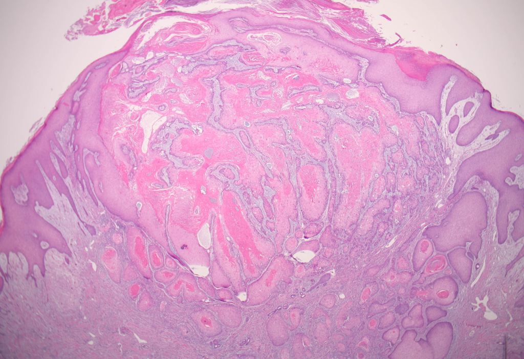
A 58-year-old man presented with a brown–red granular epulis. A periapical radiograph showed underlying bone destruction and a biopsy was reported as showing osteoclast-like giant cells in a spindle-cell background with numerous thin- walled vessels. Haemosiderin and extravasated red cells were abundant.

The clinical and radiographic findings suggest that the 58-year-old man may have a peripheral giant-cell granuloma (PGCG). This is a benign, non-cancerous tumor that often arises from the gum tissue and can cause bone destruction in the underlying jawbone.

The biopsy findings support the diagnosis of PGCG, as the presence of osteoclast-like giant cells and spindle cells in a background of abundant thin-walled vessels is characteristic of this condition. The haemosiderin and extravasated red cells seen in the biopsy are likely a result of bleeding within the lesion, which is common in PGCG.

These same features may also be observed in hyperparathyroidism. However, in hyperparathyroidism, serum calcium levels are typically elevated, while this is not the case in giant-cell granuloma. Therefore, measuring serum calcium levels can be a useful diagnostic tool in differentiating between these two conditions, especially when giant-cell granuloma features are observed.

Treatment for PGCG typically involves surgical removal of the lesion, along with the underlying affected bone. Recurrence is possible, so close follow-up and monitoring is important.

Image Based Questions – Orthodontics – Part 2

  1. IDENTIFY THE SPACE MAINTAINER = BAND AND LOOP
  1. IDENTIFY THE APPLIANCE = DISTAL SHOE SPACE APPLIANCE
  2. IDENTIFY = ACTIVE TIE BACKS ***
  3. IDENTIFY = E- CHAIN
  4. IDENTIFY = GOLD CHAIN
  5. IDENTIFY = MONOCRYSTALLINE AND POLYCRYSTALLINE CERAMIC BRACKETS
  6. IDENTIFY THE TYPE OF ELASTICS = SETTLING ELASTIC
  7. IDENTIFY THE BRACKET = DAMON BRACKET
  8. IN WHICH SECTOR IS THIS CANINE IMPACTED = SECTOR 4
  9. IDENTIFY THE INSTRUMENT = SEPARATOR PLACING PLIER
  10. IDENTIFY = SPOT WELDER
  11. IDENTIFY = SAND BLASTER AND MICROETCHER
  12. IDENTIFY = WEINGART
  13. IDENTIFY = DELA ROSA

IDENTIFY = TWEED RIBBON ARCH PLIER**