Steroids may have a less than perfect reputation in the media, but some steroids naturally occur in the body and function as a vital part of our endocrine system. For example, estrogens and testosterone serve important roles in human reproduction, while cortisol and aldosterone are integral in helping the body maintain equilibrium, especially in times of stress.
Addison’s disease is defined as the condition in which the steroid hormones produced by the adrenal cortex are either low or absent. It’s known by other names, too: chronic adrenal insufficiency, hypocortisolism, and hypoadrenalism, to name just a few.
Cortisol has many functions, including maintaining a consistent blood glucose level, reducing inflammation, and regulating water balance. Aldosterone primarily maintains sodium and water balance, which are necessary for keeping a consistent blood volume and pressure.
Both cortisol and aldosterone are produced by the adrenal glands, small endocrine glands that rest on top of both kidneys. The adrenal glands have two layers: the medulla, or inner, layer, and the cortex, the outer layer. In most cases of Addison’s disease, the cells that make up the adrenal cortex are destroyed. The destruction of these cells results in primary adrenal insufficiency, because cortisol and aldosterone are simply not produced. This is different from secondary insufficiency, in which either one or both hormones are made, but the body is unable to use them properly.
So, how does this fit into those other names for Addison’s disease? Since hypo means ‘low’ and the primary glucocorticoid is cortisol, a low amount of cortisol is ‘hypocortisolism.’ As you might guess, Addison’s is also characterized by hypoaldosteronism, low amounts of aldosterone. You’ll want to remember the roots hypo and its opposite hyper, which means high.
CAUSES
Let’s take a look at what happens to destroy the cells of the adrenal cortex. In about 80% of patients that have Addison’s, primary adrenal insufficiency is caused by an autoimmune response in which the body’s immune system mistakes its own cells or biochemicals for pathogens. Specifically, the body sees some part of a pathway used by the body for synthesis or use of cortisol and aldosterone as a threat and targets it for destruction.
Other things that cause primary adrenal insufficiency are:
Adrenaldysgenesis, a genetic disorder in which the adrenal glands aren’t formed properly
Difficulties in converting cholesteroltosteroidhormones
Infection from HIV or tuberculosis
Other diseases that impair the adrenal cortex, like adrenoleukodystrophy and cancer
CLINCAL FEATURES
Symptoms of Addison’s can also occur after a patient stops steroid use, especially if the medication taken has been taken for a long period of time or in high doses.
Sustained use of medications like hydrocortisone and prednisone can desensitize the adrenal gland to the chemical that normally stimulates cortisol production, cause its cells to shrink, and impair its ability to make natural steroid hormones. This is called a iatrogenic response, an instance where treatment causes a disorder.
The symptoms of Addison’s disease are caused by the absence of aldosterone and cortisol. If you think about the functions of these two hormones, then it’s less difficult to determine the symptoms that arise when they’re not present. Here’s where ‘hyper’ and ‘hypo’ start to come into play.
Cortisol
Keeps blood sugar levels in balance by stimulating the creation of glucose in the liver. Its absence means a drop in blood glucose, which, in turn, leads to fatigue and lethargy.
Cortisol and aldosterone work together to maintain salt and water balance, so their loss leads to orthostatic hypotension, a sudden low blood pressure that occurs on standing.
Aldosterone
Its absence also causes a drastic upset in electrolyte balance, leading to nausea, diarrhea, and vomiting. Together, these pose a real danger of causing dehydration, and, eventually, hypovolemia or low blood volume.
Because electrolytes like sodium and potassium ions are crucial for nerve and muscle function, symptoms like heart arrhythmia and nervous system changes (especially in the form of mood disorders) are common, as is a craving for salt in an effort to replace the sodium being over-excreted in the urine.
Everyone knows that there’s too much of a good thing. Even a cool glass of lemonade on a hot summer’s day can turn into a sticky mess if the person pouring it isn’t paying attention. We need the hormone cortisol to help us deal with stressful situations. In Cushing’s syndrome, the body experiences the effects of too much cortisol for too long of a period of time. The result? Cortisol, which usually mediates the effects of stress, instead ends up causing damage to the body.
Cortisol is made by the adrenal glands, a pair of glands that rest on top of the kidneys. You can think of the steps of the cortisol pathway as a baseball player who runs the bases after hitting a home run. The pathway starts in the hypothalamus, the part of the brain responsible for much of the body’s ‘autopilot’ activity – things like breathing and heart rate. When the hypothalamus receives a stimulus that the body’s under stress, it releases corticotropin-releasing hormone (or CRH). This speeds to the pituitary, where it tells the master gland to release adrenocorticotropic-stimulating hormone (or ACTH). ACTH races to the adrenal glands to release cortisol. Cortisol then goes on to do things like increase blood sugar levels, lower inflammation, and raise blood pressure.
Before we dig into what causes Cushing’s syndrome, we should clarify the relationship between Cushing’s syndrome and Cushing’s disease. A syndrome is different from a disease, which is a specific disturbance in a part of the body. Cushing’s disease is just one cause of Cushing’s syndrome. In other words, not all cases of Cushing’s syndrome are caused by Cushing’s disease.
CAUSES
Most medical professionals divide these causes into two large categories.
The first is exogenous, meaning ‘generated from without.’
Long-term use of corticosteroid medications can cause Cushing’s syndrome.
Iatrogenic steroids often prescribed for inflammatory diseases like arthritis or given by injection for pain. Cushing’s syndrome may develop if the dosage of medication is or becomes larger than the body can handle.
The second large category is endogenous, meaning ‘generated from within.’ Endogenous cases of Cushing’s syndrome are usually caused by one of three things:
The first is a pituitary adenoma, a benign tumor of the pituitary gland. An adenoma may secrete large amounts of ACTH in a way that the body can’t control. A large amount of ACTH will stimulate the adrenal gland to produce a large amount of cortisol. Here, finally, we have the specific cause of Cushing’s disease. Roughly 70% of endogenous cases of Cushing’s syndrome are caused by pituitary adenomas and therefore are classified as actual cases of Cushing’s disease.
The second endogenous cause is called ectopic syndrome. You may recall that ‘ectopic’ means ‘in an abnormal place.’ Sometimes tumors of the lungs, pancreas, or thyroid release ACTH. Again, the body does not have the means to control this unplanned ACTH, and it stimulates the adrenal glands to produce too much cortisol.
The third endogenous cause is primary adrenal disease. In these cases, a tumor of the adrenal gland (again, usually benign) goes rogue and secretes large amounts of cortisol.
Clinical Features
Common Findings:
Weight gain/ lemon on sticks/ central obesity
Increase in peripheral resistance = Hypertension
Increase in blood sugar, FBS = more than 126 mg%
Increase in cortisol levels causes insulin resistance, which increases sugar in the blood causing secondary DM
Decrease in bone calcium causes osteoporosis, calcium is needed for muscle contraction.
Cortisol effects on sex steroid receptors causing oligomenorrhoea, infertility, PCOD and hirsutism
More specific findings include:
Moon facies
Proximal myopathy
Easy bruising
Purple striae and Thin, fragile skin
Fat deposition in the face
Hypokalaemia and metabolic alkalosis are prominent, particularly with ectopic production of ACTH
Screening test – measurement of 24-h urinary free cortisol – late-night salivary cortisol measurement – 1-mg overnight dexamethasone test
Investigation of choice – low dose dexamethasone suppression test – inadequate suppression of urinary cortisol [<10 μg/d (25 nmol/d)] or plasma cortisol [<5 μg/dL (140 nmol/L)] after 0.5 mg dexamethasone every 6 h for 48 h.
Biochemical testing – Low levels of plasma ACTH levels suggest an adrenal adenoma or carcinoma – Normal or high plasma ACTH levels suggest a pituitary or ectopic source
Imaging – MRI of the pituitary – Imaging of the chest and abdomen
Treatment
Iatrogenic steroids- Taper steroids and Azathioprine
Oar=T cell ca lung- cisplatin + Trinotecam
Pituitary adenoma Cushing disease – transsphenoidal surgery
A good way to help you remember is to think of this hormone system like a house’s heating system. In this example, the thyroid gland is the furnace, the pituitary gland is the thermostat, and the hypothalamus is the person living in the house. When the house is cold, the person sets the thermostat to signal the furnace to make heat. When the house grows warmer, the system shuts off.
In hyperthyroidism, the system never shuts off, causing the release of too many thyroid hormones. For this lesson, we’ll focus on two of them: T3 (tri-iodothyronine) and T4 (thyroxine).
The thyroid needs iodine to make T3 and T4. Thyroid cells absorb iodine and combine it with the amino acid tyrosine to make these hormones. About 80% is T4, and 20% is T3.
Too much of T3 and T4 results in thyrotoxicosis. There is a subtle difference between hyperthyroidism and thyrotoxicosis – hyperthyroidism means having an overactive thyroid gland, while thyrotoxicosis is the condition of having increased levels of T3 and T4 in the blood. Thyrotoxicosis can be caused by taking too much thyroid medication, for example, or having thyroiditis (an inflamed thyroid).
Graves’ disease, caused by activating TSH-receptor antibodies, is the most common cause of thyrotoxicosis and accounts for 60–80% of cases.
Its prevalence in women is 10-fold higher than in men; its peak occurrence is at age 20–50 years.
Clinical Features
Anxiety, restless, and fidgety.
weight loss with increased appetite
frequent bowel movements
heat intolerance
excessive sweating
oligomenorrhea
Skin is warm and moist
Cardiovascular findings include tachycardia, systolic hypertension, systolic murmur, and atrial fibrillation.
Fingernails may separate from the nail bed (Plummer’s nails).
Eyelid retraction and lid lag may be present.
A fine tremor, hyperreflexia, and proximal muscle weakness also may be present.
Long-standing thyrotoxicosis may lead to osteopenia.
In the elderly, the classic signs of thyrotoxicosis may not be apparent, the main manifestations being weight loss and fatigue (“apathetic thyrotoxicosis”).
In Graves’ disease,
the thyroid is usually diffusely enlarged to two to three times its normal size
a bruit or thrill may be present.
Infiltrative ophthalmopathy (with variable degrees of proptosis, periorbital swelling, and ophthalmoplegia)
dermopathy (pretibial myxedema) also may be found;
these are extrathyroidal manifestations of the autoimmune process.
In subacute thyroiditis,
the thyroid is exquisitely tender and enlarged with referred pain to the jaw or ear
Sometimes accompanied by fever and preceded by an upper respiratory tract infection.
Solitary or multiple nodules may be present in toxic adenoma or toxic MNG.
Thyrotoxiccrisis, or thyroid storm, is rare, presents as a life-threatening exacerbation of hyperthyroidism, and can be accompanied by fever, delirium, seizures, arrhythmias, coma, vomiting, diarrhea, and jaundice.
Serum TSH is a sensitive marker of thyrotoxicosis caused by Graves’ disease, autonomous thyroid nodules, thyroiditis, and exogenous levothyroxine treatment.
Elevation of bilirubin, liver enzymes, and ferritin.
Thyroid radioiodine uptake may be required to distinguish the various etiologies: high uptake in Graves’ disease and nodular disease versus low uptake in thyroid destruction, iodine excess, and extrathyroidal sources of thyroid hormone.
The thyroid gland is found at the base of your neck and lies just below your Adam’s apple. This gland produces hormones that are important to your metabolism, known as T3 and T4.
Some T3 is secreted by the thyroid, but most is produced by deiodination of T4 in peripheral tissues.
Both T4 and T3 are bound to carrier proteins (thyroid-binding globulin [TBG], transthyretin [binds T4], and albumin) in the circulation.
Increased levels of total T4 and T3 with normal free levels are seen in states of increased carrier proteins (pregnancy, estrogens, cirrhosis, hepatitis, and inherited disorders).
Conversely, decreased total T4 and T3 levels with normal free levels are seen in severe systemic illness, chronic liver disease, and nephrosis.
Since we know that the thyroid hormones play an important role in regulating your body’s metabolism, it makes sense that a decrease in these hormones would lead to symptoms such as a loss of energy, an increased need for sleep, an intolerance to cold, a slow pulse, constipation, weight gain, and depression
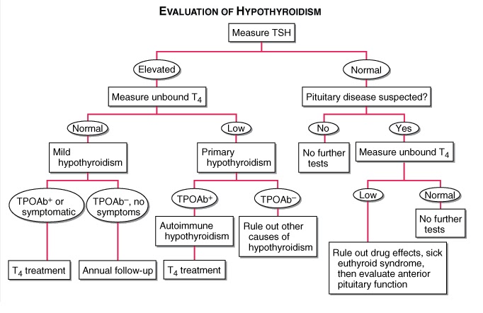
Decreased serum free T4 is common to all varieties of hypothyroidism.
An elevated serum TSH is a sensitive marker of primary hypothyroidism but is not found in secondary hypothyroidism.
Elevated cholesterol
Increased creatine phosphokinase
Anemia may be present
Bradycardia, low-amplitude QRS complexes, and flattened or inverted T waves may be present on ECG.
Treatment
Adult pts <60 years without evidence of heart disease may be started on 50–100 μg of levothyroxine (T4) daily.
In the elderly or in pts with known coronary artery disease, the starting dose of levothyroxine is 12.5–25 μg/d.
In secondary hypothyroidism, TSH levels cannot be used, and therapy needs to be guided by free T4 measurement.
Women on levothyroxine replacement should have a TSH level checked as soon as pregnancy is diagnosed, as the replacement dose typically increases by 30–50% during pregnancy. Failure to recognize and treat maternal hypothyroidism may adversely affect fetal neural development.
Therapy for myxedema coma should include levothyroxine (500 μg) as a single IV bolus followed by daily treatment with levothyroxine (50–100 μg/d), along with hydrocortisone (50 mg every 6 h) for impaired adrenal reserve, ventilatory support, space blankets, and treat- ment of precipitating factors.
It is all about increase production of growth hormone and when the long bone in the leg, called the epiphysis plate, fuses. If the epiphysis plate has not fused before puberty and there is a large amount of growth hormone being released from the pituitary gland, the person is considered to have gigantism. If the epiphysis plate has fused after puberty and there is a large amount of growth hormone being released from the pituitary gland, the person is considered to have acromegaly.
Causes
PituitarySources: Somatotrope adenomas and mammosomatotrophe adenomas
Extra Pituitary Sources: Pancreatic adenoma (islet cell tumour) and excessive GnRh production by carcinoid tumor
Clinical Features
The peak occurrence of acromegaly is at age 40–45. Now that we understand how someone is diagnosed with acromegaly, let’s take a look at what a person would look like with this condition. Because the epiphysis bone has fused, they will not get taller, but bones and organs will enlarge.
They will have big hands, feet, and forehead:
ring tightening
increased shoe or glove size
frontal bossing
Their organs and glands will enlarge and cause associated diseases:
cardiomyopathy
left ventricular hypertrophy
diastolic dysfunction
colon polyps, and colonic malignancy.
an enlarged thyroid
Increase in sebum = oily skin
Peripheral resistance will increase = hypertension
Also, they could have an increase in blood glucose because the increase of growth hormone stops insulin from carrying the glucose out of the blood in to the tissues. So the glucose (sugar) hangs out in the blood.
glucose intolerance
diabetes mellitus
Other unique features:
Thick heel pads
Deep voice
Hyperhidrosis
Skin tags
Osteoarthritis: bilateral limbs and weight gain
Spade like hand
Obstructive sleep apnea
Visual deficit
Carpal tunnel syndrome.
Oral Manifestations
Widened teeth spacing
mandibular enlargement with prognathism,
macroglossia
Overall mortality is increased approximately threefold.
Diagnosis
Insulin-like growth factor type I (IGF-I) levels are a useful screening measure with elevation suggesting acromegaly.
Investigation of choice: Due to the pulsatility of GH, measurement of a single random GH level is not useful for screening. The diagnosis of acromegaly is confirmed by demonstrating the failure of GH suppression to <0.4 μg/L within 1–2 h of a 75-g oral glucose load.
MRI of the pituitary usually reveals a macroadenoma.
Serum prolactin levels are increased in acromegaly
X-ray of Foot
Treatment
The primary treatment modality for acromegaly is transsphenoidal surgery.
GH levels are not normalized by surgery alone in many pts with macro-adenomas; in those, somatostatin analogues provide adjunctive medical therapy that suppresses GH secretion with modest to no effect on tumour size. Drugs: Octreotide, Lanreotide and Pasireotide
The GH receptor antagonist pegvisomant can be added in pts who do not respond to somatostatin analogues. Pegvisomant is highly effective in lowering IGF-I levels but does not lower GH levels or decrease tumor size.
Pituitary irradiation may also be required as adjuvant therapy but has a slow therapeutic onset and a high rate of late hypopituitarism.
Liver produces important proteins, such as albumin, which ensures you don’t look swollen, and allows for the transport of important drugs. It helps to detoxify things that may be dangerous to your health. It also helps to give you energy by generating glucose, and so much more!
What Is Bile and Bilirubin?
Other than all of the things that I just mentioned the liver is important for, it’s also important for the production of something known as bile. This is a greenish liquid produced in the liver and stored in the gallbladder, which facilitates the digestion of fat.
Bile, among other things, contains something known as bilirubin. Bilirubin is an orange-yellow pigment formed from the breakdown of red blood cells. Bilirubin is converted into other substances that eventually give urine its yellow color and feces its yellow-brown color. Who knew physiology could be so colorful and yet so disgusting?
When red blood cells break down due to age or destruction, then something known as unconjugated or indirect or water-insoluble bilirubin is released into the bloodstream. Once this unconjugated bilirubin enters the liver, the liver converts it into conjugated or direct or water-soluble bilirubin by tacking on a compound known as glucuronic acid; this is what makes bilirubin water soluble.
This newly water-soluble bilirubin is then secreted into the watery bile and, from there, into the intestines.
If all of this is making your head spin, don’t worry; we can simplify it even more. Your liver is like a giant factory. Through one end, raw ingredients come in by the truckload. In our case, that’s crates and crates of raw, hard, and unprocessed indirect bilirubin. Once inside the factory, the workers take a piece of unconjugated bilirubin and stick another ingredient right onto it, thereby making an entirely new concoction, called conjugated bilirubin. This conjugated bilirubin isn’t hard like unconjugated bilirubin; it’s slushy and can dissolve easily in water instead.
If the levels of either the conjugated or unconjugated bilirubin increase in the body, they can cause icterus, which is the more technical term for jaundice. If you are not sure of what jaundice is, then let me explain. Jaundice is the yellowing of the whites of the eyes, mucous membranes, and a person’s skin as a result of abnormally increased levels of bilirubin in the blood, aka hyperbilirubinemia.
What Causes Unconjugated Hyperbilirubinemia?
Based on what I’ve said thus far, you should be aware that the ways by which levels of unconjugated bilirubin can increase are:
if something causes indirect bilirubin levels to rise before they reach the liver
if unconjugated bilirubin isn’t being delivered properly to the liver (both of which are also known as pre-hepatic causes)
if there’s something wrong with the liver that disables the conversion mechanism to conjugated bilirubin (also known as hepatic causes)
Types of Jaundice
Pre-Hepatic
In pre-hepatic jaundice, there is excessivered cell breakdown which overwhelms the liver’s ability to conjugate bilirubin. This causes an unconjugated hyperbilirubinaemia.
Any bilirubin that manages to become conjugated will be excreted normally, yet it is the unconjugated bilirubin that remains in the blood stream to cause the jaundice.
Hepatocellular
In hepatocellular (or intrahepatic) jaundice, there is dysfunction of the hepatic cells. The liver loses the ability to conjugate bilirubin, but in cases where it also may become cirrhotic, it compresses the intra-hepatic portions of the biliary tree to cause a degree of obstruction.
This leads to both unconjugated and conjugated bilirubin in the blood, termed a ‘mixed picture’.
Post-Hepatic: Post-hepatic jaundice refers to obstructionof biliary drainage. The bilirubin that is not excreted will have been conjugated by the liver, hence the result is a conjugated hyperbilirubinaemia.
Laboratory Tests
Any patient presenting with jaundice should have the following bloods taken:
Liver function tests (LFTs), as summarised in Table 2
Coagulation studies (PT can be used as a marker of liver synthesis function)
FBC (anaemia, raised MCV, and thrombocytopenia all seen in liver disease) and U&Es
Specialist blood tests, as summarised below as part of a liver screen
Blood Marker
Significance
Bilirubin
Quantify degree of any suspected jaundice
Albumin
Marker of liver synthesising function
AST and ALT
Markers of hepatocellular injury*
Alkaline Phosphatase
Raised in biliary obstruction (as well as bone disease, during pregnancy, and certain malignancies)
Gamma-GT
More specific for biliary obstruction than ALP (however not routinely performed)
Table 2 – LFT serum markers. *as an estimate, if the AST:ALT ratio >2, this is likely alcoholic liver disease, whilst if AST:ALT is around 1, then likely viral hepatitis as the cause
Liver Screen
A liver screen can be performed for patients whereby there is no initial cause for liver dysfunction, tailored to whether acute or chronic liver failure
Viral Serology
Non-Infective Markers
Acute Liver Injury
Hepatitis A, Hepatitis B, Hepatitis C, and Hepatitis ECMV and EBV
Paracetamol levelCaeruloplasminAntinuclear antibody and IgG subtypes
Chronic Liver Injury
Hepatitis BHepatitis C
CaeruloplasminFerritin and transferrin saturationTissue Transglutaminase antibodyAlpha-1 antitrypsinAutoantibodies*
Table 3 – Acute and Chronic Liver Screens *Autoantibodies include anti-mitochondrial antibody (AMA), anti-smooth-muscle antibody (Anti-SMA), and anti-nuclear antibody (ANA), used to identify a variety of autoimmune liver conditions, such as primary sclerosing cholangitis (PSC)
Imaging
The imaging used will depend on the presumed aetiology. An ultrasound abdomen is usually first line, identifying any obstructive pathology present or gross liver pathology (albeit often user dependent).
Magnetic Resonance Cholangiopancreatography (MRCP) is used to visual the biliary tree, typically performed if the jaundice is obstructive, but US abdomen was inconclusive or limited, or as further work-up for surgical intervention.
A liver biopsy can be performed when the diagnosis has not been made despite the above investigations.
Management
The definitive treatment of jaundice will be dependent on the underlying cause. Obstructive causes may require removal of a gallstone through Endoscopic Retrograde CholangioPancreatography (ERCP) or stenting of the common bile duct.
Symptomatic treatment is often needed for the itching caused by hyperbilirubinaemia. An obstructive cause may warrant cholestyramine (acting to increase biliary drainage), whilst other causes may respond to simple anti-histamines.
Identify and manage any complications where possible. Monitor for coagulopathy, treating promptly (either vitamin K or fresh frozen plasma (FFP) is needed) if any evidence of bleeding or rapid coagulopathy, and treat hypoglycaemia orally if possible (otherwise 5% dextrose is needed).
Where patients become confused from decompensating chronic liver disease (‘hepatic encephalopathy’), laxatives (lactulose or senna) +/- neomycin or rifaximin may be used, in attempt to reduce the number of ammonia-producing bacteria in the bowel.By User:Pschemp [CC-BY-SA-3.0], via Wikimedia Commons