GRADINNG & STAGING OF SQUAMOUS CELL CARCINOMA

Squamous Cell Carcinoma (Epidermoid Carcinoma)

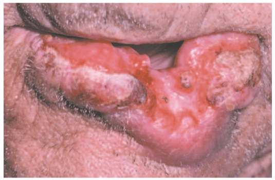
  • SCC is defined as ‘a malignant epithelial neoplasm exhibiting squamous differentiation as characterized by the formation of keratin and/ or the presence of intercellular bridges‘.
  • The epidermoid carcinoma is the most common malignant neoplasm of the oral cavity.
  • The male-female ratio is approximately 2:1 for oral carcinoma, except for carcinoma of the vermilion border of the lower lip

ETIOLOGY:-

  • The cause of oral squamous cell carcinoma is multifactorial.
  •  No single causative agent or factor (carcinogen) has been clearly defi ned or accepted, but both extrinsic and intrinsic factors  may work .
  • FACTORS:-
  • TOBACCO SMOKING
  • SMOKELESS TOBACCO
  • BETEL QUID (PAAN)
  • ALCOHOL
  • PHENOLIC AGENTS
  • RADIATION
  • IRON DEFICIENCY
  • VITAMIN-A DEFICIENCY
  • SYPHILIS
  • CANDIDAL INFECTION
  • ONCOGENIC VIRUSES
  • IMMUNOSUPPRESSION
  • ONCOGENES AND TUMOR SUPPRESSOR GENES
  • CLINICAL FEATURES:-

  • Oral squamous cell carcinoma has a varied clinical presentation, including the following:

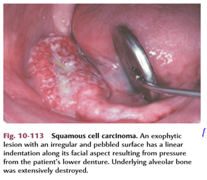
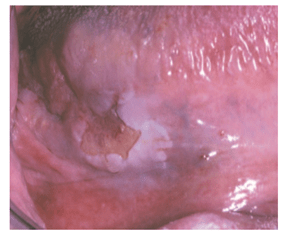
● Exophytic (mass forming; fungating, papillary, verruciform)

 ● Endophytic (invasive, burrowing, ulcerated)

 ● Leukoplakic (white patch)

● Erythroplakic (red patch)

● Erythroleukoplakic (combined red-and-white patch)

  • STAGING :-

Tumor size and the extent of metastatic spread of oral squamous cell carcinoma are the best indicators of the patient’s prognosis. Quantifying these clinical parameters is called staging the disease.

  The most popular staging protocol, the tumor-node-metastasis (TNM) system.

This staging protocol depends on three basic clinical features:

 1. T—Size of the primary tumor, in centimeters

 2. N—Involvement of local lymph nodes

 3. M—Distant metastasis

The American Joint Committee on Cancer (AJCC) designated staging by TNM Classification was used.

TNM clinical classification:-

  • HISTOPATHOLOGIC FEATUR:-
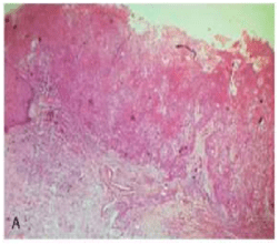
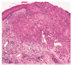
  • Squamous cell carcinoma arises from dysplastic surface epithelium.

Features are:-

  • sheets or islands of cells or cords
  • a strong infl ammatory or immune cell response
  • focal areas of necrosis
  • invading into underlying muscles, bonesor adipose tissues
  • angiogenesis
  • desmoplasia or scirrhous change
  • abundant eosinophilic cytoplasm with large, often darkly staining (hyperchromatic) nuclei
  • increased nuclearto-cytoplasmic ratio.
  • nuclear pleomorphism
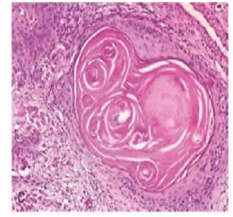
  • keratin pearls (a round focus of concentrically layered keratinized cells)

  • GRADING:-
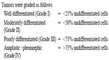
  • Histopathologic evaluation of the degree to which these tumors resemble their parent tissue (squamous epithelium) and produce their normal product (keratin) is called grading.
  • The one advantage of grading a tumour is that the grade reflects the anaplasticity of the lesion, which in turn indicates the general rapidity of growth, the rapidity of metastatic spread, the general reaction to be expected after X-ray radiation and the prognosis.
  • There are 4 classifications used to describe grading systems :-
  • 1-BRODERS CLASSIFICATION:-
  •  Broders’ system (1920) was first established on the basis of the proportion of highly differentiated cells in the tumour. Broders’ system was simple and widely used, it was a poor predictor for survival or me- tastasis.

* a system of grading tumours in which a grade 1 lesion was highly differentiated (its cells were producing much keratin), while grade 4 was very poorly differentiated (the cells were highly anaplastic and showed no keratin formation).
I-WELL DIFFERENTIATED:

-consists of sheets and nests of cells

 -cells are generally large

– intercellular bridges or tonofibrils are not demonstrated.

-The nuclei are large and demonstrate variability in the intensity of the staining reaction.

-Nuclei that stain heavily with hematoxylin are referred to as hyperchromatic.

-mitotic figures may be found(Many of these mitotic figures are atypical.)

 -the most prominent features of the welldifferentiated epidermoid carcinoma is the presence of individual cell keratinization-the formation of numerous epithelial or keratin pearls of varying size.
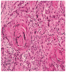
II– ModeratelyDifferentiated epidermoid carcinomas:

– lose certain features so that their resemblance to squamous epithelium is less pronounced

-The characteristic shape of the cells and their arrangement may be altered.

– The growth rate of individual cells is more rapid,

– the greater number of mitotic figures,

– even greater variation in sizes, shape and tinctorial reaction,the failure to carry out the function of a differentiated squa- mous cell, the formation of keratin.
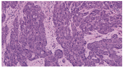
III-POORLY DIFFERENTIATED TYPE:

-bear little resemblance to their cell of origin

-will often present diagnostic difficulties because of the primitive and uncharacteristic histological appearance of malignant,

-rapidly dividing cells

– cells show an even greater lack of cohesiveness and are extremely vagarious.

2.JAKOBSS0N’s GRADING SYSTEM:-

-In 1973, Jakobsson et al developed a multifactorial

grading system which had the advantage of scoring tumour-host interactions and tumour characteristics, but eventually proved to be useful only when applied to tongue cancers.

-Parameters used in  Jakobsson’s method are:-

i- KRE- keratinization

ii-NP- nuclear pleomorphism

iii-MIT-mitosis

iv-POI- pattern of invasion

v-LPR- lympho-plasmocyticrspon
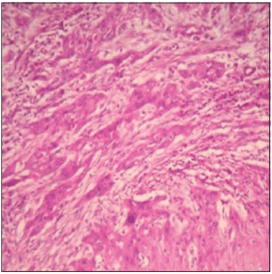

Tumor cells invading in strands and cords

-Similar findings were observed in Anneroth and Hansen’s grading  where the criteria were similar except that the parameter vascular invasion (VI) was omitted. 

3-Anneroth’s classification:

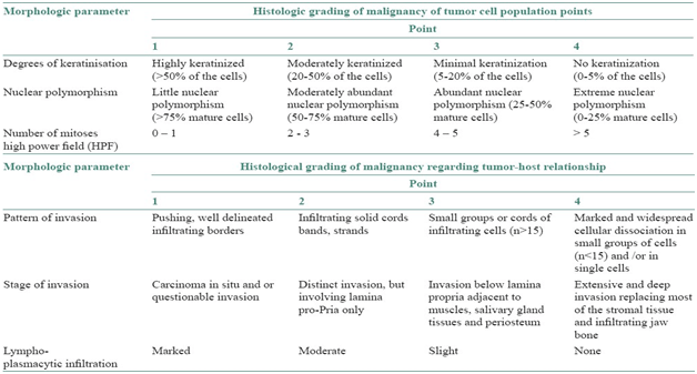
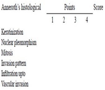
  • This system is constituted by six histological variables of equal value in the determination of the grade of malignancy, three connected with the tumor cellular population (differentiation and proliferation mitosis) and the other three connected with tumor-host relationship (pattern and stage of invasion; and cellular response).

Anneroth’s classification (multifactor grading system)

-According to the classification, three parameters reflecting tumor cell features including keratinization, polymorphism, and mitoses were evaluated in the whole thickness of the tumor and each was scored from 1 to 4. Mode of invasion and inflammatory infiltration representing tumor-host relationship were graded in the most invasive margins and scored from 1 to 4:

-Variables such as:-

i-pattern of invasion,

ii- tumor thickness,

iii-degree of keratinization,

iv-nuclear pleomorphism,

v-lymphocytic response,

vi-mitotic rate
4.BRYNE’S GRADING SYSTEM:-

-Bryne et al (1989) modified Anneroth’ s grading system and developed a malignancy grading focusing on the invasive front of the tumour.

– This method of grading appeared to be less time-consuming in the assessment of the neoplasm.


-Nevertheless, this system is not sufficiently homogeneous to allow grading parameters to be assessed individually

-This was performed at the invasive tumor front (ITF). 

-The Bryne’s grading system is more predictive for LNM as compared with the multifactorial grading systems that is, Jakobsson’s and Anneroth and Hansen’s. Broder’s grading system is of no prognostic value.

-Parameters used are:-

i-Keratinisation

ii-nuclear polymorphism

iii-mitosis

iv-pattern of invasion

v-lympho-plasmocytic response

vi-lymph nodes

REFERENCES :

1.Comparative study of various grading systems in oral squamous cell carcinoma and their value in predicting lymph node metastasis
Saleha Jamadar1, TV Narayan1, Balasundari Shreedhar2, Leeky Mohanty1, Sadhana Shenoy1
1 Department of Oral Pathology and Microbiology, The Oxford Dental College, Hospital and Research Centre, Bommanahalli, Bengaluru, Karnataka, India
2 Department of Oral Pathology and Microbiology, Career Dental College, Lucknow,Uttar Pradesh, India

2.A study on histological grading of oral squamous cell carcinoma and its co-relationship with regional metastasis

M Akhter, S Hossain, Quazi B Rahman, Motiur R Molla
Department of Oral and Maxillofacial Surgery, Bangabandhu Sheikh Mujib Medical University, Dhaka, Bangladesh

3.Shafer’s Textbook of Oral Pathology,8e

4.NEVILLE -Oral & Maxillofacial Pathology,3e

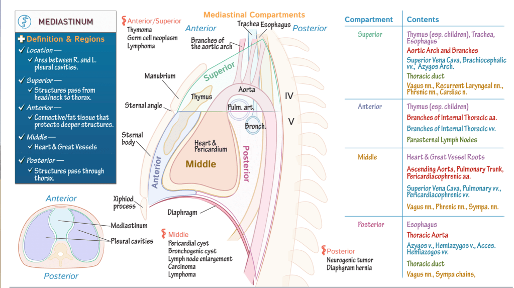
Mediastinum

Middle compartment has a sac-like shape; it houses the pericardium, heart, and roots of the great vessels.
Anterior compartment lies anterior to this, and extends from the sternal angle, superiorly, to the diaphragm, inferiorly.
Posterior compartment lies posterior to the middle mediastinum, and, like the anterior mediastinum, extends from the sternal angle to the diaphragm.
Superior mediastinum fills the space between the superior thoracic opening to the sternal angle.

Key anatomical structures

  • The thymus lies within the superior and anterior regions; recall that this structure regresses after childhood.
  • Then, return to where the root and ascending portion of the aorta arise in the middle mediastinum, and show that the aorta continues through the superior and posterior compartments. We’ve also shown the branches of the aortic arch as they extend through the superior mediastinum.
  • Next, posterior to the heart, show the opening of the left pulmonary artery as it passes to the left lung;
  • The opening of the left bronchus; indicate that the trachea extends through the superior and posterior compartments of the mediastinum.
  • The esophagus also passes through these compartments.

Pathology

  • Let’s indicate some key masses that can develop in the mediastinum; we’ll broadly categorize these by region of the mediastinum, but beware of overlap.
  • Anterior/superior mediastinum: thymoma, germ cell neoplasm, and lymphoma.
  • Middle mediastinum: pericardial cysts, bronchogenic cysts, lymph node enlargement, carcinoma, and lymphoma.
  • Posterior compartment: watch for neurogenic tumors and diaphragmatic hernias.

Summary Table

Superior compartment:

  • Thymus, esophagus, and trachea.
  • The aortic arch and its branches.
  • The superior vena cava, brachiocephalic veins, and the arch of the azygos vein.
  • The thoracic duct.
  • The vagus nerves, recurrent laryngeal nerves, phrenic nerves, and cardiac nerve.

Anterior mediastinum:

  • Thymus.
  • Branches of the internal thoracic arteries and veins, and, the parasternal lymph nodes.

Middle mediastinum:

  • The heart and the roots of the great vessels.
  • The ascending aorta, pulmonary trunk, and pericardiacophrenic arteries.
  • The superior vena cava, pulmonary veins, and pericardiacophrenic veins.
  • And, the vagus nerves, phrenic nerves, and sympathetic nerves.

Posterior mediastinum:

  • The esophagus.
  • Thoracic aorta.
  • Azygos, hemiazygos, and accessory hemiazygos veins.
  • The thoracic duct.
  • The vagus nerves and sympathetic nerves.

Larynx

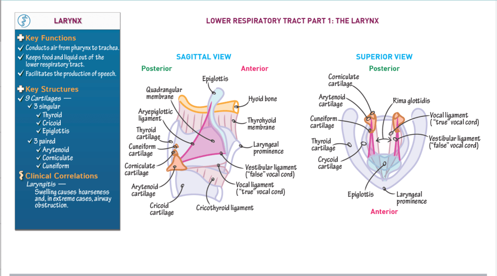
  • The larynx is the start of the lower respiratory tract.
  • Superiorly, the larynx is attached to the hyoid bone via connective tissues.
  • Three key functions of the larynx:
  • It conducts air from the pharynx to the trachea.
  • It prevents food and liquid from entering the lower respiratory tract.
  • It facilitates the production of speech.

Key anatomical structures:

Three larger, singular cartilages:

  • Thyroid cartilage – forms anterior and lateral walls; features the laryngeal prominence.
  • Cricoid cartilage – circular, forms base of larynx.
  • Epiglottis – “flap” that attaches to the internal surface of the thyroid cartilage, anteriorly, and projects posteriorly and superiorly over the opening of the larynx.
    • Upon swallowing, the flap closes off the entryway to the larynx, which prevents foods and liquids from entering the lower respiratory tract.

Three paired sets of smaller cartilages:

  • Arytenoid cartilage – articulates with cricoid cartilage posteriorly.
  • Corniculate cartilage – sits on arytenoid cartilages.
  • Cuneiform cartilage – sits anterior to corniculate cartilages; very small, wedge-shaped.

Membranes and connective tissues:

  • Thyrohyoid membrane – connects the thyroid cartilage and hyoid bone.
  • Cricothyroid ligament – extends superiorly from the cricoid cartilage to the thyroid and arytenoid cartilages.
  • Vocal ligament – thickened superior edge of the cricothyroid ligament comprises the vocal ligament; known as the “true” vocal cord because it facilitates sound production.
  • Rima glottides – opening between the vocal cords. During sound production, laryngeal muscles contract to rotate the arytenoid cartilages and alter the opening between the true vocal cords.
  • Quadrangular membrane – connects the lateral sides of the epiglottis to the arytenoid cartilages.
    • Free edges of the quadrangular membrane thicken to form the:
      Aryepiglottic ligament, superiorly
      The vestibular ligament, inferiorly

The vestibular ligament and its mucosal covering are often referred to as the “false vocal cord”; it does not participate in sound production.

Clinical correlations:

Laryngitis is inflammation of the vocal cords, which can stem from infectious or non-infectious causes (such as over-use). It typically results in hoarseness but severe swelling can block the airways.

Lungs and Pleural Membranes

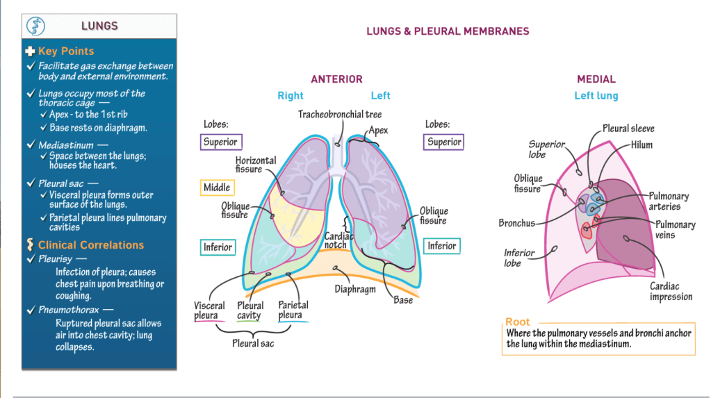
LUNGS

  • The primary function of the lungs is to facilitate gas exchange between the body and the external environment.
  • The lungs occupy most of the thoracic cage:
    * The apex of the lung, its most superior portion, reaches the first rib.
  • The base of the lung, its inferior concavity, rests on the diaphragm.
  • The mediastinum, which is the space between the lungs, houses the heart.

Right lung:

  • 3 lobes (superior, middle, inferior)
  • 2 fissures:
    • HORIZONTAL fissure separates the superior and middle lobes.
    • OBLIQUE fissure separates the middle and inferior lobes.

Left lung:

  • 2 lobes (superior, inferior)
  • 1 fissure:
    • OBLIQUE fissure separates the middle and inferior lobes.

The left lung is smaller than the right because the heart lies slightly to the left within the thoracic cage.

Specifically, the heart nestles within the cardiac impression of the left lung, which is visible as the cardiac notch in anterior view.

Membranes:

The lungs are enclosed by the double-layered pleural sac:

  • Visceral pleura forms the outermost layer of the lungs
  • Parietal pleura lines the pulmonary cavities.
  • Pleural cavity lies between the visceral and parietal pleura; it contains a small film of serous fluid that reduces friction between the layers.

Root of lung:

Pulmonary vessels and the bronchi anchor the lung within the mediastinum.

  • Medial aspects of lungs.
  • Hilum is obliquely shaped area, is lined by the pleural sleeve (aka, mesopneumonium).

Clinical Correlations:

  • Pleurisy is infection of the pleura and causes chest pain upon breathing or coughing.
  • Pneumothorax occurs when a ruptured pleural sac allows air to enter the thoracic cavity; the lungs collapse due to pressure changes.

Tracheobronchial tree

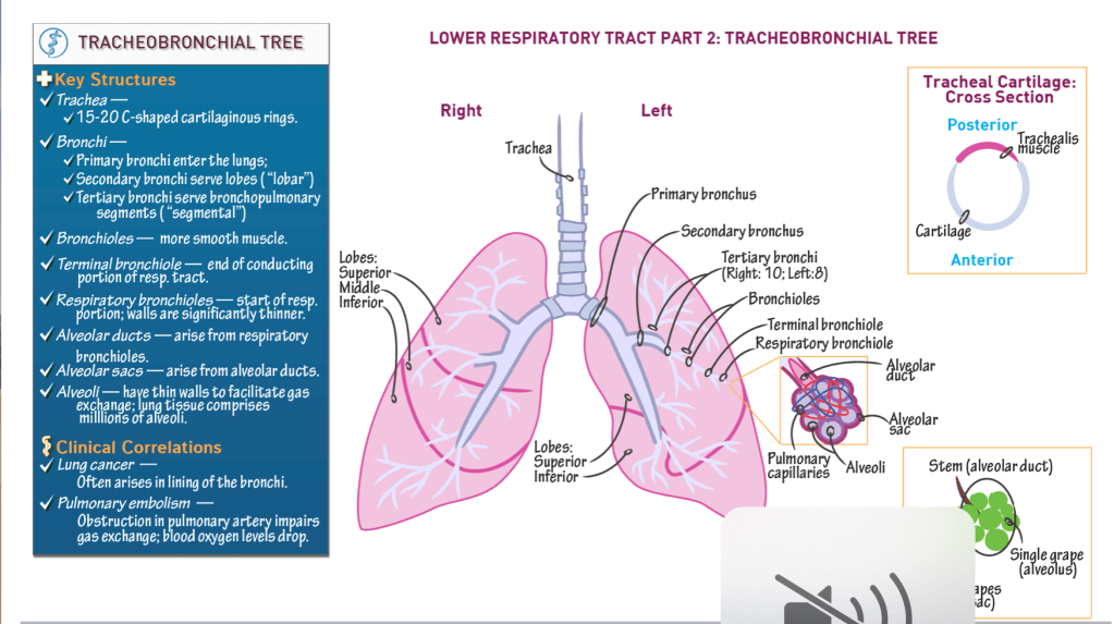
Key structures:

Trachea:

  • Cartilaginous “trunk” of the tree.
  • Comprises 15 – 20 C-shaped cartilaginous rings, are stacked vertically and connected via anular rings.
  • Trachealis posterior forms posterior wall of trachea; moves to accommodate foods passing posteriorly through the esophagus.

Bronchi:

  • Primary bronchi enter the lungs.
  • Secondary bronchi serve lobes of lung (“lobar” bronchi).
  • Tertiary bronchi serve bronchopulmonary segments (“segmental” bronchi); 10 on the right, 8-10 on the left.

Bronchioles:

  • Numerous, and narrow as they branch.
  • Have more smooth muscles in their walls, but still have cartilage in their walls.
  • Terminal bronchiole is the final passageway of the conduction portion of the respiratory system.

Respiratory bronchioles:

  • Demarcate the respiratory portion of the respiratory tract.
  • Thin walls allow some gas exchange.

Alveolar ducts:

  • Arise from respiratory bronchioles.

Alveolar sacs:

  • Terminal ends of the alveolar ducts.

Alveoli:

  • Thin-walled out-pockets of the alveolar sacs.
  • Surrounded by pulmonary capillaries.
  • Facilitate gas exchange between the respiratory and cardiovascular systems.

Lungs:

  • Hundreds of millions of alveoli.
  • Left lung = superior and inferior lobes; heart nestles into medial left lobe.
  • Right lung = superior, middle, and inferior lobes.

Clinical Correlations:

Lung cancer often originates in the bronchi.
Pulmonary embolism (aka PE) obstructs arterial supply. In a PE, gas exchange is reduced, and blood oxygen levels drop.

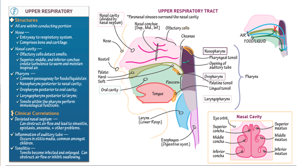
Upper Respiratory Tract

  • Begins at the nose and ends with the pharynx.

Anatomical Structures

Nose:

  • Opens the respiratory system to the external environment via the nostrils (aka, nares).
  • Comprises bone and cartilage.

Nasal cavity:

  • Posterior to the nose.
  • Separated from the oral cavity by the hard and soft palates:
    • The hard palate comprises the maxillary and palatine bones.
    • The soft palate comprises soft tissues.
  • Olfactory cells line the superior part of the nasal cavity
  • The nasal septum divides the cavity into right and left sides.
    • It comprises two vertical bony structures: the perpendicular plate of the ethmoid bone superiorly and the vomer, inferiorly.
    • A deviated nasal septum can obstruct airflow and cause sinusitis (sinus infection), epistaxis (nose bleeds), anosmia (inability to smell), and other health problems.
  • 3 nasal conchae: superior, middle, and inferior.
    • Bony projections that arise on the lateral walls of the nasal cavity.
    • The nasal conchae create meatuses (superior, middle, and inferior), which are small tunnels.
    • Movement of air around the conchae and through the meatuses creates turbulence, which helps to warm and humidify the air. Hence, the conchae are sometimes referred to as the “turbinate” bones.

Paranasal sinuses

  • Spaces within the bones surrounding the nasal cavity.

Pharynx:

  • Descends posterior to the nasal cavity, oral cavity, and larynx, and is open to each of these structures.
  • Common passageway foods/liquid and air: it serves both the respiratory and digestive systems.
  • 3 subdivisions:
    • Nasopharynx: posterior to the nasal cavity (and receives air from the nasal cavity).
      The choanae are the openings between the nasal cavity and the nasopharynx.
      Oropharynx: posterior to the oral cavity (and receives foods and liquids from the oral cavity).
      The fauces is the opening between the oral cavity and the oropharynx.
    • Laryngopharynx: posterior to the larynx (it is the final common passageway for air and food/liquid).
  • The auditory tube (aka, Eustachian or pharyngotympanic tubes) opens into the nasopharynx
    • Connects the ears and throat, which allows infection to pass between them.
    • Auditory tube inflammation occurs in otitis media (aka, ear infection), which is common amongst children.

Tonsils

  • Collections of lymphoid tissues that participate in immunological defense against bacterial and viral infections.
  • 3 pairs of extrinsic tonsils:
    • Pharyngeal tonsils reside in the superior wall of the nasopharynx.
    • Palatine tonsils are near the palate (hence, their name).
    • Lingual tonsils are at the back of the tongue (“lingual” refers to the tongue).
  • As part of an aggressive immune response, the tonsils can swell and block airflow through the nose or inhibit swallowing.
    • Chronic infection may necessitate tonsillectomy.