HISTOPATHOLOGY OF SKIN LESIONS

BY: Dr.Kriti Naja Jain :-

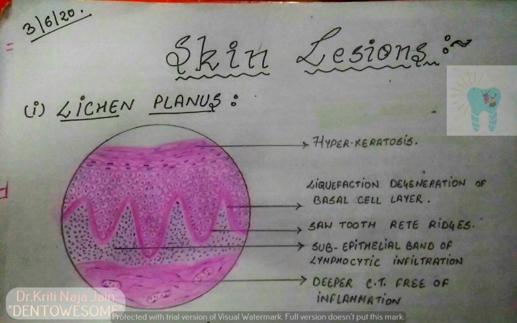
1.LICHEN PLANUS:-

*Lichen planus is a chronic mucocutaneous disorder manifested in a various forms in the oral cavity.

*The most characteristic pattern is” RETICULAR TYPE” with the interlacing white stripe called “WICKHAM’S STRIAE”.

*HISTOPATHOLOGY:-

  •   Histopathology FIRST DESCRIBED BY DUBRENILL 1906
  • later revised by Shklar in 1972
  • Hyper orthokeratinisation or hyper parakeratinisation
  • ◦Thickening of granular layer
  • ◦Acanthosis of spinous layer
  • ◦Intercellular oedema in spinous layer
  • ◦“ Saw-tooth” rete pegs
  • ◦Liquefaction necrosis of basal layer- Max Joseph spaces
  • ◦Civatte ( hyaline or cytoid) bodies
  • ◦Juxta epithelial band of inflammatory cells
  • ◦An eosinophilic band may be seen just beneath the basement membrane and represent fibrin covering lamina propria.

2.PEMPHIGUS :-

Pmphigus is a tissue specific autoimmune disease affecting the skin and mucosa. Clinical manifestations is in the from of “vesiculobullous lesions”  that rupture to form ulcer and erosions .

*Vesiculobullous lesions develop due to immune mediated acantholysis causing intraepithelial vesicle formation.

*HISTOPATHOLOGY :-

  • Formation of the vesicle or bullae within the epithelium that often results in a supra-basilar spilt or separation.
    • Following this suprabasilar spilt in the epithelium, the basal cell layer remains attached to the lamina propria, and it often appears as a row-of-tomb stones.
    • Loss of intercellular bridges and collection of edema fluid result in acantolysis within the spinus cell layer, which causes disruption of the prickle cells.
    • As a result of acantholysis, clumps of large hyperchromatic epithelial cells desquamate that are often seen lying free within the vesicular fluid, these desquamated cells are often rounded and smooth in appearance and are known as “Tzanck cells”.
  • Small number of polymorphonuclear neutrophil (PMN) and lymphocytes may be found within the vesicular fluid, but there is minimum inflammatory cell infiltration in the underlying connective tissue (unlike any other vesiculobullous lesion).

3.PEMPHIGOID :-

Pemphigoid is a vesiculobullous lesions that develop due to an autoimmune reaction directed against some components of basement membrane.

*This results in seperation of epithelium from the connective tissue with sub epithelial vesicles formation .

*Bullous pemphigoid and cicatricial pemphigoid are two different types of pemphigoid lesions.

*HISTOPATHOLOGY:-

  • The inflammatory infiltrate is typically polymorphous, with an eosinophilic predominance.
  • Mast cells and basophils may be prominent early in the disease course.
  • Electron microscope shows basement membrane attached to the connective tissue rather than overlying separated epithelium.
  • Tzanck smear shows only inflammatory cells.
  • Sub epithelial vehicle formation.
  • Intact epithelium without acantholysis.

REFERENCE:-

  1. Pic – Maji Josh 2nd edition
Hemoglobin Cooperative Binding

Cooperative Binding/Release

Definitions

Cooperative binding

  • Describes unique interactions between heme groups in hemoglobin
  • Small movement of heme group propagates through hemoglobin’s 3D structure

HEMOGLOBIN STRUCTURE

R-form (relaxed)

  • Alpha-alpha interactions: weak ionic and H-bonds form salt bridges
  • Beta-beta interactions: no interactions; move apart upon oxygenation
  • Alpha-beta dimers: strong hydrophobic interactions within each dimer

T-form (tense)

  • 3D structure changes between oxygenated and deoxygenated states

HEME SITE

T-form (tense)

  • Heme site: when O2 leaves, iron center moves out of porphyrin plane & proximal histidine moves away from iron center
  • Small movement in heme group makes O2 binding unfavorable

SALT BRIDGES

  • Salt bridges break and reform upon oxygen binding –> peptide wiggle-room
  • Alpha chain salt bridges:
    – Alpha1: arginine carboxy terminus (-) and arginine side group (+)
    – Alpha2: lysine (+) and aspartate (-)
  • Salt bridges regulate cooperativity: iron centers move into porphyrin planes

DISSOCIATION CURVE

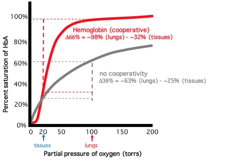
  • % oxygen saturation vs. oxygen partial pressure (torr)
  • Cooperative binding produces sigmoidal binding curve
  • After hemoglobin reaches 50% saturation: saturation increases rapidly (steepest point of curve)
  • Hemoglobin O2 affinity rapidly increases at half saturation

Hemoglobin and Myoglobin

How and why the affinity of myoglobin to combine with oxygen is ...

Globular proteins

  • Compact proteins that are approximately spherical in shape

Hemoproteins

  • Specialized proteins that have prosthetic heme group

Prosthetic groups

  • Non-protein molecules that are essential to biological function

HEME GROUP STRUCTURE

  • Porphyrin ring with iron center (Fe2+)

Fe2+ coordinates 6 bonds:

  • 1-4. Four planar nitrogen atoms (of porphyrin ring)
    1. Proximal histidine
    1. Oxygen

MYOGLOBIN

  • Skeletal and cardiac muscle
  • Reservoir for oxygen
  • Single polypeptide with 8 alpha helix segments
  • One heme group
  • Distal histidine holds oxygen in place

HEMOGLOBIN

  • Red blood cells
    – Supplies body’s tissues with oxygen
  • 4 polypeptides (instead of one): each subunit resembles myoglobin structure
    – Tetramer: with 2 alpha-beta dimers
  • Strong hydrophobic interactions: stabilize alpha-beta dimer
  • Weak ionic and H-bonds: between dimers

T-form hemoglobin

  • “Taught” or “tense” form: polypeptides restricted in movement
  • Deoxygenated form: oxygen affinity is low

R-form hemoglobin

  • “Relaxed” form: weaker ionic and H-binds between dimers
  • Oxygenated form: oxygen affinity is high

COOPERATIVE BINDING

  • Conformational change between T-form and R-form hemoglobin
  • Myoglobin does not exhibit cooperative binding: only one oxygen binding site
    – One oxygen binds hemoglobin subunit
    – Binding disrupts inter-dimer bonds: causes conformational change
    – Change in 3D structure increases oxygen affinity of remaining subunits

DISSOCIATION CURVE

  • % oxygen saturation vs. oxygen partial pressure (torr)
  • Cooperative binding produces sigmoidal binding curve
  • pO2 in body’s tissues: 30 torr
  • pO2 in lungs: 100 torr

Hemoglobin

  • Half saturated at 30 torr (body’s tissues): responds to O2 availability

Myoglobin

  • Hyperbolic curve (simpler binding pattern corresponds to single heme)
  • High affinity for O2
  • Binding properties correspond to role in oxygen in storage (not oxygen delivery)
  • Early curve: exercising muscle; plateau: muscle at rest

CLINICAL CORRELATION

Fetal hemoglobin

  • Dissociation curve to the left of adult hemoglobin
  • Greater affinity for O2: O2 transfer from maternal hemoglobin to fetus

Neural Control of Respiration

Neural Control of Ventilation - Involuntary Control ...

OVERVIEW

Medulla

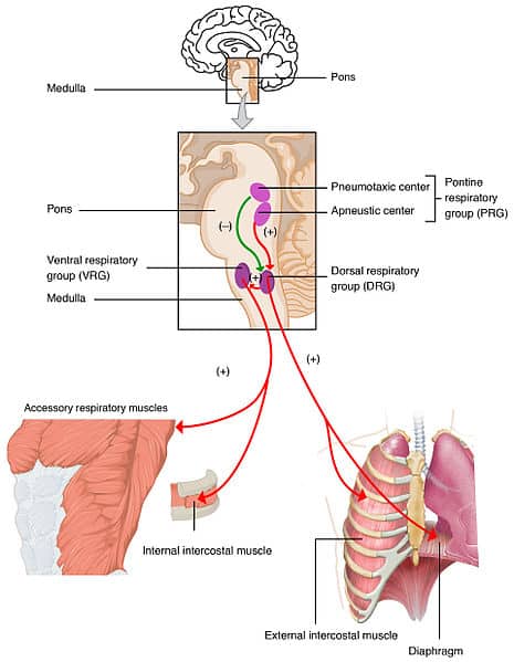
  • The medulla is the primary brainstem mediator of respiration.
  • Via the dorsal respiratory group (DRG), the dorsal (posterior) medulla controls sensory integration.
    • For its location, think: solitary tract nucleus.
  • Via the ventral respiratory group (VRG), the ventral (anterior) medulla controls motor output.
    • For its location, think: nucleus ambiguus.

Phrenic nerve

  • C3, C4, C5 supply the phrenic nerve, which innervate the diaphragm: C3, C4, C5 “keep the diaphragm alive”.

BRAINSTEM CIRCUITRY

Ventral respiratory group (VRG)

  • Within the medulla, anteriorly, lies the ventral respiratory group (VRG), which lies within the ventrolateral medulla.
    • It provides innervation for motor output.
    • It is involved in the activation of both inspiration and expiration.

Dorsal respiratory group (DRG)

  • Within the dorsal medulla (in the solitary tract nucleus), lies the dorsal respiratory group (DRG).
    • It provides sensory integration.
    • It receives sensory input related to the inspiration phase of respiration.

PERIPHERAL CHEMORECEPTORS

Peripheral chemoreceptors act on the dorsal respiratory group

  • Peripheral innervation involves the aortic bodies (shown here in the arch of the aorta) and the carotid bodies in the carotid bifurcation.
    • The carotid and aortic bodies are chemoreceptors.
    • They respond to levels of arterial oxygen and carbon dioxide levels and blood acidity.

Innervation to the brainstem respiratory center

  • Both cranial nerves 9 and 10 (we treat them jointly for simplicity) pass through the jugular foramen within the skull base across from the brainstem to innervate the dorsal respiratory group.

SECONDARY INSPIRATORY MUSCLES

Key structures

  • Anterior face, tongue, pharynx, and larynx.

Innervation

  • Cranial nerves 9, 10, and 12 innervate the secondary inspiratory muscles (again, we treat them jointly for simplicity).

VRG innervation of CNs 9 and 10

  • The ventral respiratory group acts upon these cranial nerves.

PRIMARY INSPIRATORY MUSCLES

Key structures

  • Thoracic cage, diaphragm, and intercostal muscles.

Innervation

  • The ventral respiratory group innervates C3, C4, C5 motor neurons in the anterior horn of the spinal cord gray matter, which supply the phrenic nerve, which innervates the diaphragm (again: C3, C4, C5 “keep the diaphragm alive”).

Intercostal nerves

  • Intercostal nerves innervate the intercostal muscles.

DETAILED ANATOMY OF THE DRG & VRG

Dorsal respiratory group (DRG)

  • The dorsal respiratory group lies within the solitary tract nucleus of cranial nerves 9 and 10.

Ventral respiratory group (VRG)

Simplification of VRG microanatomy

  • First, add nucleus ambiguus of CNs 9 and 10.
    • This will help us continue to recall the important of CNs 9 and 10 and thus the medulla itself in respiratory control.
  • There are many subnuclei that constitute the ventral respiratory group; we’ll only address the Bötzinger nuclei, here.
  • The Bötzinger complex lies within the superior aspect of the ventral respiratory group (some authors distinguish it from the ventral respiratory group, entirely).
  • The pre-Bötzinger complex is considered the “respiratory pacemaker.”
    • Notably, it contains mu receptors, which makes it sensitive to opioids.
    • Thus, we can see one of the ways in which opioids (such as morphine) can depress our drive to breathe.

CLINICAL CORRELATION: ONDINE’S CURSE

  • Ondine’s curse is the clinical eponym for the failure of automatic breathing during sleep.
    • It typically occurs from lower medullary or high cervical spinal cord lesions.
    • These patients are dependent on a ventilator when they sleep to survive.

PONTINE RESPIRATORY CONTROL CENTERS

Apneustic center

  • The apneustic center is a nonspecific region in the posterior lower pons.
    • It promotes apneusis: a prolonged inspiratory pause.
    • It comprises diffuse lower pontine nuclei.

Pneumotaxic center

  • The pneumotaxic center (aka the pontine respiratory group).
    • It prevents apneusis: it promotes regular breathing.
    • The pneumotaxic center comprises the medial parabrachial nucleus and the Kölliker-fuse nucleus.

Functions

  • Whereas these centers where formerly thought to be well-defined and to perform unique functions, now they are understood to be diffuse and their functions are no longer thought to be unique (the pneumotaxic center is not the only site to prevent apneusis, for instance).

BREATHING PATTERNS

We can use breathing patterns in comatose patients to localize the level of the CNS lesion, as follows:

Patterns

  • Cerebral hemispheric lesions cause Cheyne-Stokes respirations.
    – Illustrate Cheyne-Stokes respirations as periods of hyperpnea (deep breathing) with apneas (cessation of breathing)
  • Midbrain lesions cause hyperventilation.
    • Illustrate hyperventilation as rapid, deep breathing.
  • High pontine lesions cause apneustic breathing.
    • Illustrate apneustic breathing as periods of long inspiratory pauses before release of air.
  • Low pontine lesions cause cluster breathing.
    • Illustrate cluster breathing as irregular clusters of breaths.
  • Medullary lesions cause ataxic breathing.
    • Illustrate ataxic breathing as a completely irregular breathing pattern.

Limitations

  • Although these localizations are notoriously unreliable, they still give us a simple heuristic to follow when we examine comatose patients, which is essential.

Ventilation to Perfusion Ratios

Ventilation-Perfusion Ratio | Pathway Medicine

VENTILATION-TO-PERFUSION RATIOS

  • The ventilation/perfusion (V/Q) ratio is an indication of how well alveolar ventilation matches pulmonary capillary perfusion.
    • Due to gravitational forces, the V/Q ratio ranges regionally in the lung, from 3.0 at the apex to 0.6 at the base
  • Clinicians and physiologists typically use the average value for the entire lung as a reference point.
  • Healthy average alveolar ventilation rate is approximately 4 liters of air/minute
  • Blood flow rate is approximately 5 liters of blood/minute.
  • A healthy V/Q for the entire lung is 0.8.

Healthy V/Q

  • Inspired air flows through the tracheobronchial tree and to the thin-walled, sac-like alveoli
  • Pulmonary capillaries are in close physical proximity
  • When the V/Q is 0.8, ventilation and perfusion are well matched and optimal gas exchange occurs (not perfect, which would be V/Q = 1; we’ll use the average for the entire lung).
  • The partial pressures of oxygen and carbon dioxide in the alveoli and pulmonary blood flow equilibrate (the specifics of gas exchange are discussed elsewhere).

V/Q mismatches, aka, defects, occur along a spectrum.

  • Two extremes of mismatches are shunts and dead space:
    • Shunts occur when the rate of alveolar ventilation is zero; V/Q = 0
    • Dead spaces occur where the rate of blood flow is zero; V/Q = infinity
  • Notice that the normal V/Q of 0.8 is between the two extremes (a V/Q of 1 would be perfect, but we are using the average value)
  • Mismatches between 0 and 0.8 reflect defects in alveolar airflow
  • Mismatches greater than 0.8 reflect defects in pulmonary capillary blood flow

Shunt

  • Most extreme alveolar ventilation defect.
  • Occurs when air flow is blocked and the alveoli remain unventilated.
  • Unventilated alveoli cannot participate in gas exchange with the pulmonary capillaries, which remain de-oxygenated.
  • As a result, the partial pressures of oxygen and carbon dioxide of the pulmonary blood remain equal to that of mixed venous blood.

Airway obstruction is a common cause of shunts

  • Because airflow ceases, alveolar collapse, aka, atelectasis, can occur in all or a portion of the lung.
    • Without proper ventilation, the partial pressure of arterial oxygen is reduced (aka, hypoxemia).
      In the case of shunts, hypoxemia cannot be reversed by administration of concentrated oxygen because oxygen does not reach the pulmonary blood flow.

Reduced ventilation

  • Alveoli are partially ventilated, so some gas exchange occurs with the nearby pulmonary capillary.
  • When ventilation is less than perfusion, the partial pressure of pulmonary blood carbon dioxide increases because it is held within the body, and the partial pressure of oxygen decreases because the normal amount of fresh oxygen is not available.

Reduced perfusion

  • Reduced perfusion causes an increased ventilation-to-perfusion ratio.
  • The partial pressure of pulmonary blood carbon dioxide decreases, while the partial pressure of oxygen increases.

Alveolar dead space

  • Absence of blood perfusion produces a ventilation-to-perfusion ratio equal to infinity.
  • No gas exchange occurs.
  • As a result, the partial pressures of oxygen and carbon dioxide of alveolar gas remain equal to that of inspired air.
  • Pulmonary embolism is a common cause of alveolar dead space
    • To minimize “wasted” ventilation, bronchiolar constriction diverts air from non-perfused alveoli.

EMERGENCY INSTRUMENTS-II

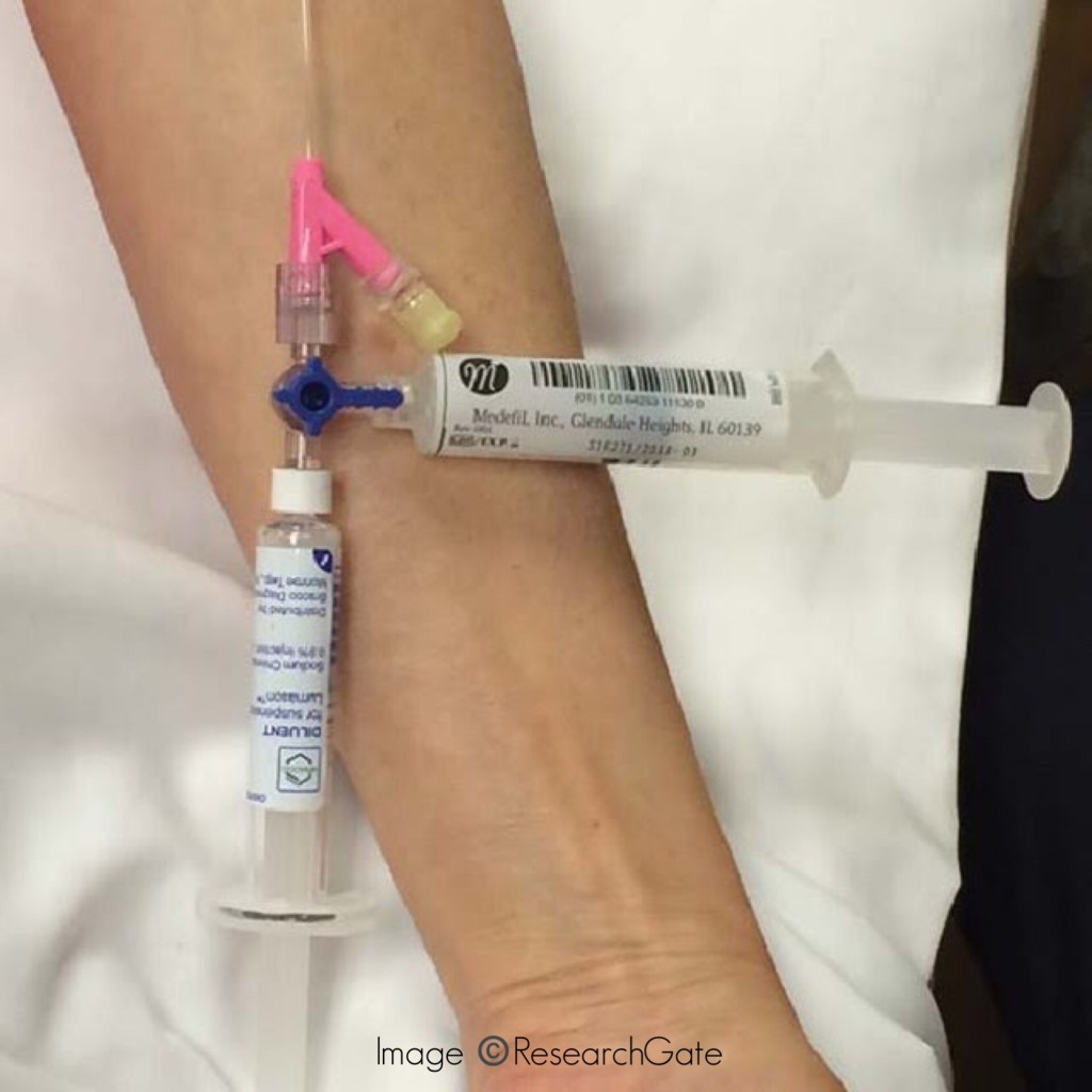
1) 3-way: It consists of two inlets & one outlet.
Uses:
  1. It is commonly connected to the IV cannula where through one inlet IV fluids pass and through the other inlet medications can be given or Central venous pressure(CVP) can be monitored.
  2. Aspirating fluid from the body cavities.
  3. Exchange transfusion.
2) IV cannulas:

Size: 14G – 24G (Smaller the no., larger the bore of the needle)

Use:

  • Venous access for longer period.
  • IV fluids
  • Drugs administration
  • Collection of blood samples

Source: Internet