
Source: Shafer’s Textbook of Oral Pathology, 8th Edition
Source: Shafer’s Textbook of Oral Pathology, 8th Edition
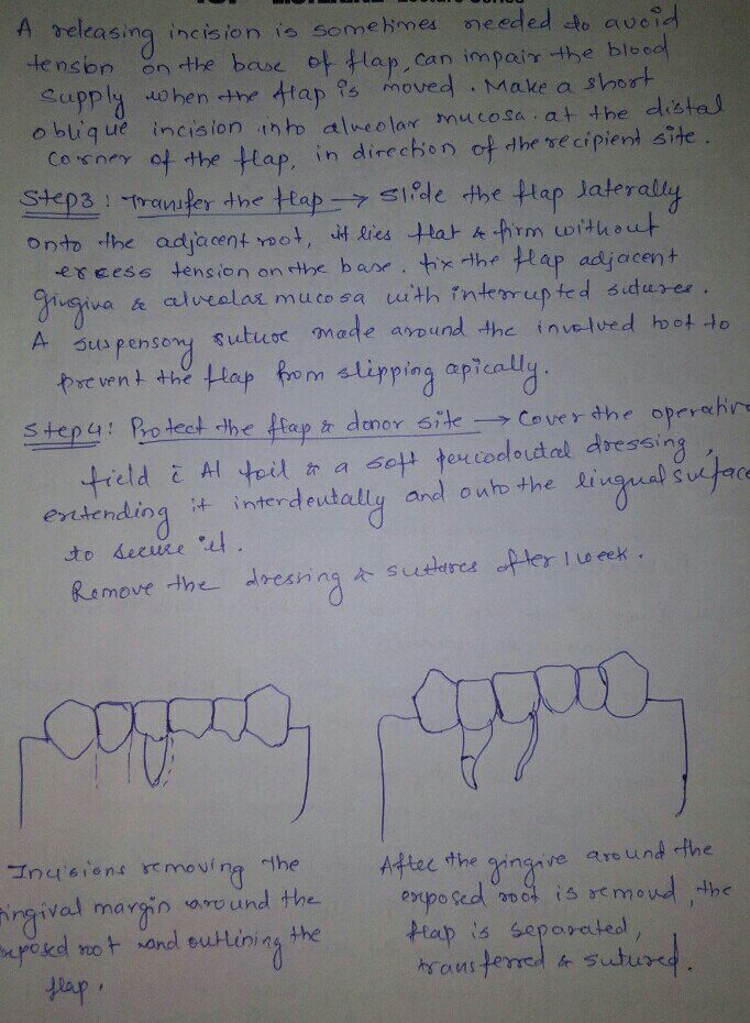
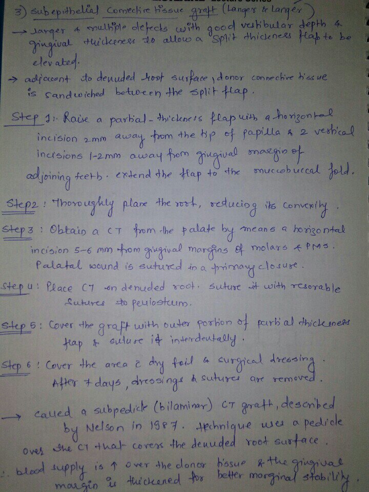
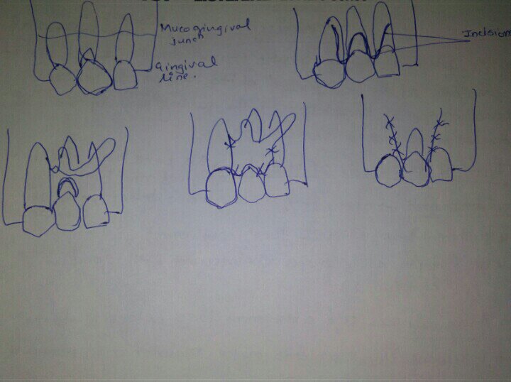
SOURCE – CARANZA TEXTBOOK
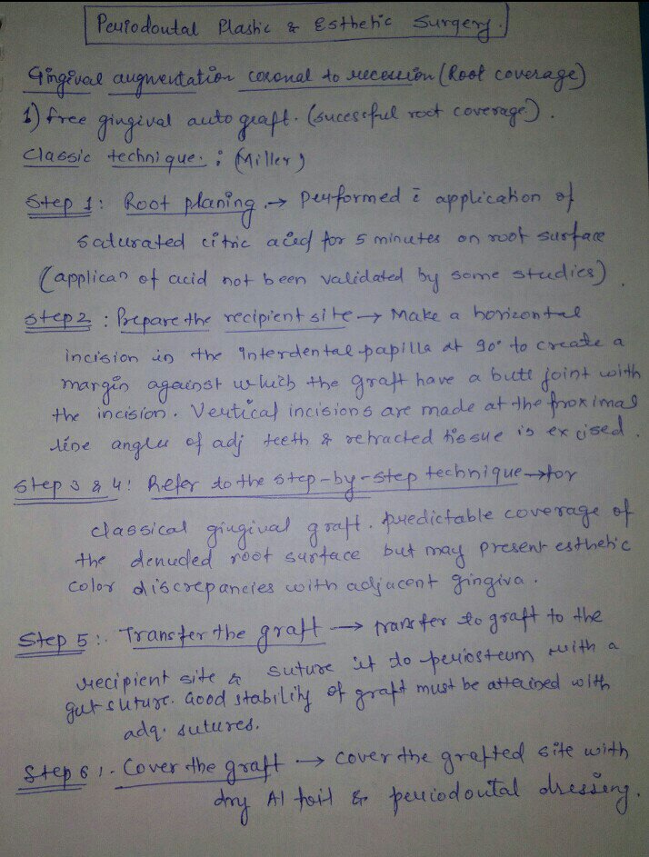
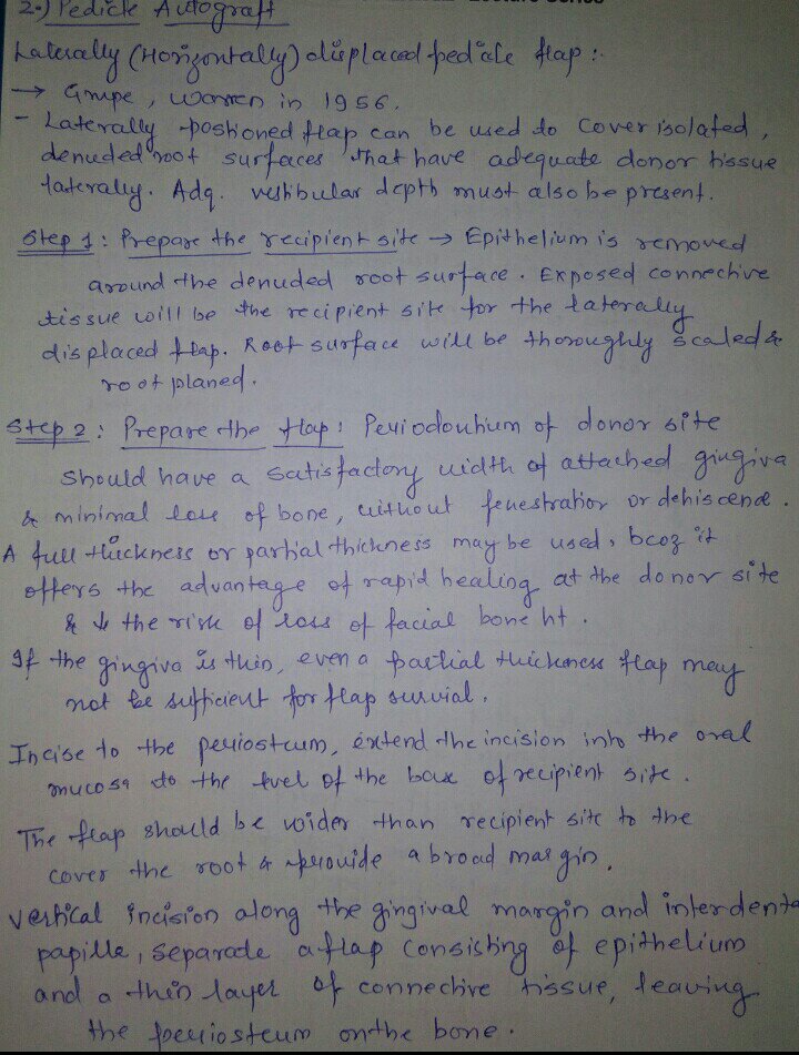
SOURCE – CARANZA TEXTBOOK
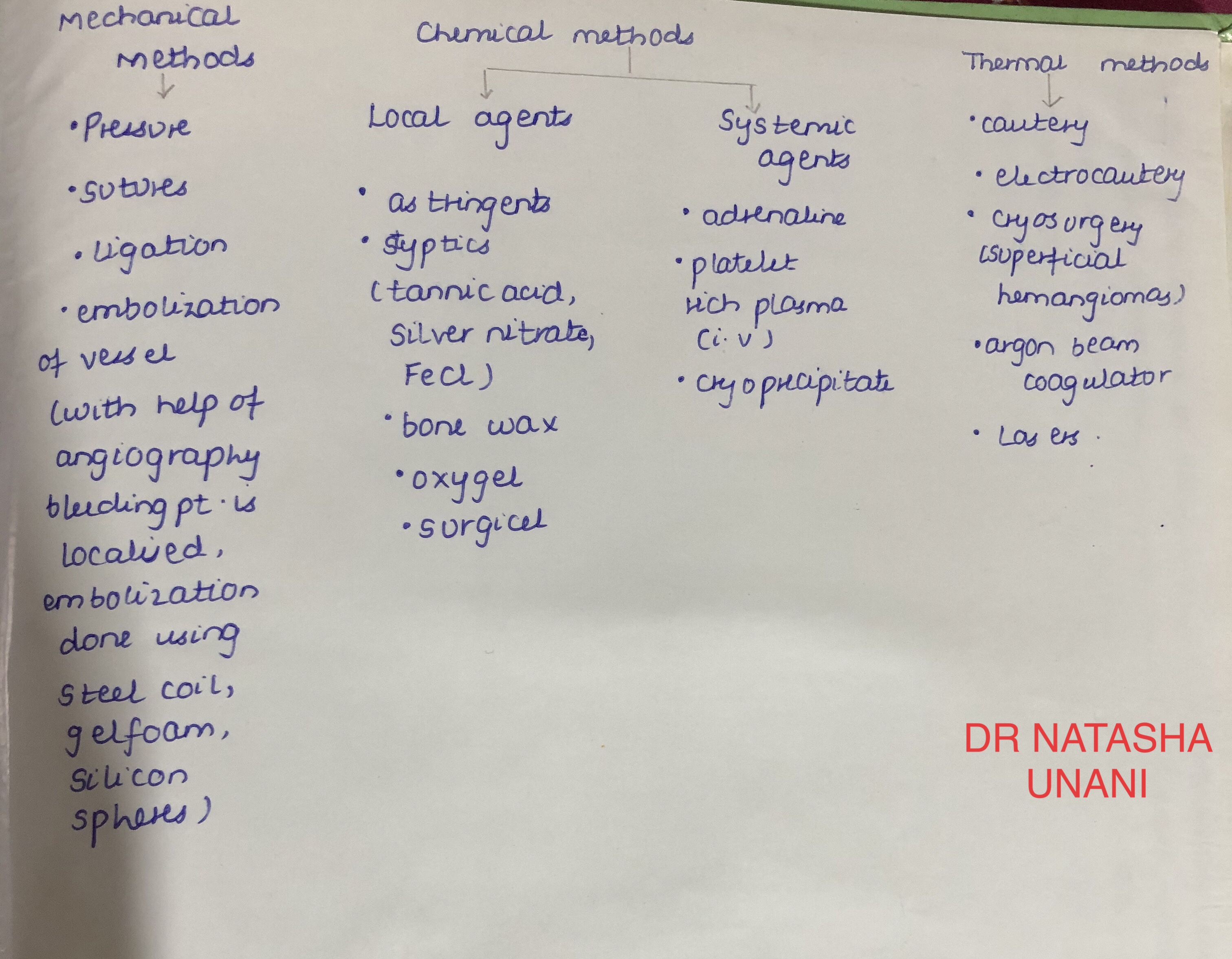
source – don’t remember, had written it a lot time ago
BY Dr. KRITI NAJA JAIN :-
1. FIBROUS DYSPLASIA :-
Def:- Fibrous dysplasia is an uncommon nonhereditary, developmental anomaly of the bone due to a defect in osteoblastic differentiation and maturation.
HISTOPATHOLOGY:


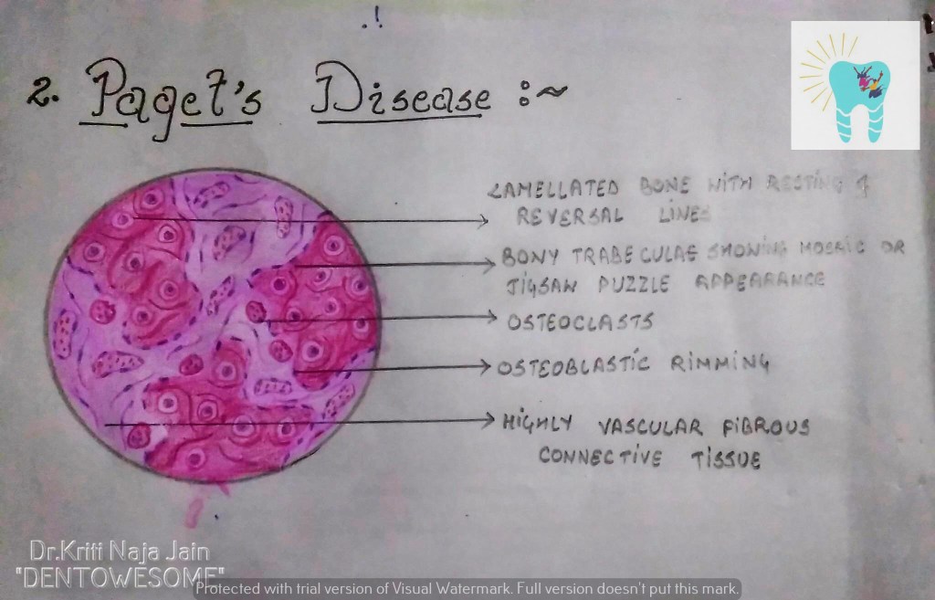
2. PAGET’S DISEASE (OSTEITIS DEFORMANS):-
Def:- Paget’s disease of bone is a condition characterized by abnormal and anarchic resorption and deposition of bone, resulting in distortion and weakening of the
affected bones.
HISTOPATHOLOGY:-

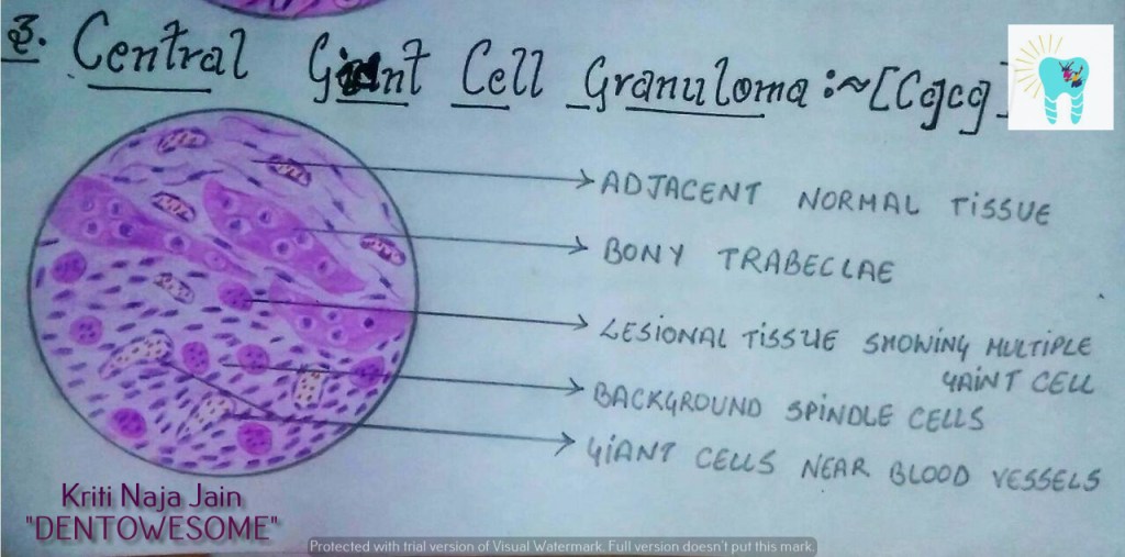
3. CENTRAL GAINT CELL GRANULOMA(GIANT CELL LESION; GIANT CELL TUMOR):-
Def :- Central giant cell granuloma (CGCG) is an uncommon, benign and proliferative lesion whose aetiology is not defined. Central giant cell granuloma is a relatively common benign intraosseous destructive giant cell lesion, which often affects the anterior part of the jawbone. By seeing clinical and radiographically , CGCG is divided into two types:-
1. Nonaggressive lesions make up most cases, exhibit few or no symptoms, demonstrate slow growth, and do not show cortical perforation or root resorption of teeth involved in the lesion.
2. Aggressive lesions are characterized by pain, rapid growth, cortical perforation, and root resorption. They show a marked tendency to recur after treatment, compared with the nonaggressive types.
HISTOPATHOLOGY:-

REFERENCE:-
1.Maji Jose 2nd edition
Source: Shafer’s Textbook of Oral Pathology, 8th Edition
TYPES OF INTERSTITIAL LUNG DISEASE
Let’s begin with interstitial lung diseases that develop in response to exposure to external agents.
Occupational
Drugs
Infections
Connective Tissue Disorders
ANCA vasculitides (ANCA = Anti-Neutrophilic Cytoplasmic Autoantibodies)
Remind ourselves that these disorders include: Granulomatosis with polyangiitis (aka, Wegener’s disease), eosinophilic granulomatosis with polyangiitis (aka, Churg-Strauss syndrome), and, microscopic polyangiitis.
Granulomatous lung diseases
Others:
Bronchiectasis is a chronic, heterogeneous disorder caused by repeated bouts of infection and inflammation that cause permanent dilation of the medium and medium-large airways.
– Damage can be focal or diffuse, depending on the cause and extent of the damage.
– Etiologies:
Obstruction, which causes focal bronchiectasis, and can be due to tumor masses or foreign bodies in the bronchi.
Infections, often bacterial; common pathogens include Pseudomonas aeruginosa, Haemophilus influenzae, and Non-tuberculosis mycobacteria.
Immune deficiencies
Autoimmune disorders; for example, bronchiectasis is associated with inflammatory bowel disease and rheumatoid arthritis.
Allergic bronchopulmonary aspergillosis; as its name suggests, this form of bronchiectasis is caused by an allergic reaction to the common mold aspergillus;
Genetic conditions, including cystic fibrosis, alpha-1-anti-trypsin deficiency, and primary ciliary dyskinesia (aka, Kartagener syndrome) – these disorders are associated with increased mucus production, neutrophilic damage, and impaired muco-ciliary functions, respectively.
**Asthma and/or COPD
– Many cases of bronchiectasis are idiopathic.
Bronchiolitis is a common affliction of children younger than two years of age; it occurs as result of respiratory infections.
Pathogenesis: A Vicious Cycle
###Bronchiolitis
Eosinophilic asthma with high levels of type-2 inflammation
Next, let’s consider some other types of asthma.
Neutrophilic, type-2-low asthma
Mixed granulocytic asthma
Paucigranulocytic asthma
Two additional asthma phenotypes that can overlap with those already listed:
Occupational, aka, work-related asthma
SOURCE – CARANZA TEXTBOOK

