

Sources – Shafer’s textbook of oral pathology , Slideshare.com
Seqeulae of wearing denture
Muhad Noorman P- Dentowesome 2020
1.DENTURE STOMATITIS
Denture-induced stomatitis is a common inflammatory reaction related to the wearing of dentures usually involving Candida (yeast) species. Less common forms of denture stomatitis may be due to mechanical trauma or a contact reaction.
Predisposing factors
Denture-related stomatitis is very common, with over 50% of denture wearers affected in some populations. It is the most common clinically important condition developing in the mouth
- Complete upper (maxillary) denture – probably due to the large contact area between denture and oral mucosa
- Acrylic dentures – Candida species seem to have a particular binding affinity for acrylic resin
- Poor dental hygiene – Candida species and Lactobacillus bacteria stick to denture surfaces and should be removed chemically and/or mechanically at least daily
- Poorly fitting dentures – mechanical trauma damages the mucosa, making it more prone to infection
- Denture age – old dentures are commonly associated with denture stomatitis probably due to poor fitting and rough surface in which Candida can hide
- Continuous wearing of denture – failure to remove at night increases the risk
- Men – are twice as likely to develop denture stomatitis than women
- Diabetes mellitus – diabetics are more prone to developing yeast infections
- Dry mouth (xerostomia) – saliva normally helps flush the mouth and clean the denture surface
CLINICAL FEATURE , Denture stomatitis usually does not cause any symptoms ,Some complain of burning sensations in mouth . But on examination the mouth lining in contact with the denture will be red and swollen sometimes with small red dots (petechial haemorrhages).
Treatment: regular oral hygiene, oral nystatin gargle, cleansing denture in nystatin solution, Not wearing denture at night.
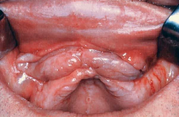
2.DENTURE INDUCED FIBROUS HYPERPLASIA aka EPULIS FISSURATUM
Denture-induced fibrous hyperplasia (DIFH) is a persistent lesion caused mostly by the prolonged wear of an ill-fitting, over-extended denture. Although the condition frequently coexists with denture stomatitis, it is a distinct entity .Develops as a reactive lesion to chronic mechanical irritation produced by the flange of a poorly fitting denture.More simply, epulis fissuratum is where excess folds of firm tissue form inside the mouth, as a result of rubbing on the edge of dentures that do not fit well. It is a benign entity.
CLINICAL FEATURE: The lesion is usually painless.The usual appearance is of two excess tissue folds in alveolar vestibule/buccal sulcus, with the flange of the denture fitting in between the two folds. It may occur in either the maxillary or mandibular sulci,although the latter is more usual.Anterior locations are more common than posterior. Less commonly there may be a single fold, and the lesion may appear on the lingual surface of the mandibular alveolar ridge.
Surgically excised, reconstruction of denture, denture rebasing can be done
3. DENTURE INDUCED ULCER
Traumatic ulcers caused by dentures with overextended or unbalanced occlusion are seen in about 5% of denture wearers.Overextention of flanges,sequestration of bony spicules, illfitting, sharp boders are cause of Traumatic ulcer.
CLINICAL FEATURE: Superficial, Painful, shallow ulceration often with erythematous halo and greyish membrane covering the ulcer.
Symptomatic relief by topical analgesic,steroid oinments and repair of denture.
4. INFLAMMATORY PAPILLARY HYPERPLASIA OF PALATE
Inflammatory papillary hyperplasia (IPH) is a benign lesion of the palatal mucosa. It is usually found in denture-wearers but also has been reported in patients without a history of use of a maxillary prosthesis use.The lesion almost exclusively involves the hard palate
CLINICAL FEATURES:
Inflammatory papillary hyperplasia is usually asymptomatic. It presents as a cluster of individual papules or nodules that may be erythematous, somewhat translucent, or normal in surface coloration. Mucosa is erythematous and has a pebbly or papillary surface. Many cases are associated with denture stomatitis.
Often the entire vault of the hard palate is involved .Has a female prediliction.
This may be aided by use of topical antibiotic or antifungal therapies. Small lesions are also typically treated with mouthrinses such as chlorhexidine mouthrinse at 0.12% or antifungal mouthrinse/ gels. Larger lesions recquired surgical excision.
References: Neville Textbook of Oral pathology.
Shafers textbook of pathology. Cover photo image: Internet
Composition and Functions of Blood
By Dr Musaddika Shaikh Dentowesome @drmusaddikashaikh
Blood :- It is body fluid in body that delivers necessary substance like oxygen,nutrients to cell and transports metabolic waste away from cell.

- Total blood volume :- 5-6litres 80ml/kg body weight
- Viscosity :- 4-5 litres
- Ph :- 7.4
- Specific gravity :- 1.040-1.060
Composition :-
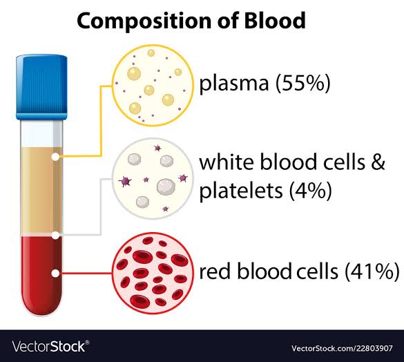
Anti coagulated sample of blood allow to stand in a tube,it settles cells and plasma.
A) Cells :- Cellular element is 45% of total blood volume called packed cell volume. 1. RBC/Erythrocytes :- Normal count 5million cu.mm. 2. WBC/Leucocytes :- Normal count 4000-11000 cu.mm. 3. Platelets/Thrombosis :- Normal count 1.4-4 lakh cu.mm
B) Plasma :- It is clear,straw colored with 55% of total volume. It contains 91% water+9% organic+1%inorganic substances. a) Inorganic substance:- 1. Na+,Ca+,HCO3(extracellular) 2. k+,Mg,Po4 (intracellular). 3. Ferrous and ferric oxide. b) Organic substance :- 1. Plasma protein. 2. Sugar. 3. Fat 4. Hormone
Non protein nitrogenous substance :- 1. Urea :- 20-40% mg% 2. Uric acid :- 2-4% mg% 3. Creatine :- 1-2% mg% Other substance are fat, phospholipids, glucose.

Functions :-
- Respiratory :- blood transprt oxygen from lungs to tissues and CO2 from tissues to lungs.
- Nutritive :- absorb food material,glucose,amino-acid,fatty acid and vitamins.
- Excreatory :- blood transport metabolic waste.
- Heamostatic function :- to regulate normal cell function by heamostasis
- Regulate body temperature :- regulate body temperature by buffer system.
- Chemical :- regulate negative feedback machanism and defence system for inflammation.
Reference :- Books Human physiology for bds A.K Jain. Google website texasheart.org , britannica.com , vectorstock.com
Haemoglobin
By Dr Musaddika Shaikh Dentowesome @drmusaddikashaikh
Haemoglobin :- It is protein found in red blood cells that carries oxygen in blood and gives blood red colour
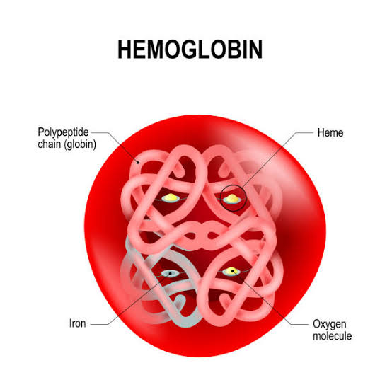
Structure :-
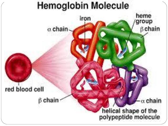
- Heame is porphin called iron protoporphyrine 9th
- Iron is ferrous form which combine with oxygen
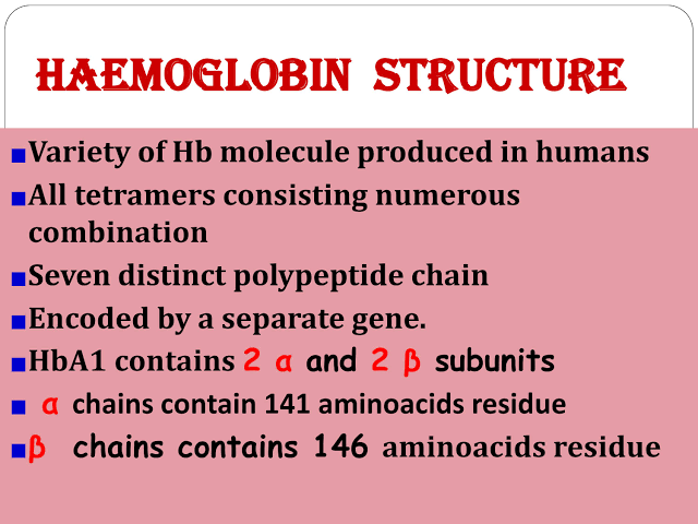
- Globin :- It is protein 4 polypeptide chain of alpha and beta which contains Mb amino acid each polypeptide attach are heam group
- Hemoglobin react with oxygen in 0.01 sec
- Molecular weight of hemoglobin 64,458 g/mol
- Oxyheamoglobin :- hemoglobin affinity for oxygen by Ph, temperature, concentration
- Carbaamino-heamoglobin :- carbon dioxide react with heamoglobin to form carbaamino-heamoglobin
- De-oxegenated heamoglobin :- oxygen removed from heamoglobin
- Carboxy-heamoglobin :- carba+monooxide reacts with heamoglobin
- Metheamoglobin :- reduced or oxygenated heamoglobin exposed to drugs is oxidised ferrous to ferric acid metheamoglobin
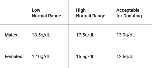
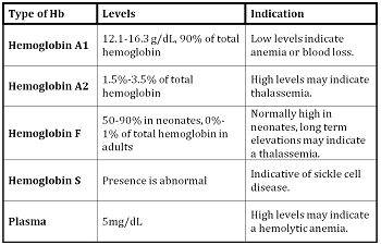
Normal values of heamoglobin :-
Types :-
Synthesis :-
- Role of protein :- Low protein reduces heamoglobin
- Role of minerals :- It helps in formation of heame
- Role of Cu :- It promotes absorption, mobilization and utilisation of iron
- Role of cobalt :- It enhances absorption in GI tract
- Role of vitamin :- Vit C,Vit B12,folic acid helps in synthesis of nucleic acid
Functions :-
- Transport of oxygen from lungs to tissues
- Transport of carbon dioxide from tissues to lungs
- Act as acid balance buffer
- It gives red colour to blood
- Genetic resistance to malaria etc
- Source of physiological active catabolites
Reference :- Book Human physiology for bds A.K Jain Google website study.com,learnpick.in, redcrossblood.org
Coagulation of Blood
By Dr Musaddika Shaikh Dentowesome @drmusaddikashaikh
Defination :-
Arrest or prevention of bleeding by physiological process is called hemostasis
Factors :-
- F1. Fibrinogen
- F2. Prothombin
- F3. Thromboplastin
- F4. Calcium
- F5. Labile
- F6. Not known
- F7. Stable
- F8. Antihaemophillic
- F9. Christmas
- F10. Stuart
- F11. Plasma thrombin antecedent
- F12. Hegman
- F13. Fibrin stabilizing
Mechanism :-
Injury vessel walls – Formation of clot – Seal off damage blood vessel – Prevent blood loss
Series of event :-
Constriction of blood of blood vessel – Temporary haemostatic plug by platelet – haemostasis
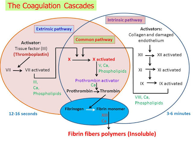
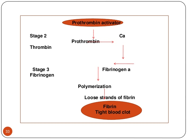
Factors responsible for coagulation :-
- Fibrin formation :- by soluble plasma fibrinogen due to f3,f4,f6
- Thrombin formation :- inactive prothrombin into thrombin by prothrombin activator
- Prothrombin activator :- by extrinsic and intrinsic factor
- Clot retraction :- conversion of prothrombin to thrombin within 15-30min
- Role of Calcium :- it act as catalyst in cascade reaction
Why does not blood clot in circulation? Smoothness of endothelial line prevents adhesion. Negatively charged particles repells cloting factor. Natural anticoagulant eg :- heparin in circulation
Reference :- Book Human physiology for bds A.K Jain Google website slide player
Urea Cycle
By Dr Musaddika Shaikh Dentowesome @drmusaddikashaikh
Urea cycle :-
- It is end product of protein metabolism.
- It is also known as urea Cycle or ornithine cycle or Krebs hensleit cycle.
- Nitrogen of amino acids get converted to ammonia which is toxic to body.
- Ammonia gets converted into urea and detoxified.
- Urea is mainly synthesized in liver and transported to kidney for excreation.
- Urea synthesis in 5steps cyclic process with 5 distinct enzymes.
- First two enzymes – mitochondria and Rest – cytosol
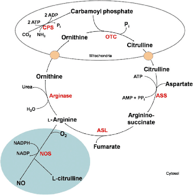
Formation of Carbamoyl Phosphate :- Carbamoyl Phosphate synthase 1st present in mitochondria catalyses condensation of NH ions with CO2 to form carbamoyl phosphate. This step consumes 2ATP and is irreversible.
Formation of citrulline :- It is synthesized from carbamoyl phosphate synthetase 1st and Ornithine by transcarbomyle. Ornithine is regenerated and used in urea cycle.
Formation of Arginosuccinate :- It condenses citrulline with aspartate to produce Arginosuccinate. 2amino group of urea is involved in reaction. This step requires ATP which is cleaved to AMP.
Formation of Argentine :- Arginosuccinate breakes to form argenine and fumarate.
Formation of Urea :- Arginase is final enzyme which cleaves argenine to yeild urea and orthinine. Orthinine enters mitochondria for its reuse in urea cycle.
Reaction :-NH4+CO2+ASPARTATE+3ATP = UREA+FUMARATE+2ADP+2PIi+AMP+Ppi
Reference :- Biochemistry book U. Satyanarayan, U Chakrapani. Google website
Glycolysis
By Dr Musaddika Shaikh Dentowesome @drmusaddikashaikh
Glycolysis :- It is sequence of reaction converting glucose into lactose and pyruvate with the production of ATP
- It is also know as EMBDEN MEYENHOFF Pathway.
- It takes place in cell of body.
- The enzyme are present in cytosomal fraction.
- It occurs in presence of oxygen (aerobic) and absence of oxygen (anaerobic).
- Lactate is end product of anaerobic pathway.
- Pyruvate is end product of aerobic pathway forming CO2 and H2O.
- It is major pathway for synthesis of ATP.
- It is essential for brain.
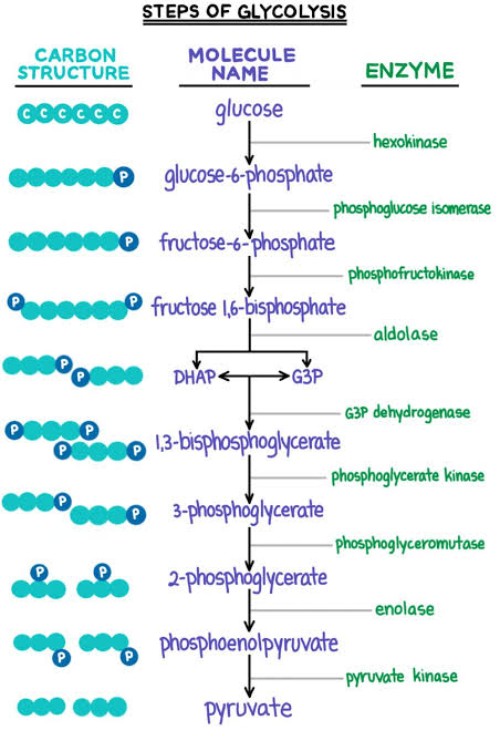
Reaction :- Glucose+2ADP+2Pi = 2Lactase+2APP
Significance :-
- It is formed by active skeletal muscle major precursor for gluconeogenesis
- Lactate is dead end in glycolysis
- It is carried from skeletal muscle through blood and handed over to liver , where it is oxidized to Pyruvate
Reference :- Books Human Physiology for bds A.K Jain Google website expii
Plasma Proteins
By Dr Musaddika Shaikh Dentowesome @drmusaddikashaikh
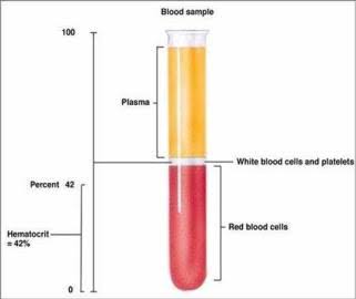
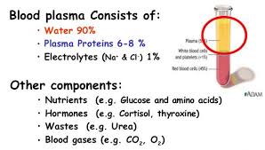
Plasma proteins :- They are also know as blood proteins. They are proteins which are present in blood plasma
Constituents :-
Origin :-
- Embryo from Mesenchymal cell
- Adult albumin from liver
- Globulin from tissue, macrophages,plasma cells, lymphocytes
Types :-
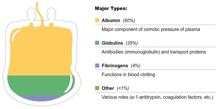
- Albumin :- 1.control collodial osmotic pressure 2.transport anion,cation,drugs,hormones,enzymes,billirubin
- Globulin :- regulate iron absorption and transport
- Fibrinogen :- helps in clotting
- Hepatoglobin :- prevent loss of iron in urinary excretion
Variation in plasma concentration :- Decrease – haemorrhage causes loss of plasma protein. Fibrinogen, albumin,globulin are regenerated by restoration effect in few days. Decrease of albumin causes during pregnancy,chronic liver disease, malnutrition,fasting, malabsorption,burn,nephrosis. Increase – loss due to burn or dehydration
Functions :-
- Helps in coagulation of blood
- Helps in maintaining colloidal osmotic pressure by urea,Na+,glucose
- Helps in maintaining Hydrostatic Pressure across cavity wall
- Helps in maintaining viscosity of blood
- Helps in Hypoproteinemia
- Helps in immune function
Reference :- Book Human physiology for bds A.K Jain Google website studyblue.com, Philadelphia.edu.jo
AMYLOIDOSIS
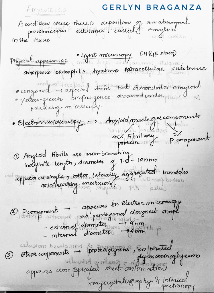
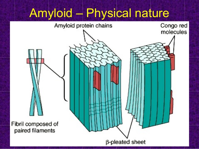
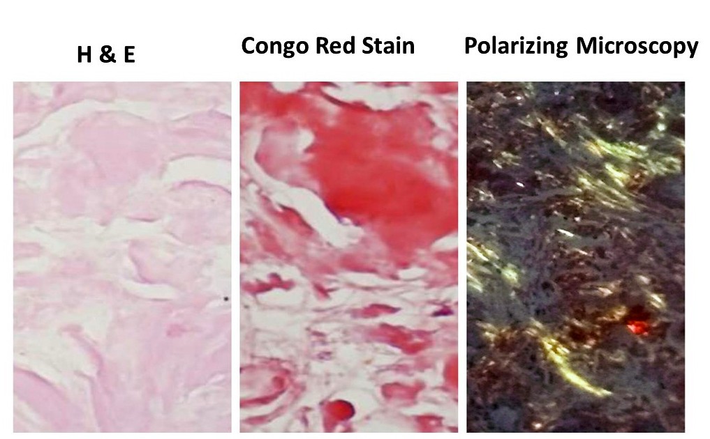
What is amyloidosis ?
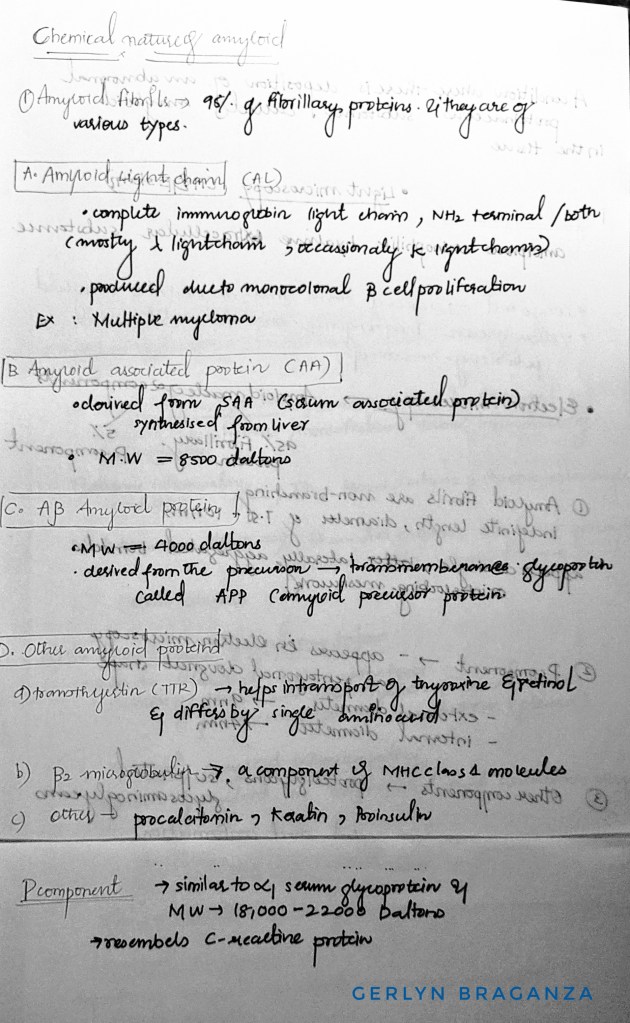
CHEMICAL NATURE OF AMYLOIDOSIS
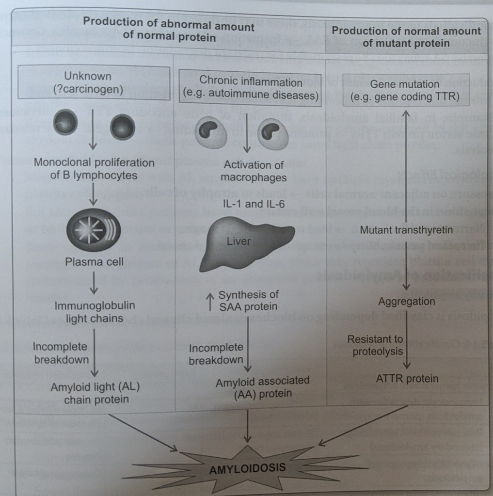
PATHOGENESIS –
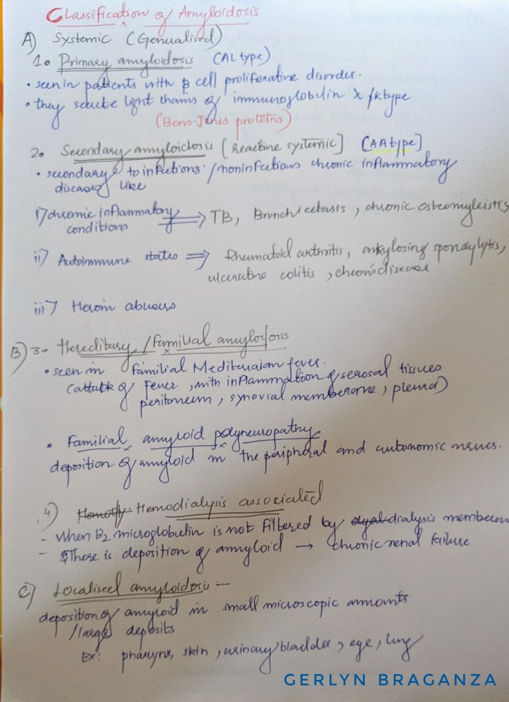
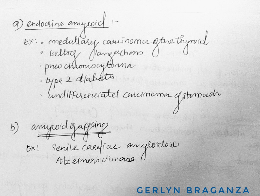
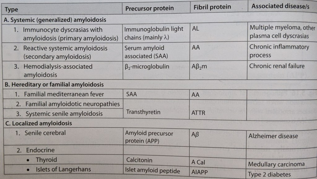
CLASSIFICATION OF AMYLOIDOSIS
Main organs involved-
Secondary amyloidosis– kidneys , liver, spleen, lymph nodes, adrenals , thyroid
Primary amyloidosis– heart , skin , peripheral nerves, tongue, respiratory tract
Source – textbook of pathology ( harsh mohan ) and pathology preparatory manual by ramdas nayak
Pictures – Google
CERVICOFACIAL EMPHYSEMA
Muhad Noorman P- Dentowesome 2020
During impacted teeth extraction process,Surgical managment of facial fractures with high speed rotary instruments like airotor, blowing of compressed air into root canal , airotor in middle face fractures with use of air-pushing machine such as airotor or even an air syringe, Due to air leakage inside the tissues can cause the phenomenon of confined air within tissues called Cervicofacial Emphysema and is the result of air trapping, so use the micromotor handpiece instead of the airotor, because it is a dangerous condition._The treatment is only to reassure the patient that it will go away after 3-4 days. However rarely can cause Venous Air Embolism, mediastenal emphysema and rarely pneumothorax.
Clinically presented as unilateral swelling of face/neck with or without dysphagia resembling Angioedoema,however crepitus on palpation is a positive diagnostic feautre.
Another type is PNEUMOPAROTID,entrapment of air in parotid duct either accidental or by profession in trumphet blowing artist etc.. presenting with painful swelling in face over parotid region with froathy saliva.and ctepitus on palpation.
References: Shafer’s Textbook of Oral Pathology

