ROOT CANAL SEALERS 🦷

These are used in conjugation with semi solid or solid obturating materials to establish an adequate seal of root canal system.This is a brief note on the various root canal sealers ,composition ,salient features and brand names of some sealers which would prove helpful in clinical setup.

Classification :(according to Grossman)

1)ZnOE based sealers:

i. Grossman’s Formula

ii. Roth’s 801

iii. Tubliseal

iv. Wachs Sealer

2)CaOH based sealers:

i. Sealapex

ii. Apexit

3)Glass ionomer based sealers:

4)Resin based sealers:

i. AH Plus

ii. AH 26

iii. Epiphany

iv. Diaket

ZnOE based sealer-

Composition:

Base ingredient of powder -ZnOE,

Liquid usually consists -Eugenol alone or in combination with other liquids such as Canada balsam.

Features:

.Hardens in 2 hours approx at 37 degree celsius & under 100% relative humidity.

.Setting time in canal is less.(10-30 mins).

. Tissue tolerance is satisfactory with little inflammation & no inhibition of repair.

CaOH based sealers-

Composition:

Base consists -Zinc oxide, CaOH, butyl benzene, sulfonamide & Zinc stearate

Catalyst consists – resin,isobutyl salicylate, barium sulfate,titanium dioxide & aerosol

Features:

.antimicrobial

.osteogenic-cementogenic

.better healing capability

Glass ionomer based sealer-

Feature : dentin bonding ability but are unpopular due to difficulty in removal from root canal walls after treatment.

Resin based sealer-

Composition:

Paste A -Epoxy resin ,Calcium tungstate,zirconium oxide,silica,iron oxide

Paste B – Admantaneamine-N,calcium tungstate,zirconium oxide,silica-silicone oil

Features:

. Dentinal adhesion (strong adhesive properties).

.Contracts slightly while hardening.

.Good sealing ability.

.Bio compatibility to periapical tissues.

.Moderate antimicrobial activity.

.Long working time and easy for manipulation.
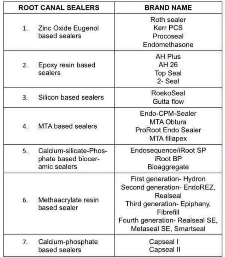
List of various root canal sealers and brand names

Sources: Grossman’s endodontic practice (13 th edition ),Instagram – @__dentistars__
CONSIOUS SEDATION

Source – pinterest

Conscious Sedation

Conscious sedation is a level of CNS depression where a patient does not lose consciousness but is able to communicate and cooperate during the procedure/treatment.

Indications

•Uncooperative patients.

• Anxious patients.

• Emotionally compromised patients.

Conscious sedation should be avoided in:

• Chronic obstructive pulmonary disease.

• Pregnancy

• Prolonged surgery.

• Psychoses

Drugs used

Drugs used

  1. Benzodiazepines
  • Diazepam is the most commonly used drug for conscious sedation.
  • Small doses (1-2 mg) of diazepam is administered intravenously slowly. It can also be administered orally.
  • Midazolam is a short-acting BZD given intravenously. Temazepam is given orally. It is safe and has better patient compliance.
  1. Nitrous oxide + oxygen: Nitrous oxide is given by inhalation route along with 100% oxygen.
  1. Propofol( i.v. infusion), fentanyl (i.v.), etc. can also be used for conscious sedation.

Precautions

• Written informed consent should be obtained from the patient prior to the procedure.

• Conscious sedation should be administered by trained personnel.

• Constant monitoring of the vital signs should be done during and after the procedure.

• The procedure should be documented. Postoperative instructions should be in written form.

• Equipment and emergency drugs should be kept ready to tackle any emergency • Patient should be escorted by an attendant.

Source- textbook of pharmacology for dental students – Tara V Shanbhag

THUMB SUCKING HABIT

THUMB & DIGIT SUCKING :
• Digit Sucking is defined as the placement of the thumb/one or more fingers in varying depths into the mouth.
• It may be practiced even during intra-uterine (IU) life.
• Presence of this habit is quite normal till 3½ – 4 yrs.

ETIOLOGY:
A number of theories have been put forward to explain why thumb sucking occurs. The following are some of the accepted ones:

  1. Freudian theory: This theory was proposed by Sigmund Freud in the early part of this century.
    • He suggested that a child passes through various distinct phases of psychological development of which the oral and the anal phases are seen in the first three years of life.
    • In the oral phase, the mouth is believed to be an oro-erotic zone.
    • The child has the tendency to place his fingers or any other object into the oral cavity.
    • Prevention of such an act is believed to result in emotional insecurity and poses the risk of the child diversifying into other habits.
  2. Oral drive theory of Sears and Wise: Sears and Wise in 1950 proposed that prolonged suckling could lead to thumb sucking.
  3. Benjamin’s theory:
    • Benjamin has suggested that thumb sucking arises from the rooting or placing reflex seen in all mammalian infant.
    • Rooting reflex is the movement of the infant’s head and tongue towards an object touching his cheek.
    • The object is usually the mother’s breast but may also be a finger or a pacifier.
    • This rooting reflex disappears in normal infants around 7 – 8 months of age.
  4. Psychological aspects: Children deprived of parental love, care and affection are believed to resort to this habit due to a feeling of insecurity.
  5. Learned pattern: According to some authors, thumb sucking is merely a learned pattern with no underlying cause or psychological bearing.

CLASSIFICATION:

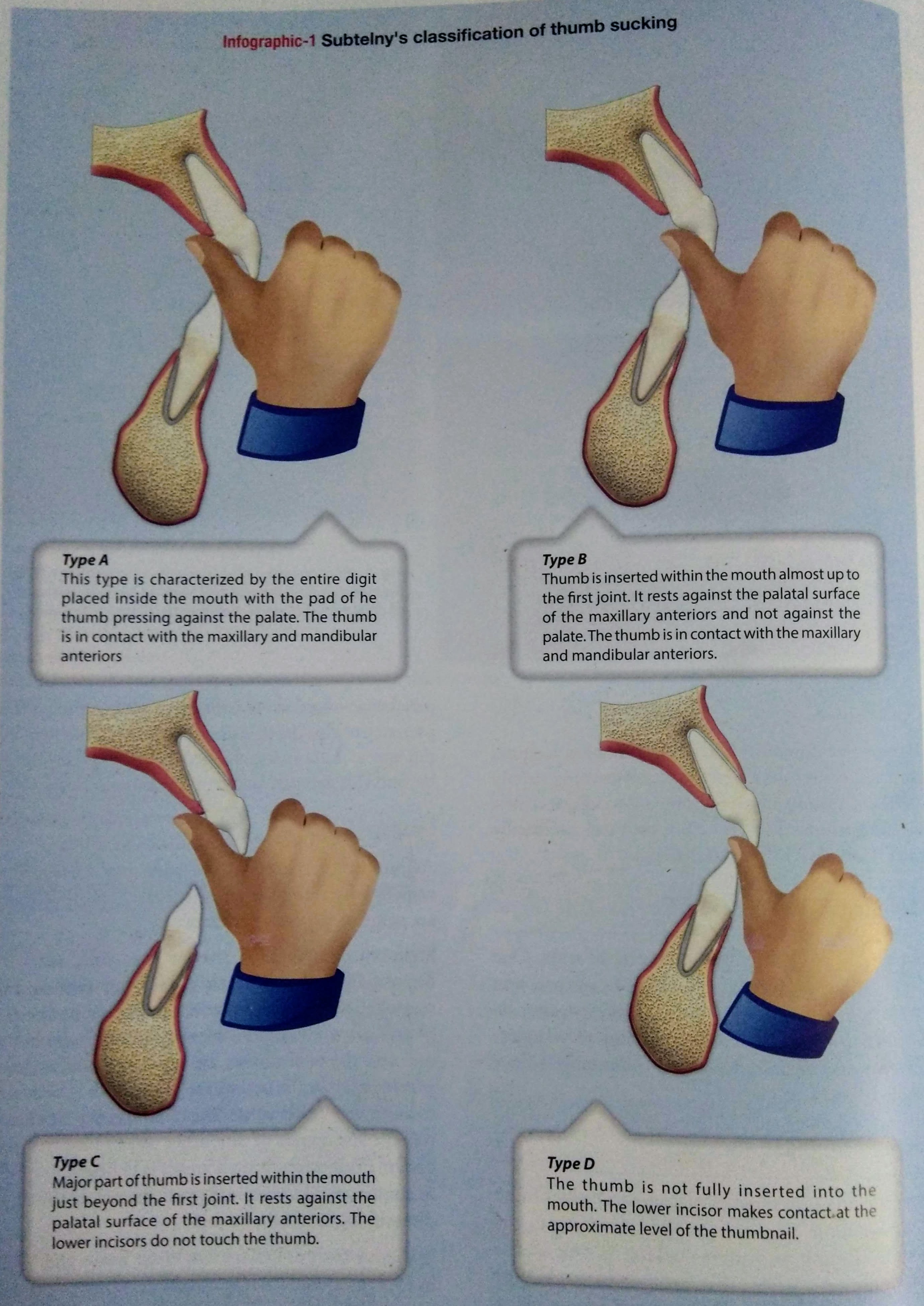
SUBTELNY’ S CLASSIFICATION OF THUMB SUCKING

EFFECTS :
The severity of malocclusion caused by thumb sucking depends on:

DURATION: Amount of time spent indulging in the habit.

FREQUENCY : The number of time the habit is activated in a day.

INTENSITY: Vigour with which the habit is performed.
The following are some effects:

  1. Effects on Maxilla
    • Constricted maxilla due to lowered tongue position + buccinator action, while sucking.
    • V-shaped narrow palate.
    • Posterior cross-bite may occur.
    • Increased SNA angle.

2. Effects on Maxillary Teeth:
• Spacing in maxillary anterior teeth.
• Increased maxillary arch length.
• Labial flaring of maxillary anterior teeth.

3. Effects on Upper Lip :
• Short, hypotonic incompetent upper lip

4. Effects on Thumb:
• Presence of clean nails & callus on fingers.

5. Effects on the Lower lip:
• Hyperactive lower lip with increased mentalis activity.

6. Effects on the Mandibular Teeth:
•
Retroclination of lower teeth.

7. Effects on Inter-arch Relationship:
• Increased overjet due to flaring of maxillary incisors
• Spacing of maxillary anterior teeth.
• Increased maxillary arch length.
8. Effects on Tongue:
• Lowered tongue position.
• Increased chance of developing tongue thrust habit.


MANAGEMENT:

  1. PSYCHOLOGICAL APPROACH:
    • Parents should be counseled to provide child with adequate love, affection and spend quality time with the child.
    • Success of any habit interception largely depends on the subject’s willingness to be helped to discontinue his/her habit.

Dunlops Theory of Beta Hypothesis
States that the best way to break a habit is by conscious purposeful repititions, i.e the child should be made to sit in front of a large mirror &asked to observe himself as he indulges in the habit.

  1. POSITIVE REINFORCEMENT:
    • Keeping track of the habit free days and rewarding the child can give the child a sense of pride.

3. MECHANICAL AIDS:
• Reminding appliances that assist the child who is willing to quit the habit but is not able to do as the habit has entered a subconscious level.
• Following types of habit louis used as mechanical reminding aids in treatment of thumb sucking.
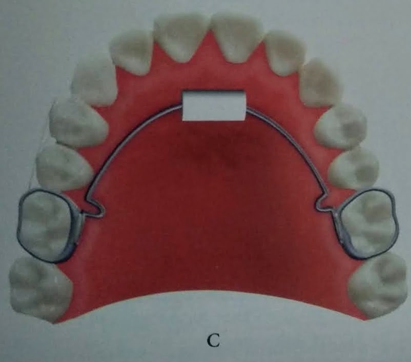
REMOVABLE HABIT BREAKER
Passive removable appliances, that consist of a crib &is anchored to the oral cavity by the means of clasps on the posterior teeth.

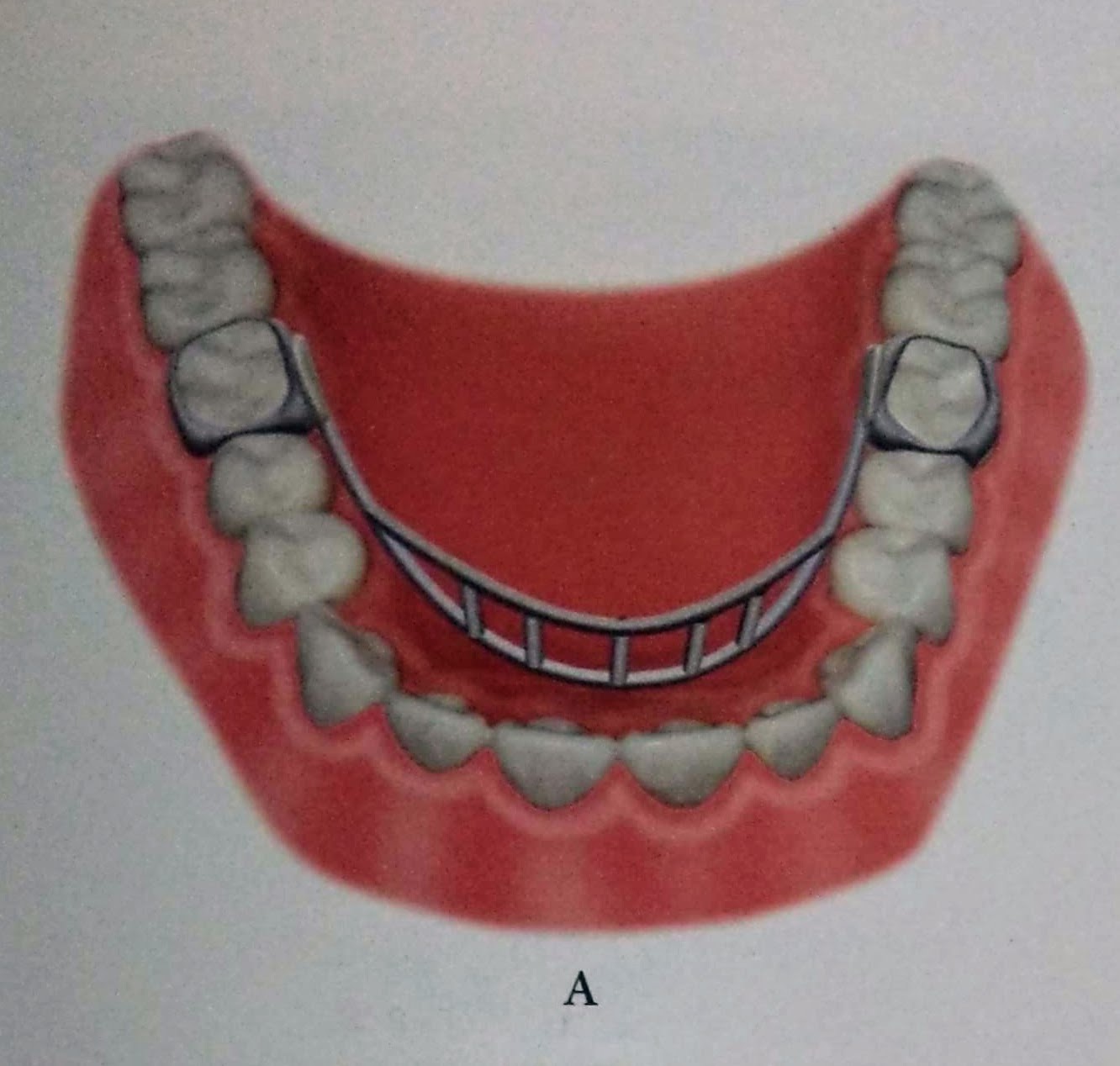
FIXED HABIT BREAKER
Heavy gauge stainless steel wire can be designed to form a frame, that is soldered to bands in the molars.

4. CHEMICAL APPROACH:

Use of the bitter tasting/foul smelling preparation placed on the thumb, that is sucked can make the habit distasteful.

  • Pepper dissolved in volatile medium.
  • Quinine
  • Asafoetida

REFERENCES:

  • ORTHODONTICS:The Art & Science,S.I BALAJHI (7th edition)
  • dryasmin.ae

Pyrexia of Unknown Origin ( Part -1 )

Written by - Dr.Urusa I Inamdar

Definition:

  1. Fever higher than 38.3 c ( 101F ) on several occasions.
  2. Illness of more than 3 weeks duration.
  3. No diagnosis made after 1 week of inpatient investigation.

Common causes not be missed for Fever which is not subsiding after 1 week of treatment:

  • enteric fever
  • Tuberculosis
  • viral fever
  • Leptospirosis
  • Scrub typhus
  • urinary tract infection
  • intraabdominal abscess
  • hepatitis A and B

Common etiologies of Fever of Unknown Origin

  • Infections
  1. Miliary TB
  2. Enteric Fever
  3. Intraabdominal abscess
  4. TB meningitis
  5. Brucellosis
  6. EBV/CMV
  7. Complicated UTI
  • Neoplastic disorders
  1. Lymphoma
  2. Hepatoma/Liver metastasis
  3. Colon cancer
  4. Myeloproliferative disorders
  5. Renal cell carcinoma
  6. CNS tumors
  • Rheumatic /inflammatory disorders
  1. Still’s disease
  2. Temporal arteritis
  3. Rheumatic arthritis
  4. SLE
  5. Sarcoidosis
  6. Polyarticular gout
  7. Polymyalgia rheumatica
  • Miscellaneous
  1. Drug fever
  2. Alcoholic cirrhosis
  3. Sub-acue thyroiditis
  4. PTE
  5. Inflammatory bowel disease

Classification of Fever of Unknown Origin

  • Classic

duration of more than 3 weeks.

Evaluation of atleast 3 outpatient visits or 3 days on hospital.

  1. Infection
  2. Malignancy
  3. Collagen vascular disease
  • Nosocomial

patient hospitalized more than 24 hrs but no fever or incubating on admission.

Evaluation of atleast 3 days.

  1. Clostridium difficile enterocolitis
  2. Drug induced
  3. Pulmonary embolism
  4. Septic thrombophlebitis
  • Immune deficient

Neutrophil count less than 500 per mm3.

Evaluation of atleast 3 days.

  1. Opportunistic bacterial infection
  2. Aspergillosis, Candidiasis, Toxoplasmosis
  3. Herpes virus
  • HIV associated

Duration of more than 4 weeks for outpatients.

More than 3 days for inpatient.

HIV infection confirmed.

  • Cytomegalovirus
  • Mycobacterium
  • PCP

Reference

  • Gsk – webevent console
  • Dental notes
  • Davidson’s – Principles and practice of Medicine

PREVENTIVE RESIN RESTORATIONS

:
• Preventive resin restoration (PRR) is a thin, resin coating applied to the chewing surface of molars, premolars and any deep grooves of the teeth.
• They are a natural extension of the use of occlusal sealants.
• It consists of an enamel sealant with a resin filling.
• If caries is present in one area or parts of pits and fissures, that particular area of caries is restored and fissures are protected with sealants.
• It integrates the preventive approach of the sealant therapy for caries susceptible pit & fissure with therapeutic restoration incipient caries with composite resin that occur on the same occlusal surface.
• They are the conservative answer to conventional extension for prevention” philosophy of Class I amalgam cavity preparation.

Preventive resin restoration(PRR) consists of a thin, resin coating applied to the occlusal surface of molars, premolars and deep grooves.
Most decays begin in the deep grooves. Thus, teeth with this condition are difficult to clean and becomes more susceptible to caries.
PRR protects the tooth by sealing the deep grooves and creating a smooth, easy to clean surface.
By this way, the teeth is protected from decay for several years, provided they are checked for thinning and wear at regular dental check-ups.

  • Dental caries is due to the imbalance between loss & gain of
  • minerals from a tooth surface.
  • The loss of minerals from our teeth occurs from the bacteria from the foods and producing acids, whereas the tooth gains minerals from our saliva and fluoride that is present within our mouth which over a period of time becomes a tooth decay.
  • Fissure sealants are a preventive treatment that is part of the minimal intervention dentistry approach to dental care.
  • This approach facilitates prevention and early intervention, in order to prevent or stop the dental caries process before it reaches the ends stage of the disease.

CLASSIFICATION:

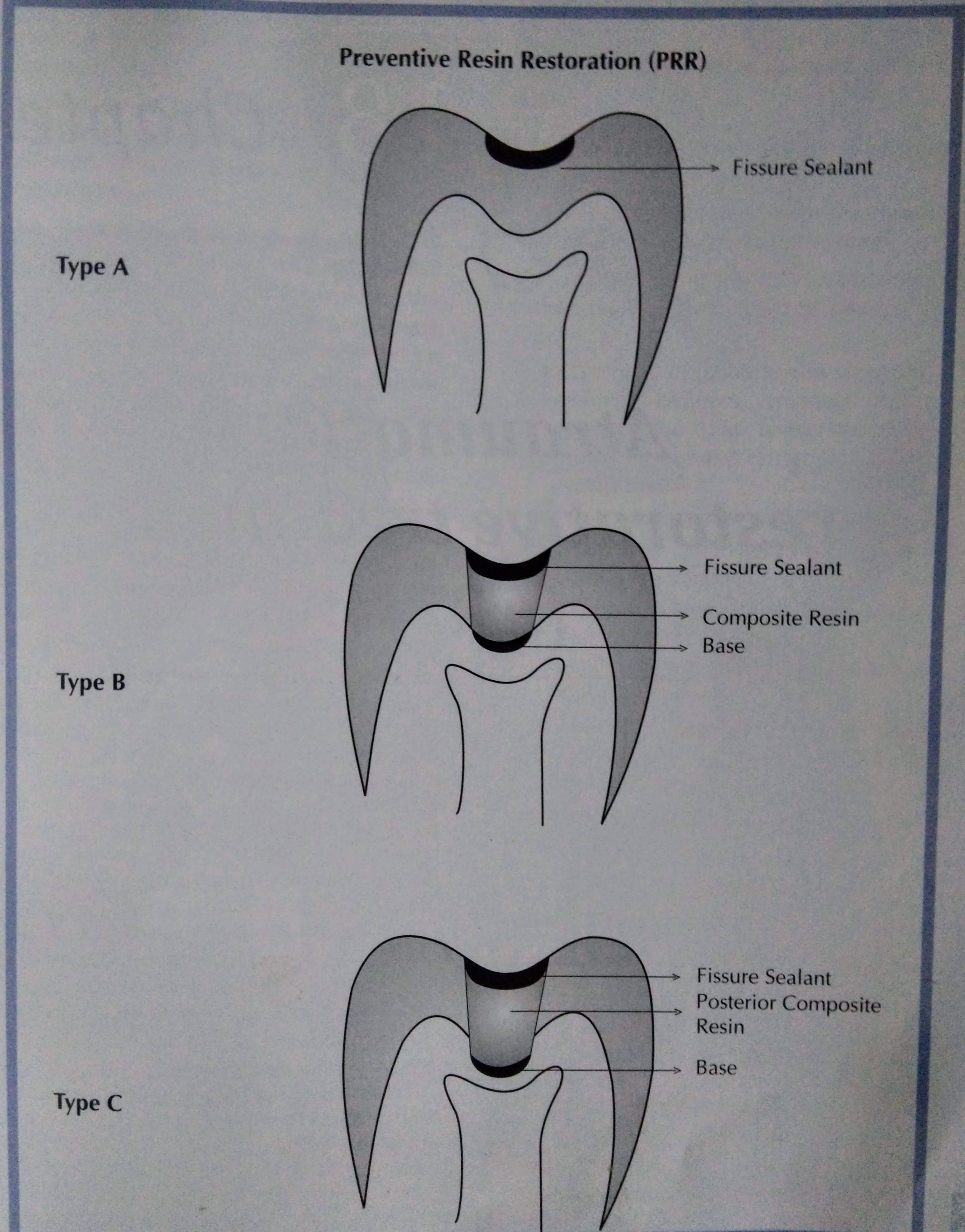
There are three types of preventive resin restoration based on the extent & depth of carious lesion as determined by exploratory preparation.

Simonsen (1978) has classified them:

TYPE A:

• Suspicious pits & fissures where caries removal is limited to enamel
• Local anesthesia is not required.
• A slow speed 4 or 2 round burs. used to remove decalcified enamel
• Sealant is placed

TYPE B:

• Incipient lesion in dentin that is small & confined.
• No local anesthesia is needed.
• An appropriate base is placed in areas of dentin exposure, composite resin is placed & the remaining pit & fissure are covered with a sealant.

TYPE C:
• More extensive dentinal involvement & requires restorations with posterior composite material
• Appropriate base is placed over the dentin.
• Pits & fissures are covered with sealant.
• Local anesthesia is required.

PROCEDURE:

CONCLUSION:
• Regular maintenance and sealant addition when necessary is important in long-term caries protection after sealant placement.
• Much better effectiveness data will result if sealants are used on teeth with a true predilection to caries.
• Better materials and better use of bonding agents with sealants will improve overall effectiveness on all teeth,particularly on those teeth now thought of as difficult to seal.
• Use of sealants has proved to have good results.
• For prevention of dental caries in pit and fissure, sealants were introduced.
• There is evidence suggesting effectiveness of sealants.
• Sealants prevent bacteria growth which causes caries.
• Biomaterials to seal pit and fissure should present with the simple application method, biocompatibility, low viscosity and good surface retention and low solubility.
• To improve this biomaterial, more laboratory should be developed.

REFERENCES:

  • Essentials of Public Health Dentistry, Soben Peter (6th Edition).
  • FENESTRA 2002-18, Dr. Bruno Jacquot(Bruno.Jacqout@odonto.u-nancy.fr)

DENTINOGENESIS IMPERFECTA

🔹 Introduction:

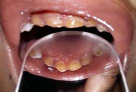
Clinical picture showing dentinogenesis imperfecta involving maxillary teeth

👉🏻 The affected teeth (both dentitions) are grey to yellow brown with broad crowns and constriction at cervical area resulting in tulip shape.

👉🏻 The enamel is easily broken, exposure of dentin ➡️ accelerated attrition.

👉🏻 Normal non-mineralized pulp chambers and canals

👉🏻 This condition is inherited in an autosomal dominant pattern, as a result of mutations on chromosome 4q21, in the dentine sialophosphoprotein gene (DSPP).

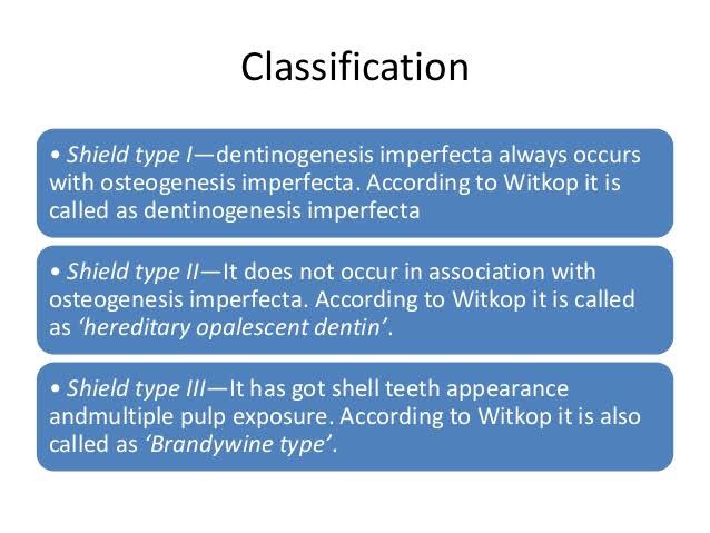
🔹Classification:

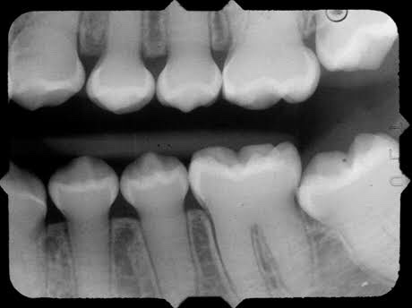
🔹Radiographic Features:

🔹Histopathological Features:

👉🏻 Mesodermal disturbance.

👉🏻 Dentin composed of irregular tubules with large areas of unclassified matrix. The tubules are larger in diameter & less numerous.

👉🏻 Odontoblasts – dentinal matrix not layed properly & they are entrapped within this matrix.

CHEMICAL FEATURES:
• Increased water content (60 times the normal)
• Decreased inorganic content

🔹Treatment: Cast Metal crowns & Jacket crowns

Dr. Mehnaz Memon🖊


References:

  1. Shafer’sTextbook Of Oral Pathology (7th Ed)
  2. Image Source: International Journal of Medicine Research; Dr G’s Toothpix; Google