Source Carranza textbook
Source Carranza textbook
Source Carranza textbook
Source Carranza textbook
• Also known as PARTSCH II or CYSTECTOMY.
• Enucleation is the surgical removal of the entire cystic lining in to-to.
By definition, it means shelling out of the entire cystic lining without rupture.
This surgical procedure leaves behind a hollow cavity in bone covered by oral mucoperiosteum.
This gets filled up with blood clot which eventually organizes to form healthy bone.
INDICATIONS:
• Is the treatment of choice for removal of cysts of jaws+ should be employed with any cyst of the jaw, that can be safely removed without sacrificing underlying structure.
ADVANTAGES:
DISADVANTAGES:
1.Relatively radical procedure.
2.Chances of devitalising the adjacent teeth.
3.Chances of fracture of the jaw.
4.Risk of creation of oroantral/oronasal communication
TECHNIQUES:
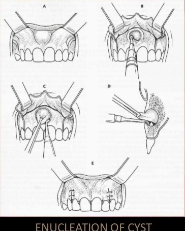
Enucleation with Primary Closure:
• Small cysts can be removed under local anaesthesia
• large cyst should be taken out under general anesthesia, as they are close to vital structures and blood vessels.
• After achieving adequate anesthesia, a mucoperiosteal incision is made such that the incision rests on sound bone.
• Mucoperiosteal flap is reflected taking care not to perforate rate the cystic lining.
• If the bone is perforated by the cyst, the lining will be adherent to the periosteum and will be difficult to reflect it.
• The cystic lining is exposed and now carefully teased away from the bone.
• It is relatively easy to separate the cystic lining from the bone because there is a layer of fibrous tissue between the two which is easily separabl.
• In case of infected cysts or an odontogenic keratocyst. the cystic lining is friable and more difficult to remove entirely.
• Every attempt is made to remove the entire cystic lining in a single piece without perforating it.
• This ensures complete removal.
• After the cyst is removed completely, the cavity in irrigated thoroughly, hemostasis ensured, sharp bone debris are filed and the flap replaced and sutured.
Enucleation with Open Packing :
• In case of a large cyst which was previously infected closure may not be possible
• After enucleation, the wound is then packed with me impregnated with bismuth iodoform paraffin paste (BIPP) or Whitehead’s varnish.
Enucleation with Peripheral Osteotomy:
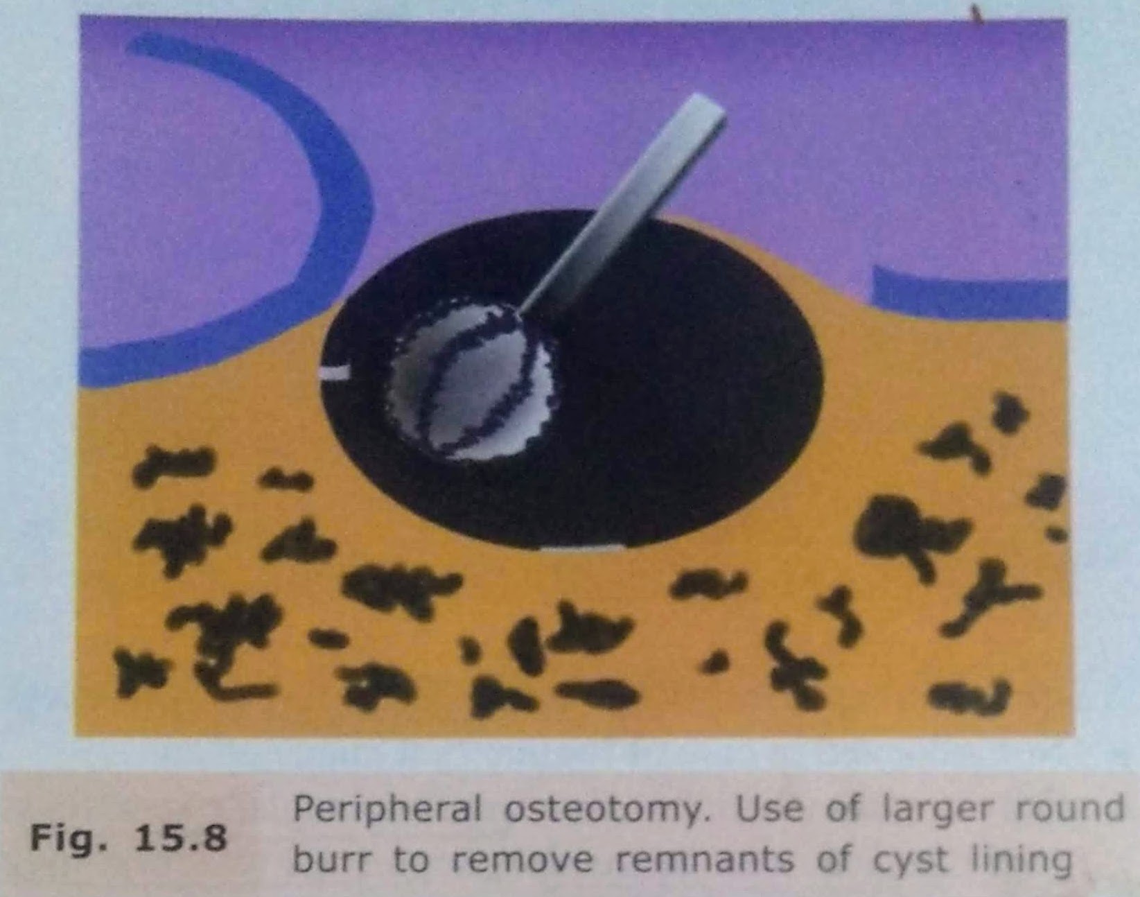
Enucleation with Bone Curettage:
• After enucleation, if there is a doubt that a part of the line has been left behind, it can be curette out.
• A bone curette is used to scrape the band remove any remaining lining
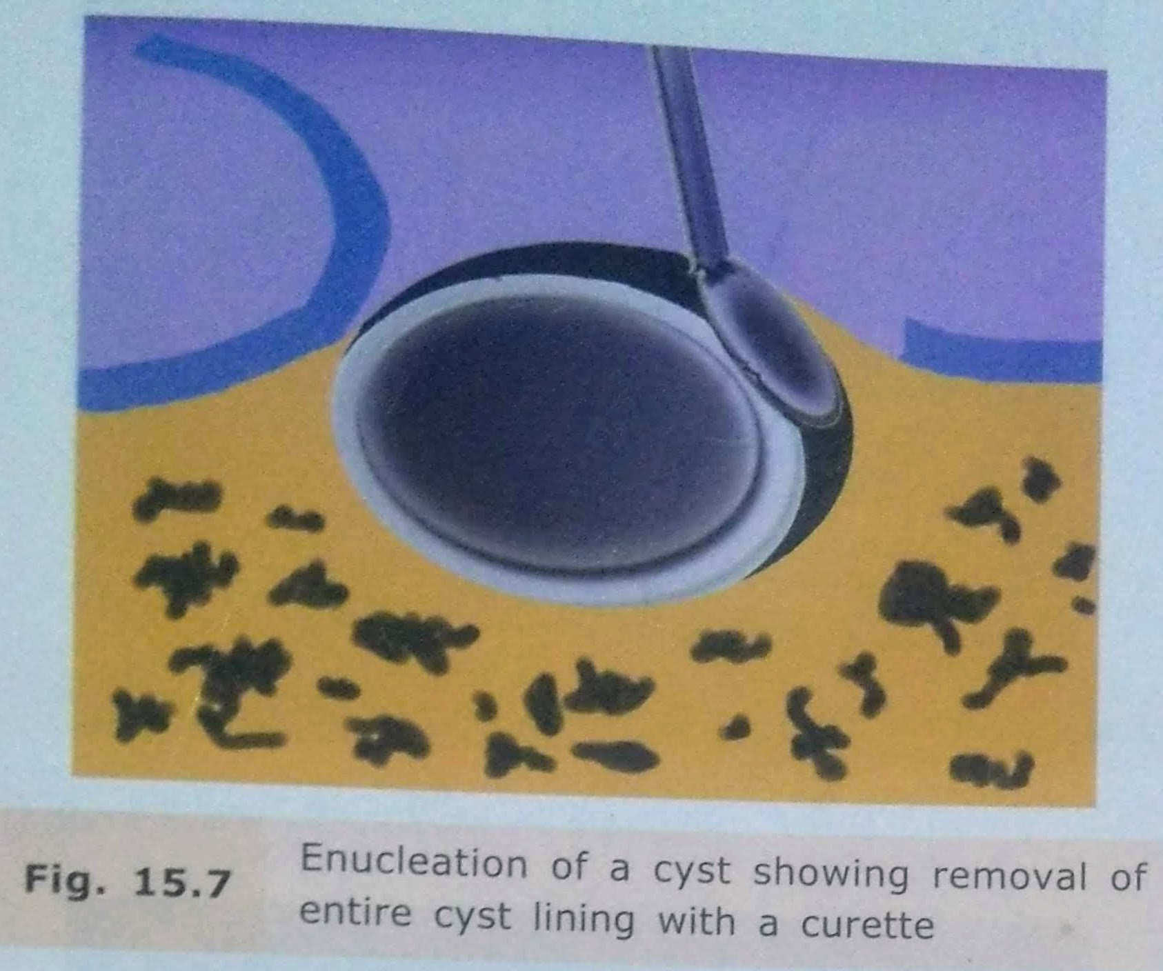
Enucleation with Peripheral Osteotomy
• In this procedure, instead of using a curette, a large round bur may be used to remove around 1-2 mm of bone around the entire periphery of the cystic cavity.
• This is done to ensure that any remaining epithelial cells present in the east wall or body cavity are removed.
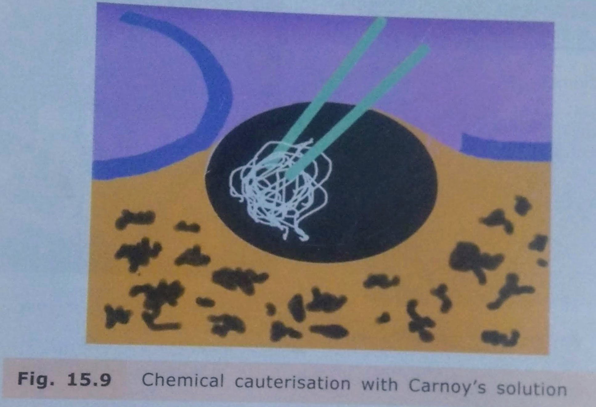
Enucleation with Chemical Cauterisation:
• It is indicated mainly in cases of odontogenic keratocyst.
• After enucleation, to remove any remaining lining of the cyst chemical cauterising agent Carnoy’s solution is applied along the walls of the cystic cavity.
• It is left for about 5-7 minutes and then irrigated thoroughly with saline.
• This solution chemically cauterizes any remaining cells of the system.
Enucleation with Bone Grafting
• Bone grafting with autogenous cancellous bone grafts can be done in case of large bony defects.
• The bone graft obliterates the cavity and stimulates osteogenesis.
• There is, however, a risk of wound breakdown and infection of the bone graft which may lead to failure.

COMPLICATIONS:
• Risk of bone fracture due to cyst.
• Inferior dental nerve involvement.
• Management of teeth related to cyst.
• Oroantral Communication.
• Hematoma formation.
• Infection.
• Dead Space: At surgical procedure, a hollow dead space is formed. It is usually filled up with blood clot and eventual bone formation.
However, there are chances of infection of the clot, breakdown of suture line and pus discharge.
REFERENCES:
• Textbook of Oral & Maxillofacial Surgery, Chitra Chakravarthy (2nd Edition)
• SlideShare
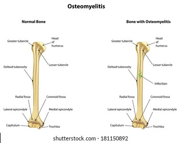
Definition: Osteomyelitis is defined as inflammation of the bone and marrow.
Classification
🔻Primary: Where bone is the primary solitary focus of disease .
🔻Secondary: Develops as a complication of any systemic infection.
Any infection (bacteria, viruses, parasites, fungi) may cause osteomyelitis but infections certain pyogenic bacteria and mycobacteria are the most common
It is usually caused by bacteria.
• Most common pathogens are Staphylococcus species (aureus in 80-90% of the cases).
🔻Other organisms: Klebsiella, Neisseria gonorrhoeae, Haemophilus influenzae Escherichia coli, Pseudomonas, and Salmonella species
🔻Escherichia coli in patients with genitourinary tract infections or intravenous drug abusers.
Neonatal period: Haemophilus Influenzae and group B streptococci.
Patients with sickle cell disease: Salmonella infection.
• Mixed bacterial infections: It is due to direct spread or surgery or open fractures.
• In about 50%, no organisms can be isolated.
Causative organisms may reach the bone through the bloodstream, directly or extend from a contiguous site.
• Source of organisms may be a focus of infection anywhere in the body (e.g skin pustule or infected teeth and gums, intestinal mucosa).
• Minor injuries to the mucosa (vigorous chewing of hard foods, brushing of teeth), or minor infections of the skin, release these organisms into the blood → causing temporary bacteremia → reach the bone.
•In children (5 to 15 years) and drug addicts (infected needles), it develops in the long bones.
Location of infection: It varies with age.
• Neonate: Metaphysis, epiphysis, or both.
•Adult: Epiphysis and subchondral regions
•Children with hematogenous spread: Metaphysis of long bones (knee, ankle, and hip).
The sequence of events and morphological features in osteomyelitis are described together.
• Transient bacteremia: Mild injury or trauma can initiate bacteremia by organisms (eg.Staphylococcus aureus).
•Infection reaches metaphysis long bone: Because in the metaphysis capillaries form loop – which slows the blood flow provides time for bacteria to penetrate blood vessel walls and establish infective foci within the marrow.
• Inflammatory Reaction: Once in bone, the bacteria grow and induce an acute inflammatory reaction with exudates.
• Necrosis of bone: Exudate increases the pressure on the adjacent vessels and further decreases the blood supply produces bone necrosis. The necrotic areas coalesce and allow further bacterial proliferation.
•Formation of sequestrum: Bacterial infections and pus spreads into the cortex and collects beneath the periosteum and may lift the periosteum and reduce the blood supply to the affected region.
The fragment of dead necrotic piece of bone, which is embedded in the pus, is known as a sequestrum.

🔻The pus penetrates the periosteum and leads to a soft-tissue abscess may penetrate the skin form a draining sinus.
Hole formed in the bone during the formation of a draining sinus is known as cloaca
🔻The sinus tract may become epithelialized and may remain open, continually draining pus, necrotic bone, and bacteria.
After first week, chronic inflammatory cells become more numerous and the cytokines released stimulates osteoclastic bone resorption and deposition of reactive bone in the periphery.
Reactive new bone forms a sheath around the necrotic (segment of devitalized infected bone) sequestrum. This reactive new bone formed is known as involucrum.
• Brodie abscess: It is a distinctive form of subacute pyogenic osteomyelitis.
It may be due to inadequate treatment of infection by less virulent organisms.
• Sclerosing osteomyelitis of Garre – characterized by extensive newborn formation which obscures the underlying structure of the bone and typically develops in the jaw.
•Septicemia: From infection in the bone, organisms may disseminate through the blood stream and cause septicemia.
• Acute suppurative arthritis: Infection may spread through the articular surface into a joint producing suppurative arthritis may lead to destruction of the articular cartilage and permanent disability. It is more common in infants.
•Pathologic fractures.
•Squamous cell carcinoma: It may arise from the epithelialized sinus tract, rarely sarcoma of bone may develop.
• Secondary amyloidosis.
• Chronic osteomyelitis: It may develop due to delay in diagnosis, extensive bone necrosis, and inadequate therapy.
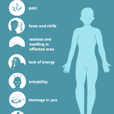
🔻 present with malaise, fever, chills , leukocytosis and throbbing pain over the affected area
🔻 Diagnosis-
Radiography- lytic focus of bone destruction
Blood cultures are positive
Biopsy and bone culture
source -textbook of pathology for dental students Harsh mohan
Written by : Dr. Urusa I Inamdar
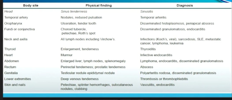
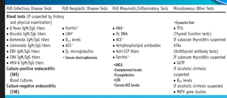
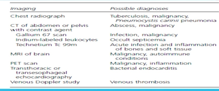
In the majority of cases, treatment other than supportive care should not be commenced until a diagnosis is obtained. Early use of antipyretics or antimicrobial may delay diagnosis. The mortality rate for PUO is less than 10%.
If infective endocarditis is suspected, then the patient should be admitted to a hospital and empiric intravenous antibiotics should be commenced after three sets of blood cultures have been collected.
The recommended empiric regimen of infective endocarditis in Australia is gentamycin , benzylpenicillin and flucloxacillin; however this may vary with different patient factors and should be discussed with an infectious disease specialist.
Osteoporosis is a disease of bone which characterized by low bone mass that leads to porosity of bones.
🔻characterised by porus bone – thinning of the cortex and trabaculae
🔻 increase in the risk of fractures – hip ,wrist and spine
Types:
Osteoporosis may be:
•Localised : It is usually due to disuse and is seen as a complication of some other disease.
For example, local immobilization following fracture due to other causes.
• Generalized: Involves the entire skeleton. It may be primary or secondary.
–Primary osteoporosis occurs without any known cause.
These include senile postmenopausal.
-Secondary osteoporosis develops due to a large variety of conditions.
These include endocrine disorders (hyperparathyroidism, hyperthyroidism, etc.),
gastrointestinal disorders (e.g. malabsorption, vitamin D deficiency, malnutrition),
drugs (e.g. corticosteroids),
tumors (multiple myeloma)
reduced physical activity,
hormonal disturbances
and other causes (e.g. immobilization).
Age : Peak bone mass is achieved between 25 and 35 years of age and gradually declines from the age of 50 years onwards.
Sex: It is generally seen in elderly and women. Genetic/hereditary factors also play an important role .
🔸Ageing-due to decrease in the osteoblastic activity and decrease in the growth factors.
🔸Hormones–decreased estrogen, cytokines like IL-1 and IL-6,which increase the osteoclastic activity.
🔸Decrease in the physical activity
🔸Genetic factors
🔸Nutritional status- low calcium intake
Entire skeleton is involved in postmenopausal and senile osteoporosis but certain regions are more severely involved than others.
The involved bones are thin and brittle.
Microscopically, it is composed of the lamellar bone which is discontinuous and has thin trabeculae.


Depend on the bones involved.
Commonly involved areas are vertebral bodies and femoral neck
Loss of bone mass predisposes the bone to fracture which results in bone pain.
Fractures are most common in the vertebra in the thoracic and lumbar region, neck of the femur and Colles’ fracture (fracture of distal radius).
Femoral neck
• Pulmonary embolism and pneumonia as a complication of fractures of the femoral neck, pelvis or spine.
Vertebral fracture
• When fractures of vertebrae are multiple, it may lead to lumbar lordosis and kyphoscoliosis (forward bending).

🔸plain X-ray – not reliable till 30-40%of bone mass isn’t lost
🔸Dual energy X-ray absorptiometry and quantitative computed tomography
🔸Bone biopsy
Exercise
Appropriate calcium and vit d intake
Agents like bisphosphonates
Source – textbook of pathology for dental students – harsh mohan and pathology preparatory manual for dental students Ramdas Nayak


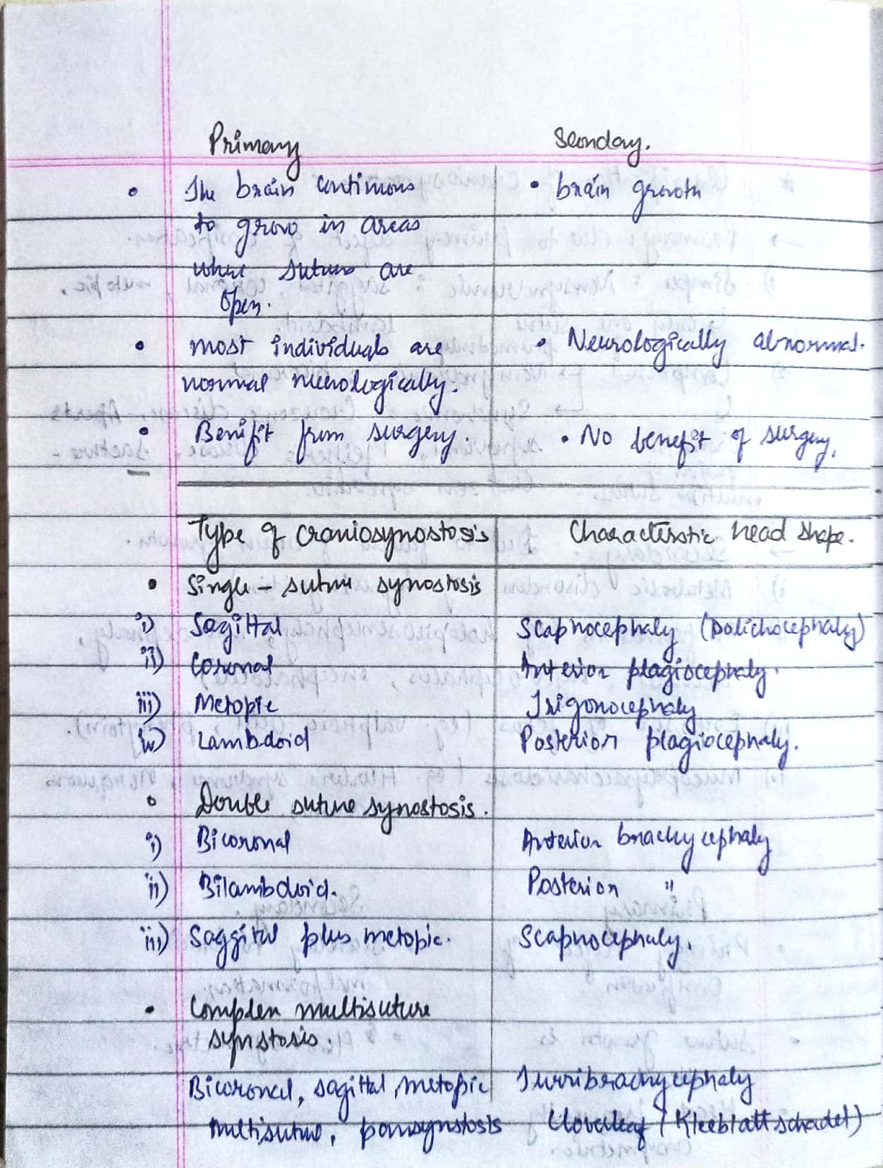
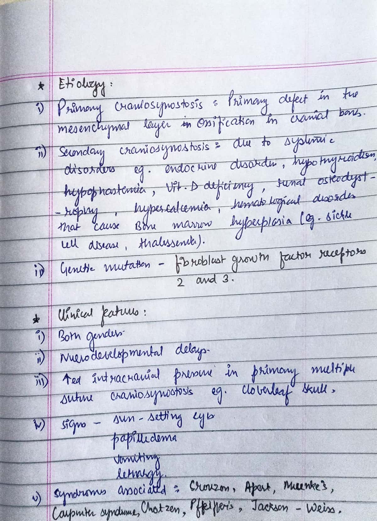
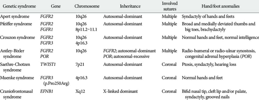
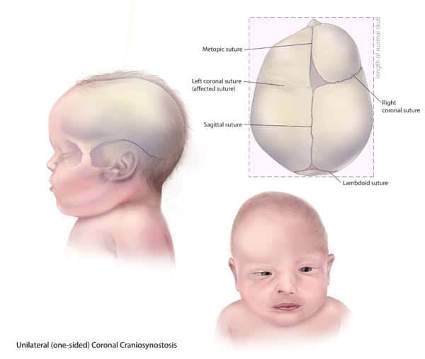









Sources – Shafer’s textbook of oral pathology , slideshare.com
Image source – Google images , researchgate.net
Chemical alteration of the drug in a living organism is called biotransformation.
The metabolism of a drug usually converts the lipid-soluble and unionized compounds into water-soluble and ionized compounds.
They are not reabsorbed in the renal tubules and are excreted. If the parent drug is highly polar (ionized), it may not get metabolized and is excreted as such.
Sites: Liver is the main site for drug metabolism: other sites are GI tract, kidney, lungs, blood, skin and placenta.
1.Active drug to inactive metabolite: This is the most common type of metabolic transformation
Phenobarbitone – Hydroxy Phenobarbitone
Phenytoin- P-Hydroxy Phenytoin
2.Active drug to active metabolite
Codeine – Morphine
Diazepam — Oxazepam
3. Inactive drug to active metabolite
Prednisone -Prednisolone
Levodopa- Dopamine
MARSUPIALIZATION:
• PARTSCH(1892) described a type of compression procedure for treatment of cyst.
• Also k/a PARTSCH I OPERATION & CYSTOTOMY.
• In this procedure a window/fenestrations made in the bone and cystic content is evacuated.
• The cyst lining is left behind.
• Once the cyst contents are evacuated, the intracystic pressure reduces.
• Hollow cavity is packed till it gets obliterated by bone over a period of time.
• The cystic lining then becomes continuous with normal oral mucosa.
INIDICATIONS:
• In extremely large cysts.
• Risk of cyst opening into maxillary sinus/nose due to surgical removal of complete lesion.
• In very young patients, where it will permit eruption of enclosed tooth,underlying developing tooth.
• Patient with poor general condition for allowing minimal surgical procedure.
• In cases, where surgical procedures may cause pathological fracture of jaws.
TECHINQUE:
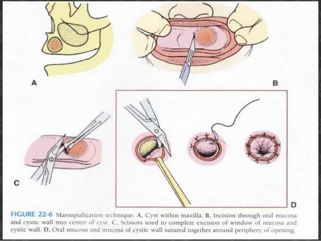
1) Anaesthesia
2) Aspiration
3) Incision: Circular, oval or elliptical. Inverted U Shaped incision with broad base the buccal sulcus. Mucoperiosteum is reflected in this case.
4) Removal of bone
5) Removal of cystic lining specimen
6) Visual examination of residual cystic lining
7) Irrigation
8) Suturing of cystic cavity.
• Cystic lining sutured with the edge of oral mucosa.
• In Y Shaped incision the mucoperiosteal flap can be turned into cystic cavity covering the margin.
• The remaining is sutured to oral mucosa.
9) Packing- Prevents food contamination & covers wound margins. Done with ribbon gauze soaked with WHITEHEAD VARNISH
COMPOSITION
• Benzoin – 10g
• iodoform-10g
• Storax -7.5g Balsam of Tolu -5g
• Solvent ether to 100ml
Pack removed after 2 weeks.
10) Maintenance of cystic cavity
Instruct the patient to clean and irrigate the cavity regularly with oral antiseptic rinse with a disposable syringe.
11) Use of plug:
• Prevents contamination, Preserves patency of cyst orifice.
• Plug should be stable, retentive and safe design.
• Should be made of resilient material (to avoid irritation) like acrylic.
12) Healing:
• Cavity may or may not obliterate totally. Depression remains in the process.

ADVANTAGES:
• Once the cystic lining is evacuated, there is a tendency of the cystic lining to contract. This stimulates endosteal bone formation.
• Marginal ingrowth of normal mucoperiosteum occurs as the cystic lining shrinks. It provides with additional bone regeneration factors.
• Not much surgical skill is required.
• No risk of oroantral/oronasal fistula.
• No damage to adjacent vital structures.
• No risk to adjacent vital tooth.
DISADVANTAGES:
• Entire pathological tissue is left behind.
• High chances of recurrence.
• As the bony cavity is large, healing and filling up with normal bone takes longer time.
• Use of cyst plug is required with repeated cleansing.
• Time consuming & repeated appointment for patients.
REFERENCES:

