APICOECTOMY


• It is the surgical resection of the apex of the root.
• It is the procedure done in case of root canal treatment failure.
• If an infection does not subside even after root canal treatment,it may concern enquire a surgical procedure.
• In this procedure, the apical region of root is visualised by reflecting a flap and performing an osteotomy.


INDICATIONS :
• Aberrant Anatomy: Dilaceration of root apex do not allow endodontic restoration of apex.
• Obliteration of apex by secondary dentin.
• Iatrogenic repair: A broken endodontic file which cannot be retrieved by conventional means.
• Apex perforation.
• Improper apical seal which cannot be removed.
• Increased drainage of pus from root canal will not allow adequate apical seal.
• Open apex.
• Non healing periapical granuloma.
• Fracture of apical third of root.
• Periapical cyst/granuloma.

CONTRAINDICATIONS:

  1. Local Contraindications:
    • Poor periodontal status of tooth.
    • Grossly decayed tooth.
    • Inadequate tooth length.
    • Acute infection.
    • Traumatic occlusion.
    • Uncooperative patients.
    • Close proximity of root apex to vital anatomic structures such as Maxillary antrum & Nasal floor.
  2. Systemic Contraindications:
    • Poor medical status of diabetes,Bleeding disorders.etc

STEPS IN ENDODONTIC SURGERY:

  1. Cleaning of the area involved with antiseptic solutions.
  2. Local anaesthesia.
  3. Design of mucoperiosteal flap & reflection of flap.
  4. Bone removal for access to root tip.
  5. Root tip resection & curettage.
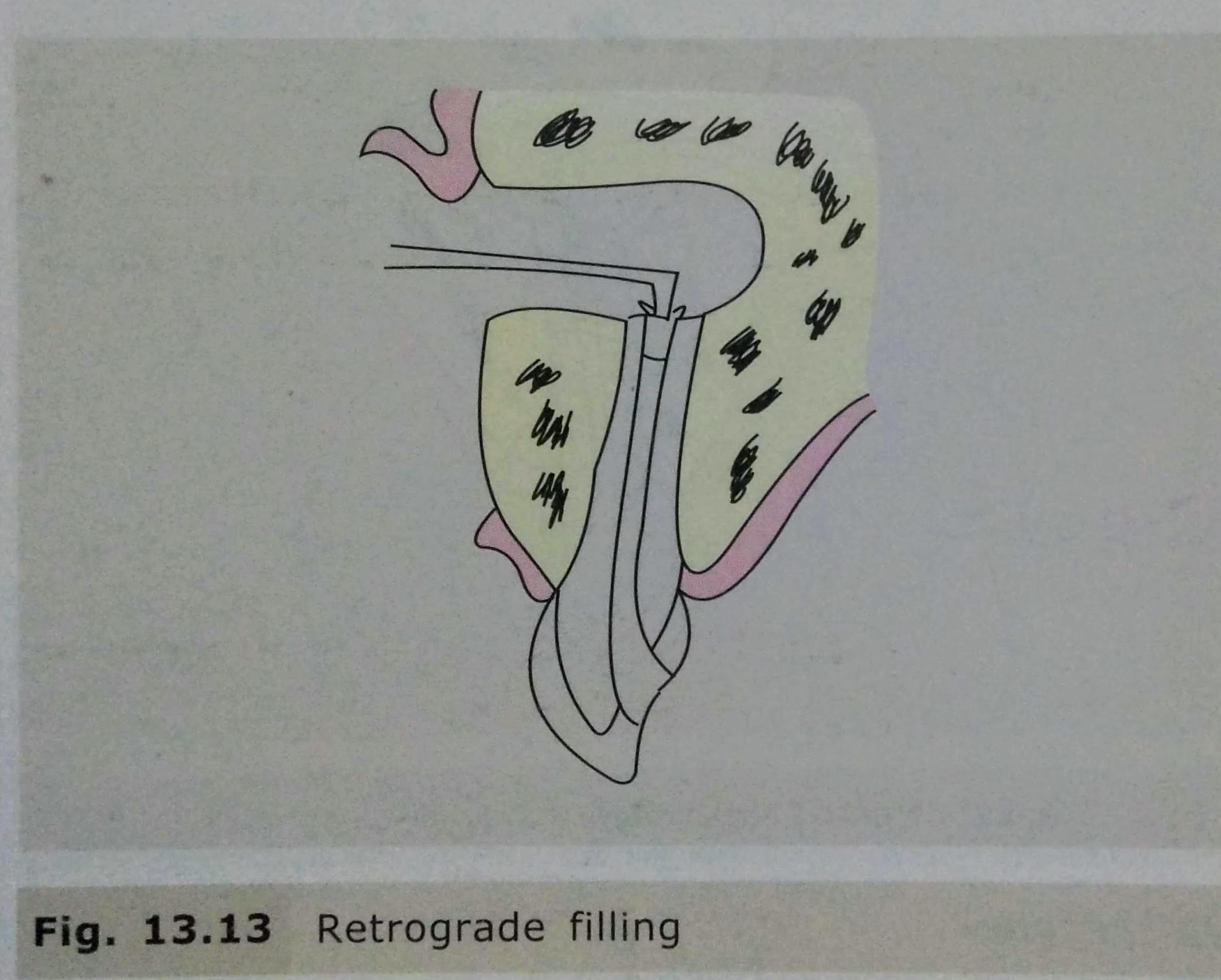
  6. Retro preparation & retrograde filling.
  7. Suturing & Follow up.

COMPLICATIONS:
• Mobility of tooth/adjacent tooth.
• Haemorrhage.
• Nasal perforation.
• Oroantral Fistula.
• Mental Nerve Damage.
• Inferior Alveolar Nerve damage.

REFERENCES:
• Textbook of Oral & Maxillofacial Surgery, Chitra Chakravarthy (2nd Edition)
• DentaGama.com
Maxillary Posterior teeth arrangement

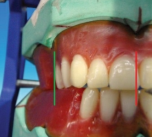
Steps in placement of maxillary first premolar

Placement of the maxillary first premolar in all three planes

side view

The buccal cusp tip of the maxillary first premolar is in contact with occlusal plane (black line), and palatal cusp 1 mm short of the occlusal plane

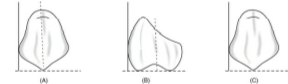
occlusal view

buccal cusp corresponds with the canine tip, and
the palatal cusp corresponds to previous drawn reference line of the mandibular residual ridge line

front view

When viewed from:
Front: Long axis parallel towards the vertical axis.
Side: Long axis parallel towards the vertical axis.
Occlusal plane: Buccal cusp is in contact with occlusal plane
and palatal cusp 1 mm short of occlusal plane

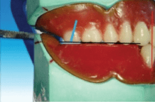
Steps in placement of the maxillary second premolar

Placement of the maxillary second premolar in all three planes

scoop out the wax
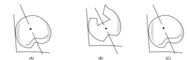
occlusal relation
side view

The buccal surface of the maxillary second premolar is barely visible when the patient smiles when viewed from the front
front view

The buccal cusp of the maxillary second premolar corresponds with the buccal cusp of maxillary first premolar (blue line), and the palatal cusps of both the premolars correspond to the previous drawn line

When viewed from:
Front: Long axis parallel towards the vertical axis.
Side: Long axis parallel towards the vertical axis.
Occlusal plane: Both buccal cusp and palatal cusp is in contact
with occlusal plane

Steps in placement of maxillary first molar

Placement of the maxillary first premolar in all three planes

side view
front view

the mesio- and distobuccal cusps correspond with
the buccal cusp of second premolar (blue line) and the mesio- and distopalatal cusps correspond to the previous drawn line (yellow line).

When viewed from:
Front: Long axis slopes buccally.
Side: Long axis slopes distally.
Occlusal plane: Only mesiopalatal cusp is in contact with
occlusal plane

Steps in placement of maxillary second molar

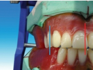
The long axis of the maxillary second molar (blue line) is distally
inclined in relation to the vertical axis (red line) when viewed from the front .

All the cusps are short of the occlusal plane (black line).

The maxillary second molar is slightly buccally inclined when viewed from the front. The buccal surface of the maxillary second molar is barely visible when viewed from the front

front view

When viewed from:
Front: Long axis slopes buccally more steeply than first molar.
Side: Long axis slopes distally more steeply than first molar.
Occlusal plane: Only mesiopalatal cusp is nearest to occlusal
plane

Source – S. Lakshmi preclinical manual of prosthodontics

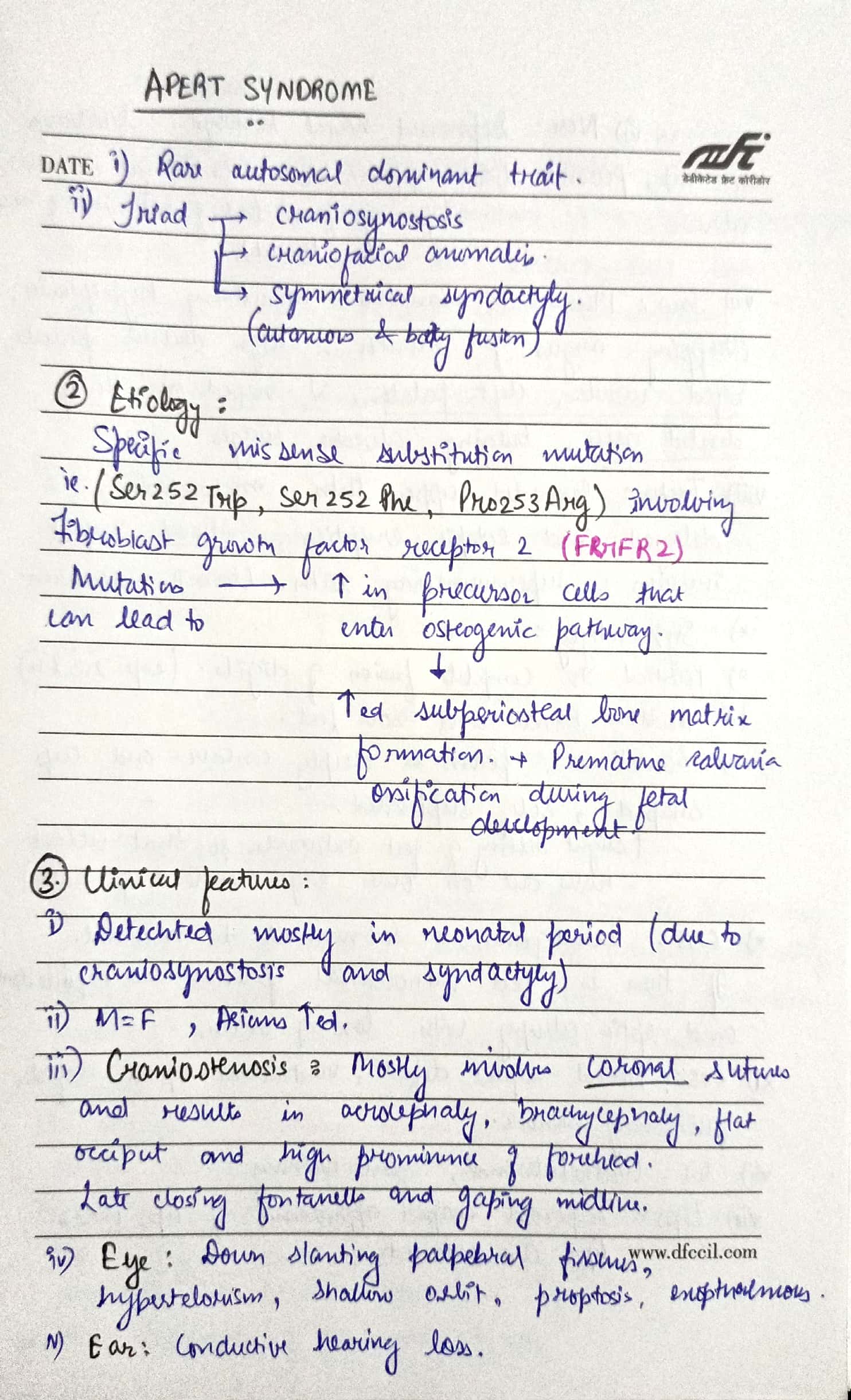
APERT SYNDROME

🔖 Acrocephalosyndactyly. The condition is autosomal dominant i.e, one copy (out of 2) of the defective gene is sufficient to cause the abnormality in the offspring.

🔹Characteristics:

  • Craniosynostosis (premature fusion of the skull bones)
  • Craniofacial anomalies
  • Syndactyly (Fusion of fingers and toes)

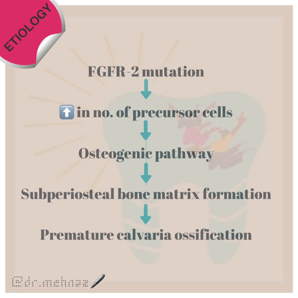
🔹Etiology:

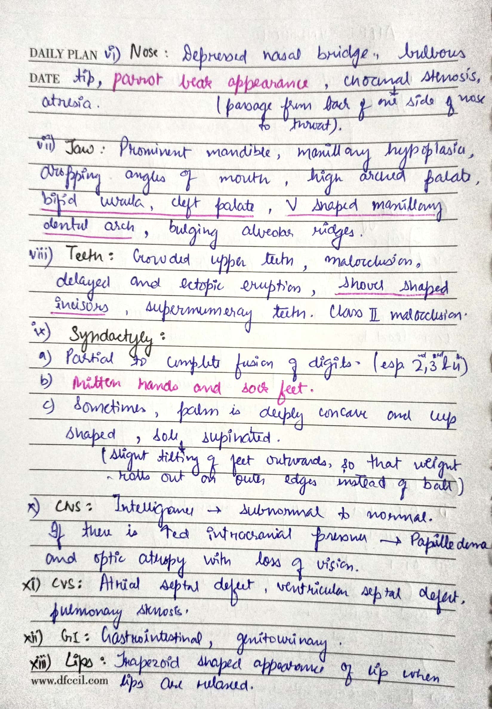
🔹What are the Symptoms and Signs of Apert Syndrome?

The various clinical features include:

  1. Asians affected
  2. Acrocephaly, Brachycephaly, flat occiput & prominent forehead.
  3. Late closing fontanels
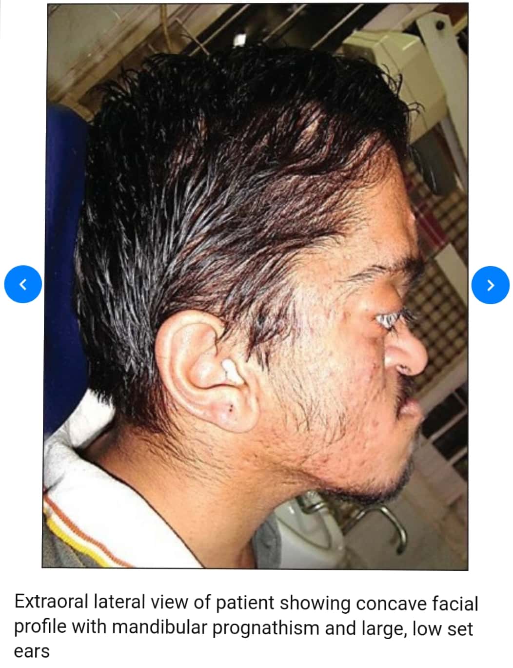
  4. Low set ears, hearing loss
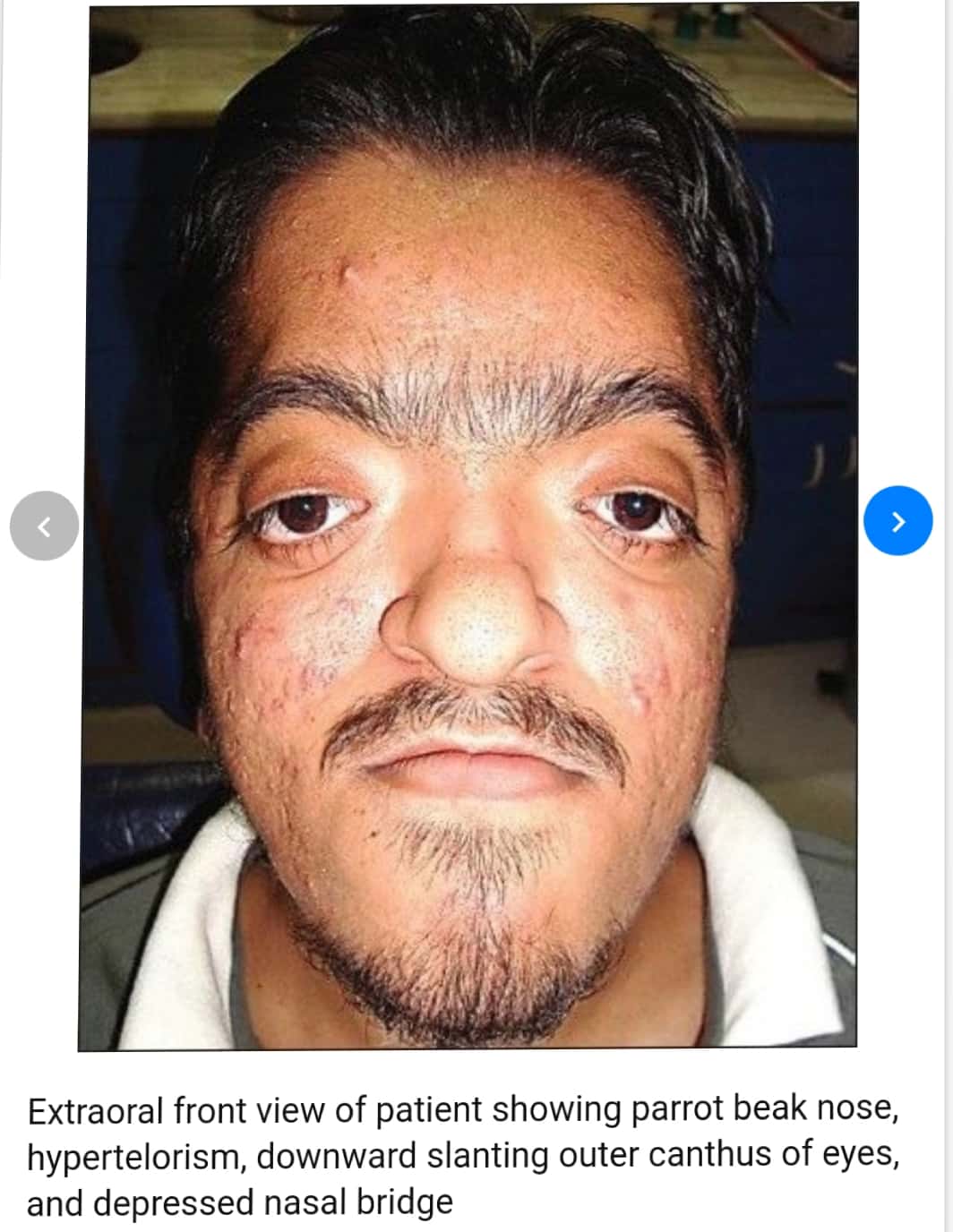
  5. Eyes: Down slanting of palpebral fissures, Widely spaced eyes(Hypertelorism), Shallow orbits, Abnormally bulging eyes (Exophthalmos)
  6. Nose: Depressed Nasal bridge, short, wide with bulbous tip, Parrot beaked appearance, Atresia
  7. Jaw:
  • Prominent Mandible
  • Maxillary hypoplasia
  • Drooping angles of mouth
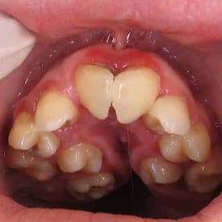
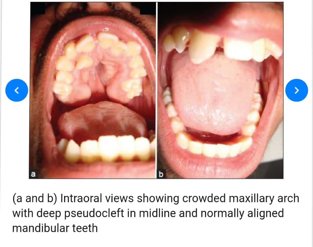
  • High arched palate
  • Bifid uvula
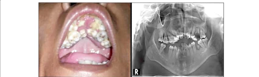
  • Cleft palate
  • Crowded upper teeth
  • Malocclusion
  • Delayed & ectopic eruption
  • Shovel shaped incisors
  • Supernumerary teeth
  • V-shaped maxillary dental arch
  • Bulging alveolar ridges

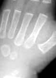
8. Partial to complete fusion of digits: 2-4th digits – MITTEN HANDS & SOCK FEET; Sole – supinated

9. Intelligence – Normal

10. ⬆️ intracranial pressure – optic atrophy, papilledema

11. Hyperhidrosis

12. Cardiovascular system: Atrial Septal defect, Ventricular septal defect, Patent ductus Arteriosus

🔹How do you Treat Apert Syndrome?

Standard Therapies

The treatment of Apert syndrome aims at addressing the specific symptoms that may be present in the particular individual. Treatment is usually symptomatic and supportive.

  • Craniosynostosis and associated hydrocephalus in some cases may give rise to an abnormally increased pressure within the skull (intracranial pressure) and on the brain. In these cases, early surgery (within 2 to 4 months after birth) becomes necessary to correct the defects in the skull and facial bones.
  • Insertion of a tube (shunt) to drain excess cerebrospinal fluid (CSF) away from the brain and into another part of the body like the abdomen where the CSF can be absorbed can be done to relieve associated hydrocephalus (fluid accumulation in the brain).
  • Early repair and reconstructive surgery may also be done in some infants with Apert syndrome to address craniofacial abnormalities.
  • Other defects such as those of heart, eye and ear defects may also need correction.

Dr. Mehnaz Memon🖊


References:

  1. https://www.medindia.net/amp/patientinfo/apert-syndrome.htm
  2. Shafer’s textbook of Oral Pathology – 7th Ed.

Root Canals

Written by : Dr. Urusa I Inamdar

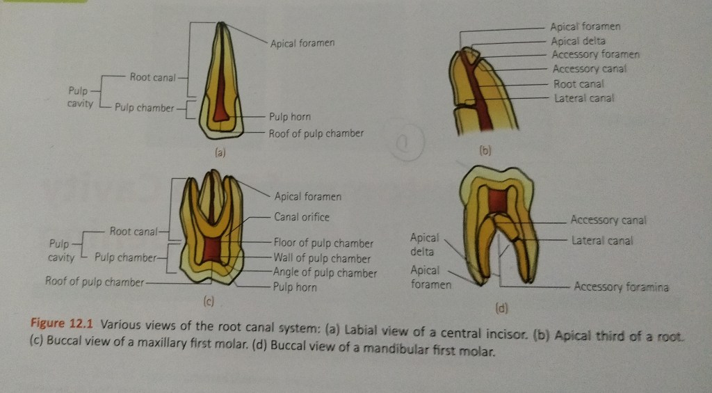
It is the portion of the pulp cavity from the canal orifice to the apical foramen.

It is divided into 3 sections :

  • Coronal
  • Middle
  • Apical
  1. Accessory canals or lateral canals : lateral branching of the main root canal generally occurring in the apical third or furcation area of a root.
  2. Lateral canal : accessory canal that branches to the lateral surface of the root and may be visible on a radiograph.
  3. Apical foramen : aperture at or near the apex of a root through which the blood vessels and nerves of the pulp enter or leave the pulp cavity.
  4. Accessory foramina : openings of the accessory and lateral canals in the root surface.

A straight root canal extending the entire length of the root is uncommon. Either a constriction is present before the apex is reached or , as is often the case , a curvature is present.

The curvature may be :

  • A straight canal extending with minimal apical curvature.
  • A gradual curvature of the canal with a straight apical ending.
  • A gradual curvature of the entire canal.
  • A sharp curvature of the canal near the apex.

A curvature of about 20° in a narrow root canal may be difficult or even impossible to negotiate with endodontic instruments, whereas a curvature of even 30° may be negotiated if the root canal is wide.

Success in negotiating a narrow , curved canal depends on following :

  • Degree of curvature.
  • Size and constriction of the root canal.
  • Size and flexibility of the endodontic instrument blade.
  • Skill of the operator.

The various classification proposed are as follows :

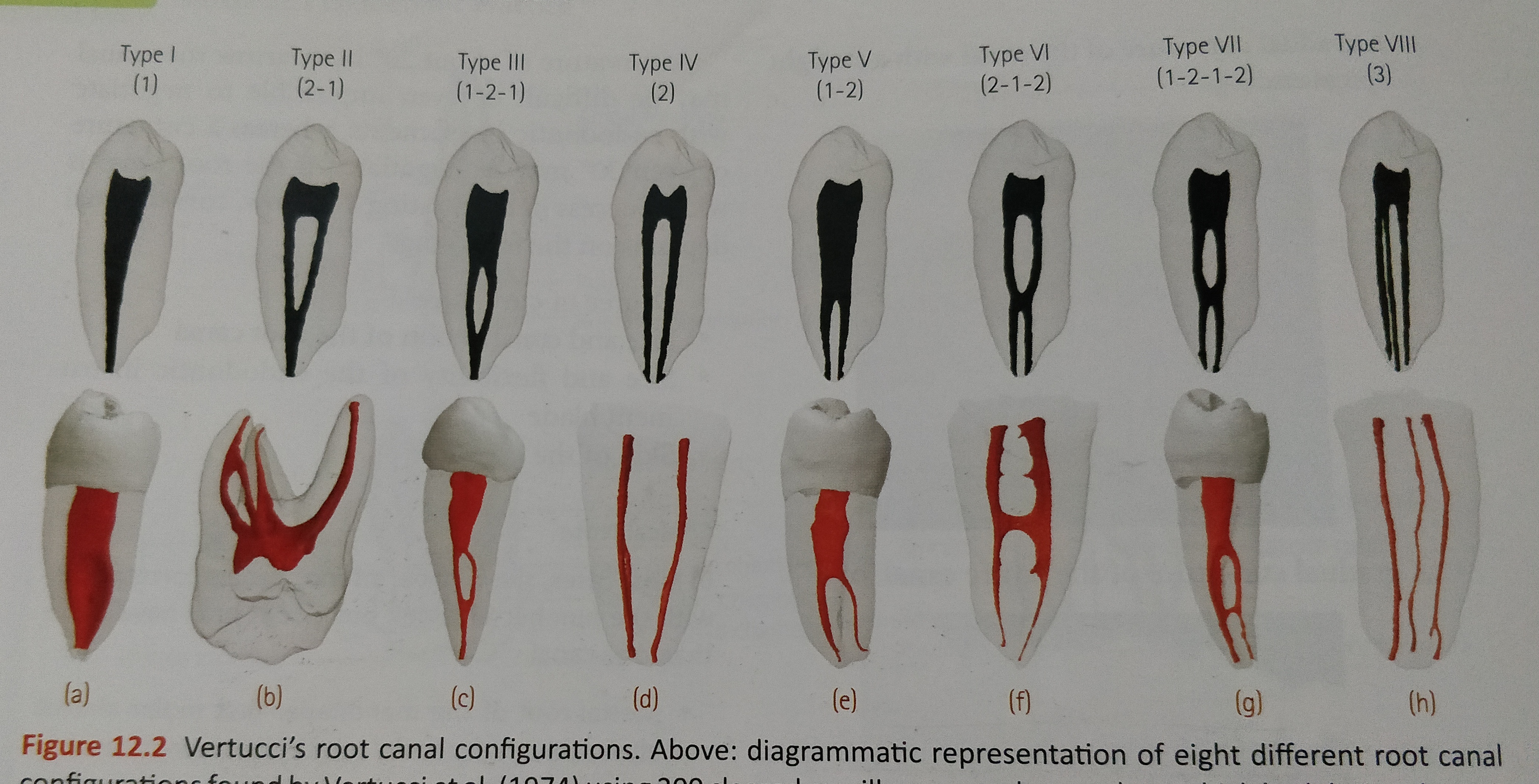
  • Vertucci’s Classification:
  1. Type I : Single canal extends from the pulp chamber to the apex (1)
  2. Type II : Two separate canals leave the pulp chamber and join short of the apex to form one canal (2-1)
  3. Type III : One canal leaves the pulp chamber and divides into two in the root , the two then merge to exit as one canal (1-2-1)
  4. Type IV : Two separate distinct canals extend from the pulp chamber to the apex (2)
  5. Type V : One canal leaves the pulp chamber and divides short of the apex into two separate distinct canals with separate apical foramina (1-2)
  6. Type VI : Two separate canals leaves the pulp chamber , merge in the body of the root , and redivide short of the apex to exit as two distinct canals (2-1-2)
  7. Type VII : One canal leaves the pulp chamber, divides and then rejoins in the body of the root , and finally redivides into two distinct canals short of the apex (1-2-1-2)
  8. Type VIII : Three separate distinct canals extend from the pulp chamber to the apex (3)
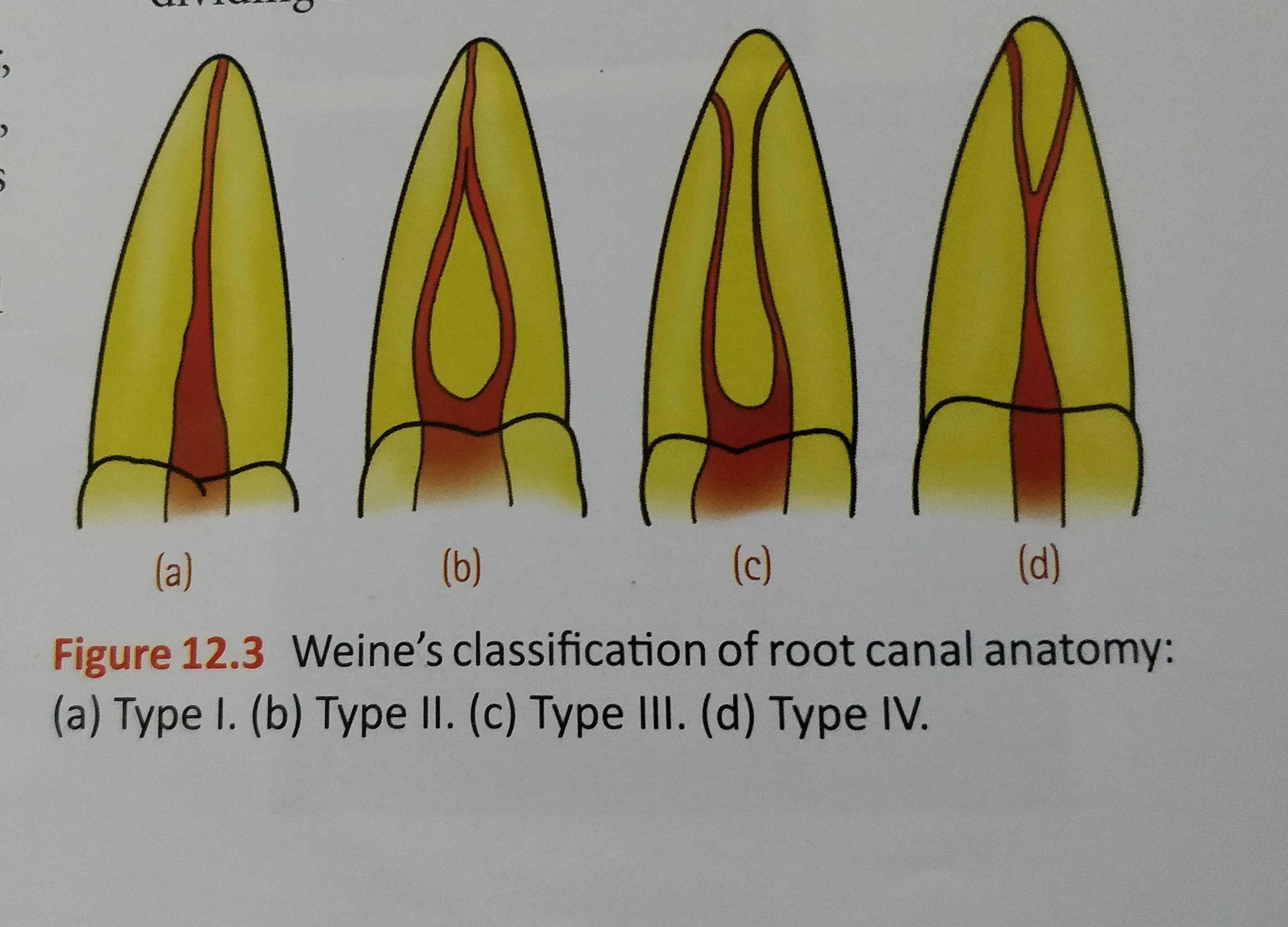
  • Weine’s classification :
  1. Type I : Single canal from pulp chamber to apex
  2. Type II : Two canals leaving from the chamber and merging to form a single canal short of the apex
  3. Type III : Two separate and distinct canals from chamber to apex
  4. Type IV : One canal leaving the chamber and dividing into two separate and distinct canals
  • Classification based on canal cross – section:
  1. Round (circular)
  2. Oval
  3. Long oval
  4. Flattened (flat/ribbon)
  5. Irregular

References:

  • Dental notes
  • Grossman’s Endodontic Practice (13th edition)

Prosthesis


Prosthesis may be defined as an artificial replacement of a missing part of the human body

Types of prosthesis

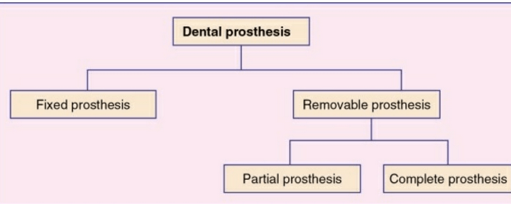
i. Dental prosthesis
ii. Maxillofacial prosthesis
iii. Ancillary prosthesis

Dental prosthesis

An artificial replacement of one or more teeth and associated dental/alveolar structures.

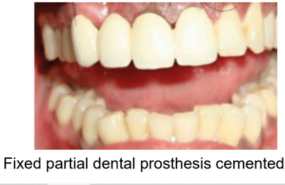
Fixed dental prosthesis

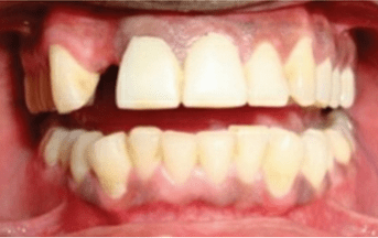
Any dental prosthesis that is cemented, screwed or attached to the retained natural teeth or roots.

missing upper lateral incisor

Removable prosthesis

Any dental prosthesis that replaces some of the missing teeth in a partially edentulous arch

Removable Partial Dentures | Partial dentures, Dental braces, Dental

Removable partial denture prosthesis (RPDP)


The prosthesis that replaces some of the teeth in a partially edentulous arch and that can be removed from the mouth by the patient.

It can be a simple removable partial denture fabricated in acrylic resin called temporary partial
denture.

A removable partial denture fabricated in cast metal alloy and acrylic resin is called cast partial denture

removable partial dentures

Removable complete denture prosthesis

The prosthesis that replaces the entire dentition and associated structures of maxilla and mandible

Complete Dentures - What's Your Strategy? - Spear Education

Maxillofacial prosthesis

The prosthesis that is used to replace a part or all of any stomatognathic or craniofacial structures.

Examples of the maxillofacial prosthesis are auricular
prosthesis, orbital prosthesis, nasal prosthesis and facial prosthesis
.

Endolite Silicone Maxillofacial Prosthesis, Endolite India Limited ...

Implant supported prosthesis

The prosthesis that is used to replace the missing teeth , retained by implant supported prosthesis.

Implant supported dentures. Types of prosthetic restorations

Ancillary prosthesis

The type of dental prosthesis used in prosthodontics for a very short term of usage

example- splints ,stents and guides

Instant Hydrostatic Splint
hydrostatic splint

source – preclinical textbook of prosthodontics by S Lakshmi and image source- google and S Lakshmi

Endotoxins and Exotoxins

Endotoxins –

🔻They are Lipopolysaccharide in nature and former integral part of the gram negative bacteria cell wall.

🔻 They are Heat stable.

🔻Form integral part of the cell wall; released only on disruption of bacterial cell.

🔻Weakly antigenic; antitoxin is not formed but antibodies against polysaccharide are raised.

🔻Cannot be toxoided.

🔻No enzymatic action.

🔻Non-specific action of all endotoxins.

🔻Low potency

🔻Non-specific in action.

🔻Usually produce fever.

🔻Produced by Gram-negative bacteria.

🔻 Massive gram negative septicemia may cause a syndrome of endotoxic shock characterized by fever, leukopenia, thrombocytopenia ,profound fall of blood pressure and circulatory collapse  to death.

Exotoxins

🔻Protein (polypeptides) M.W. 10,000 to 900,000.

🔻 Heat labile (more than 60°)

🔻Actively secreted by living cells into medium.

🔻 Highly antigenic, stimulates formation of antitoxin which neutralises toxin.

🔻Converted into toxoid by formaldehyde.

🔻Enzymatic in action.

🔻Specific pharmacological effect for each exotoxin.

🔻Very high potency.

🔻Highly specific for particular tissue eg. tetanus toxin for CNS.

🔻Don’t produce fever in host.

🔻Produced mainly by Gram-positive bacteria and also by some Gram-negative bacteria.

Source- textbook of microbiology for dental students c p baveja