
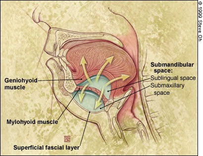
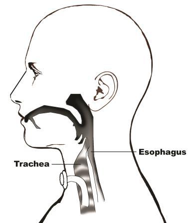
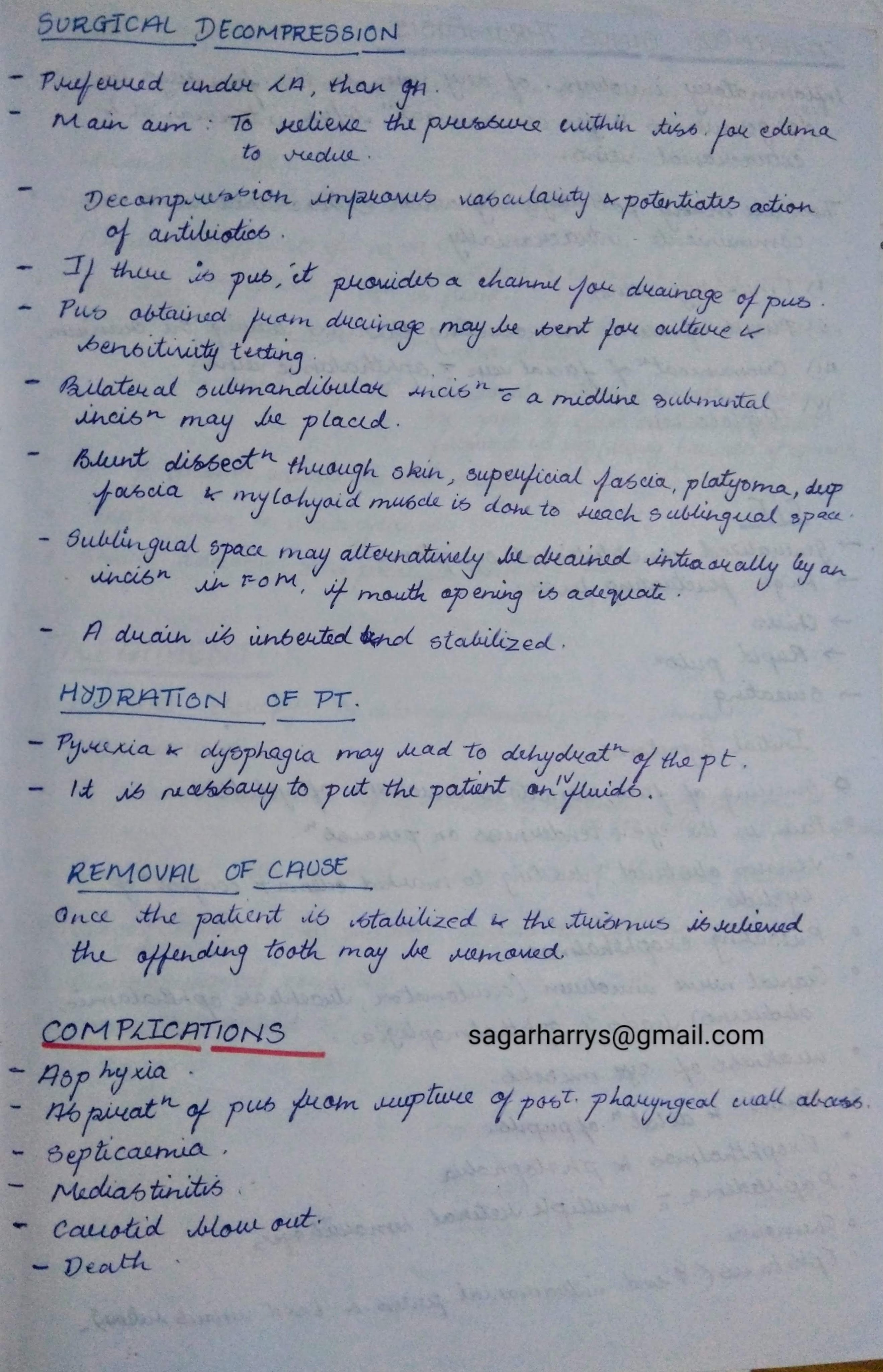
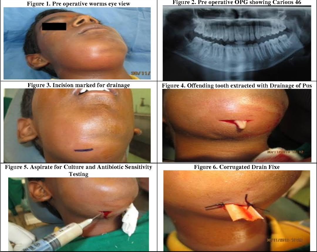
LUDWIG`S ANGINA
REFERENCES:
- Textbook of Oral & Maxillofacial Surgery,Chitra Chakravarthy (2nd Edition)
- AAFP.org
- orosurgery.com
- seminarscholar.com
- craighospital.com





REFERENCES:

Oral cancer is a disease with very poor prognosis because it is not recognised and treated when small and early.
INCIDENCE.
-Squamous cell (epidermoid) carcinoma comprises 90% of all oral malignant tumours and 5% of all human malignancies.
-The peak incidence in the UK and the USA is from 55 to 75 years of age, whereas in India it is from 40 to 45 years of age.
-Oral cancer is a very frequent malignancy in India, Sri Lanka and some Eastern countries, probably related to habits of betel-nut chewing and reversed smoking .
– There is a definite male preponderance.
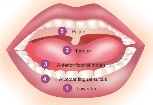
-It can occur anywhere in the mouth but certain sites are more commonly involved.
-These sites, in descending
order of frequency, are: the lips (more commonly lower),
tongue, anterior floor of mouth, buccal mucosa in the region
of alveolar lingual sulcus, and palate.
ETIOLOGY.
As with other forms of cancer, the etiology of squamous cell carcinoma is unknown. But a number of etiological factors have been implicated:
Strong association:
i) Tobacco smoking and tobacco chewing causing leukoplakia is the most important factor .
ii) Chronic alcohol consumption.
iii) Human papilloma virus infection, particularly HPV 16, 18 and 33 types.
Weak association:
i) Chronic irritation from ill-fitting denture or jagged teeth.
ii) Submucosal fibrosis as seen in Indians consuming excess of chillies.
iii) Poor orodental hygiene.
iv) Nutritional deficiencies.
v) Exposure to sunlight (in relation to lip cancer).
vi) Exposure to radiation.
vii) Plummer-Vinson syndrome, characterised by atrophy
of the upper alimentary tract.
The most common molecular alterations in oncogenes
seen in squamous cell carcinoma of the oral cavity are in
p16, p53, cyclin D, p63, PTEN, and EGFR.
MORPHOLOGIC FEATURES.
Grossly, squamous cell carcinoma of oral cavity may have the following types
i) Ulcerative type—is the most frequent type and is
characterised by indurated ulcer and firm everted or
rolled edges.
ii) Papillary or verrucous type—is soft and wart-like growth.
iii) Nodular type—appears as a firm, slow growing submucosal nodule.
iv) Scirrhous type—is characterised by infiltration into
deeper structures.

*All these types may appear on a background of leukoplakia or erythroplasia of the oral mucosa.
Enlarged cervical lymph nodes may sometimes be present.
Histologically.
– squamous cell carcinoma ranges from well-differentiated keratinising carcinoma to highly undifferentiated neoplasm . -Changes of epithelial dysplasia are often present in the surrounding
areas of the lesion.
–Carcinoma of the lip and intraoral squamous carcinoma are usually always well-differentiated
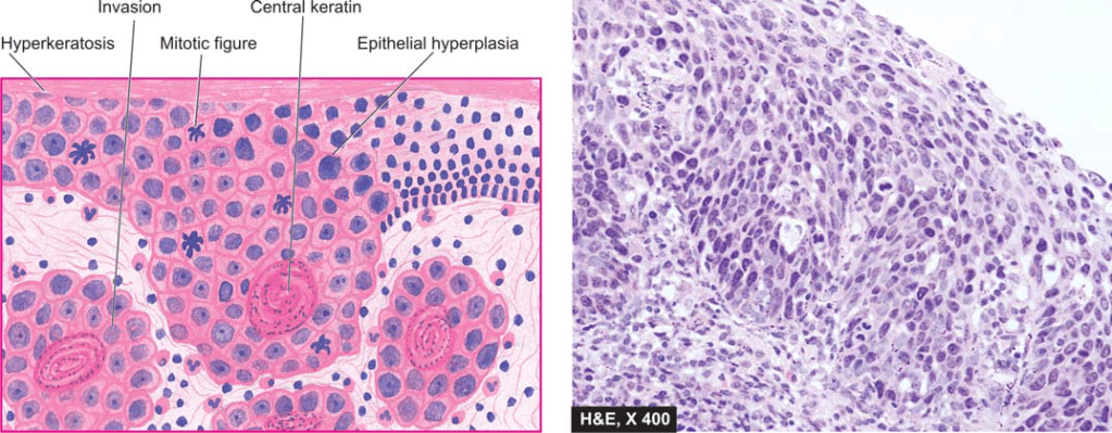
source -textbook of pathology for dental students harsh mohan
A chronic, autoimmune, sub-epidermal blistering skin disease that rarely involves mucous membrane.

Age: Elderly (>60 years)
Skin Lesions:
Oral Manifestations:
Vesicles appear gingivally👇🏻
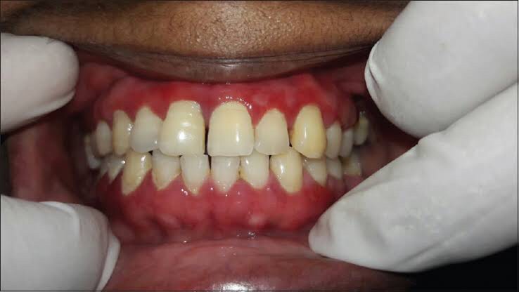
Erythematous & desquamate as result of minor frictional trauma
👉🏻 Oral lesions comprise of bullae/vesicle that rupture to form erosions and ultimately leave out ulcerations
👉🏻 Other sites:
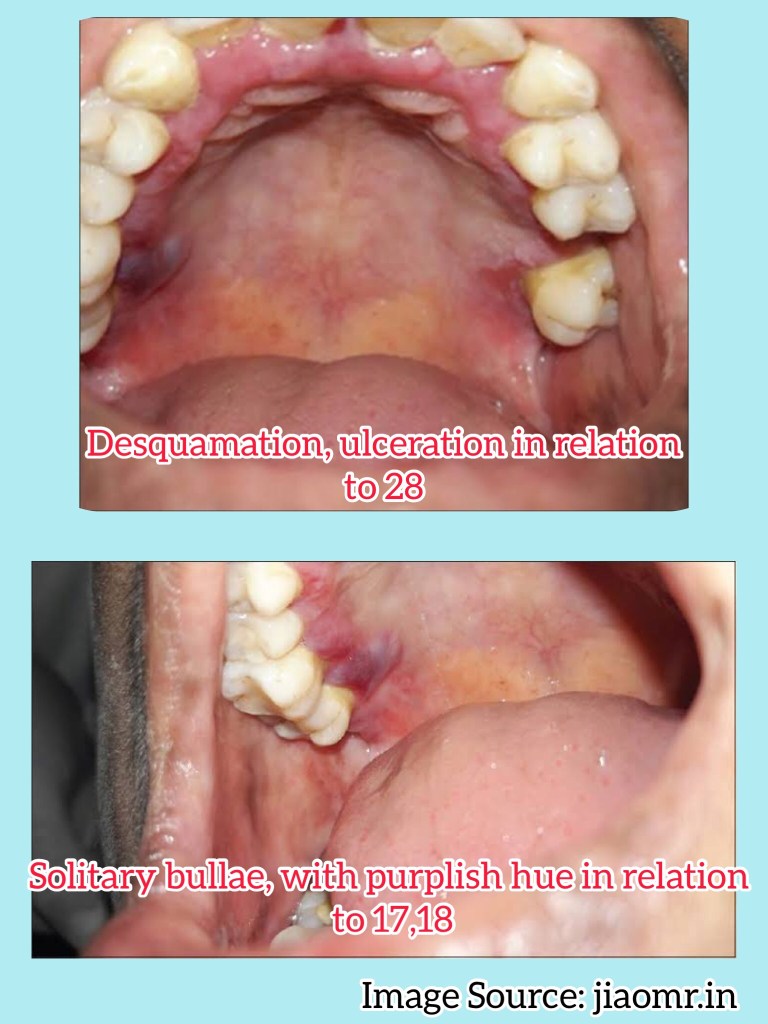
Apart from evaluating history, clinical presentation, histopathological analysis is carried out followed by direct immunofluorescence study for the differential diagnosis and confirmation of the condition.
👉🏻Histopathology:
👉🏻Direct immunofluorescence is found to be the gold standard test. Deposition pattern of different types of immunoreactants differentiates the various immune-mediated diseases. Direct immunofluorescence shows presence of IgG and C3 deposits along the basement membrane zone.
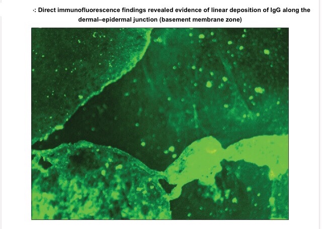
👉🏻Electron Microscopy: In bullous pemphigoid (BP), the 180 kD antigen (BPAG2) was shown by immuno-EM to be a transmembrane molecule and to possess an autoantibody binding site outside the cell, suggesting a major pathogenic role for the BPAG2 in blister formation.
• Mucous membrane pemphigoid can be differentiated from BP by its predominant involvement of mucosal surfaces and positive Nikolsky’s sign.
• Lichen planus pemphigoides is clinically differentiated by the presence of lichen planus lesions in addition to tense blisters.
• Nikolsky’s sign is present in case of pemphigus and cicatricial pemphigoid, but not in the case of BP.
👉🏻Treatment is based on the degree of cutaneous and oral involvement. Mostly, topical steroid (clobetasol propionate) gives satisfactory result in case of smaller area of skin involvement, whereas larger area of skin involvement and recurrent cases are treated satisfactorily with systemic steroids and immunosuppressive agents.
👉🏻Recommended dosage for oral prednisolone is 0.3–1.25 mg/kg body weight/day, controls disease within 1–2 weeks, followed by which the dose is tapered. Dexamethasone (100 mg in 500 mL 5% dextrose i.v. over 2–3 h for three consecutive days) is the preferred steroid for pulse therapy, either administered alone or in combination with cyclophosphamide.
Other drugs for treating BP include new antibody modulators, rituximab 375mg/m2weekly over 4 weeks and omalizumab subcutaneously 300–375 mg for every 6 weeks.
👉🏻Higher doses of systemic corticosteroids seem to be associated with higher mortality rates, which led to the addition of corticosteroid-sparing agents to the treatment of BP. The most frequently used immunosuppressive agent is azathioprine (0.5–2.5 mg/kg body weight/day). Others being cyclophosphamide, methotrexate, cyclosporine A, combination tetracycline/minocycline along with nicotinamide, and more recently, mycophenolate mofetil, a DNA synthesis inhibitor, and methotrexate, a folate antagonist.
👉🏻IVIg – A dose of 1–2 g/kg for five consecutive day cycle of 0.4 g/kg/day, although a 3-day cycle may be used in cases that are nonresponsive to conventional therapy.
Dr. Mehnaz Memon🖊
References:
DEFINITION- Leukoplakia (white plaque) may be clinically
defined as a white patch or plaque on the oral mucosa, exceeding 5 mm in diameter, which cannot be rubbed off nor can be classified into any other diagnosable disease.
–the term ‘leukoplakia’ is reserved for epithelial thickening which may range from completely benign to atypical and to premalignant cellular changes.
INCIDENCE–
1. It occurs more frequently in males than
females.
2.The lesions may be of variable size and appearance.
3. The sites of predilection, in descending orderof frequency, are: cheek mucosa, angles of mouth, alveolar mucosa, tongue, lip, hard and soft palate, and floor of the mouth.
4.In about 4-6% cases of leukoplakia, carcinomatous
change is reported.
ETIOLOGY-
1. It has the strongest association with the use of tobacco in various forms, e.g. in heavy smokers (especially in pipeand cigar smokers) and improves when smoking is discontinued, and in those who chew tobacco as in paan, paan masaala, zarda, gutka etc.
2.The condition is also known by other names such as smokers keratosis and stomatitis nicotina.
3. Other etiological factors implicated are chronic friction such as with ill-fitting dentures or jagged teeth, and local irritants like excessive consumption of alcohol and very hot and spicy foods and beverages.
4. A special variety of leukoplakia called ‘hairy leukoplakia’ has
been described in patients of AIDS and has hairy or corrugated surface but is not related to development of
oral cancer.

MORPHOLOGIC FEATURES.
1.Grossly, the lesions of leukoplakia may appear white, whitish-yellow, or redvelvety of more than 5 mm diameter and variable in appearance.
2. They are usually circumscribed, slightly elevated, smooth or wrinkled, speckled or nodular.
Histologically, leukoplakia is of 2 types:
-The subepithelial tissues usually show an inflammatory infiltrate composed of lymphocytes and plasma cells.
– The extent and degree of the epithelial changes indicate the degree of severity of the epithelial dysplasia.
-Usually, mild dysplasia may revert back to normal if the offending etiologic factor is removed, whereas severe dysplasia indicates that the case may progress to carcinoma.

source – textbook of pathology for dental students harsh mohan
Written by : Dr. Urusa I Inamdar
Ribbon shaped or thin connecting structure between two root canals.
Kim et al. have classified the isthmus into following categories:

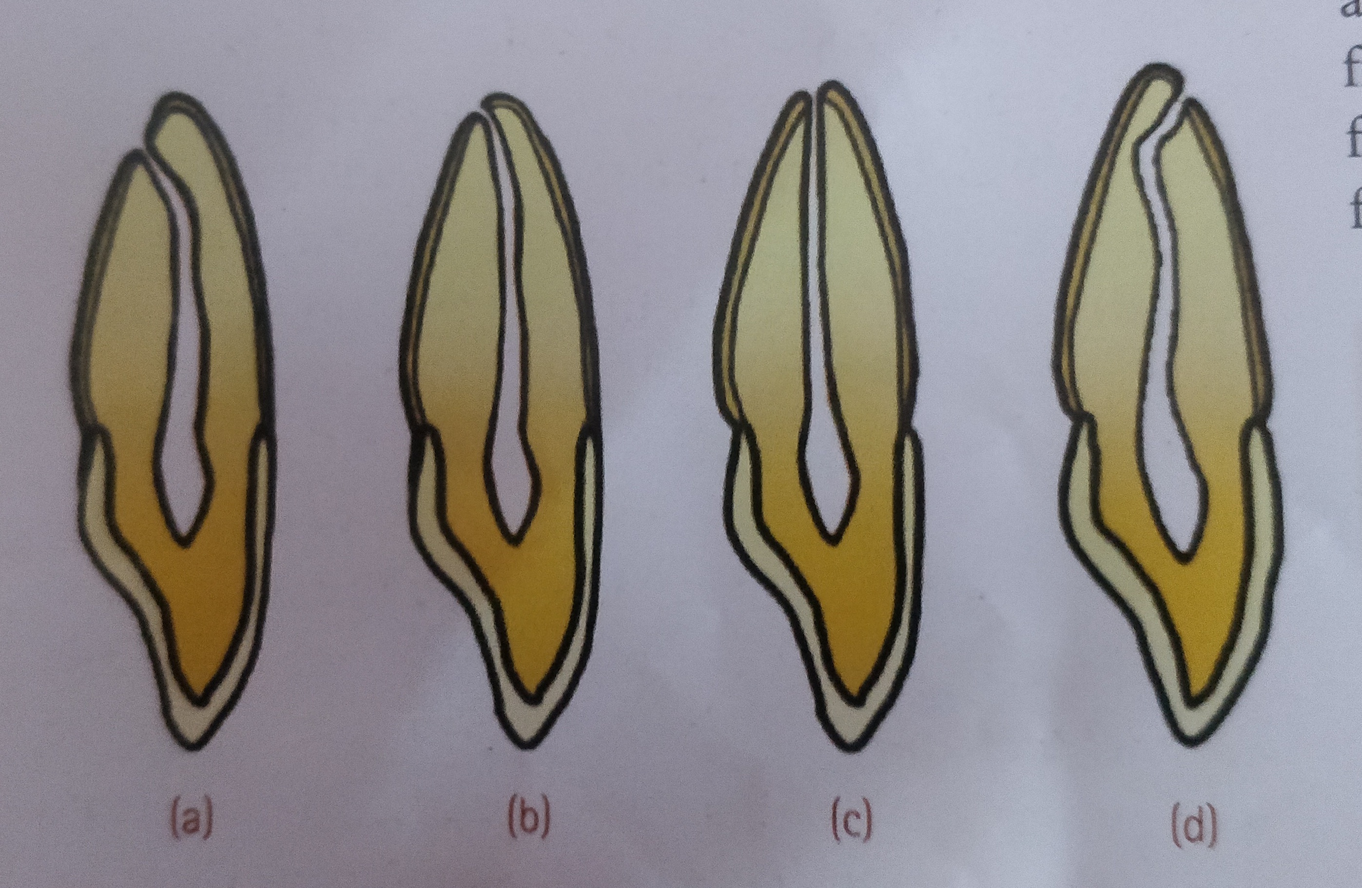
In young , incompletely developed teeth , the apical foramen is funnel shaped , with the wider portion extending outward . The mouth of the funnel is filled with periodontal tissue , which is later replaced by dentin and cementum.
As the root develops , the apical foramen becomes narrower .
It is not necessary to shape , clean , or fill root canals to their anatomic apices , but rather to the cementodentinal junction , which usually lies within the canal just short of the apex .
The apical foramen is not always the most constricted portion of the root canal. Constrictions can and do occur before the extremity of the root ks reached. Apical constrictions are found 0.5-1.0 mm away from the root apex.
The apical foramen is not always located in the centre of the root apex . It may exit on the mesial , distal , labial or lingual surface of the root , usually slightly eccentrically.
In few cases , the apical foramen has been found as much as 2-3 mm away from the anatomic apex.
The root canal obturation should end approximately 0.5-1.0 mm short of the anatomic root apex .
The periodontal vessels curve around the root apex of a developing tooth and often become entrapped in Hertwig’s epithelial root sheath , resulting in the formation of lateral canals and accesory foramina during calcification .This phenomenon frequently occurs in the apical third of the root .
Lateral canals may also occur in the area of bifurcation or trifurcation of multirooted teeth.
With increasing age , the accesory foramina diminish in number because of calcification of their contained soft tissue.
Transplants from one individual to another member of the same species (‘allografts’) are recognised as foreign and rejected.
Gorer (1930) identified the antigens responsible for allograft rejection in inbred mice that led to the discovery of the major histocompatibility complex (MHC).
The MHC in humans is known as the human leukocyte antigen (HLA) complex.
Histocompatibility antigens mean cell surface antigens that evoke immune response to an incompatible host resulting in allograft rejection.
These alloantigens are present on surface of leucocytes in man and are called human leucocyte antigens (HLA) and the set of genes coding for them is named the HLA Complex.
The HLA complex of genes is located on short arm of chromosome 6 and is grouped in three classes
Class I
HLA-A, HLA-B and HLA-C
Class II
HLA-DR, HLA-DQ and HLA-DP (All of these are present within HLA-D region of HLA complex.)
Class III ( Complement loci encode for C2, C4 and Factor B of complement system and tumour necrosis factors (TNF) alpha and beta)
A locus is the position where a particular gene is located on the chromosome.
HLA loci are multiallelic i.e. the gene present on the locus can be any one of several alternative forms (alleles).
Each allele determines a distinct antigen. There are 24 alleles at HLA-A locus and 50 at HLA-B. HLA system is very pleomorphic. Every individual inherits one set of HLA-genes from each parent.
The MHC class I antigens are present on the surface of all nucleated cells. They are involved in graft rejection and cell mediated cytolysis. The cytotoxic T cells (CD8) recognise MHC class I antigens for their action.
They have a very limited distribution and are principally found on the surface of macrophages, monocytes, activated T-lymphocytes (CD4) and B-lymphocytes. They are primarily responsible for the graft-versus-host response and the mixed leukocyte reaction (MLR).
Class III genes encode C2, C4 complement components of the classical pathway and properdin factor B of the alternative pathway.

Source – textbook of microbiology for dental students – harsh mohan
Pathologic changes in gingivitis are associated with the presence of oral microorganisms attached to the tooth and perhaps in or near the gingival sulcus.
STAGE I GINGIVITIS: THE INITIAL LESION
The first manifestations of gingival inflammation are vascular changes consisting of dilated capillaries and increased blood flow. These initial inflammatory changes occur in response to microbial activation of resident leukocytes and the subsequent stimulation of endothelial cells. Clinically, this initial response of the gingiva to bacterial plaque is not apparent.
Changes can also be detected in the junctional epithelium and perivascular connective tissue at this early stage. For example, the perivascular connective tissue matrix becomes altered, and there is exudation and deposition of fibrin in the affected area. Also, lymphocytes soon begin to accumulate. The increase in the migration of leukocytes and their accumulation within the gingival sulcus may be correlated with an increase in the flow of gingival fluid into the sulcus.
The character and intensity of the host response determine whether this initial lesion resolves rapidly, with the restoration of the tissue to a normal state, or evolves into a chronic inflammatory lesion. If the latter occurs, an infiltrate of macrophages and lymphoid cells appears within a few days.
STAGE II GINGIVITIS: THE EARLY LESION
The early lesion evolves from the initial lesion within about 1 week after the beginning of plaque accumulation.Clinically, the early lesion may appear as early gingivitis, and it overlaps with and evolves from the initial lesion with no clear-cut dividing line. As time goes on, clinical signs of erythema may appear, mainly because of the proliferation of capillaries and increased formation of capillary loops between rete pegs or ridges . Bleeding on probing may also be evident.1 Gingival fluid flow and the numbers of transmigrating leukocytes reach their maximum between 6 and 12 days after the onset of clinical gingivitis.
The amount of collagen destruction increases 70% of the collagen is destroyed around the cellular infiltrate. The main fiber groups affected appear to be the circular and dentogingival fiber assemblies. Alterations in blood vessel morphologic features and vascular bed patterns have also been described.
PMNs that have left the blood vessels in response to chemo- tactic stimuli from plaque components travel to the epithelium,
cross the basement lamina, and are found in the epithelium, emerg- ing in the pocket area. PMNs are attracted to bacteria and engulf them in the process of phagocytosis . PMNs release their lysosomes in association with the ingetion of bacteria.Fibroblasts show cytotoxic alterations, with a decreased capacity for collagen.
Meanwhile, on the opposite side of molecular events, collagen degradation is related to matrix metalloproteins (MMPs). Different MMPs are responsible for extracellular matrix remodeling within 7 days of inflammation, which is directly related to MMP-2 and MMP-9 production and activation.
STAGE III GINGIVITIS: THE ESTABLISHED LESION
Over time, the established lesion evolves, characterized by a predominance of plasma cells and B lymphocytes and probably in conjuncation with the creation of a small gingival pocket lined with a pocket epithelium.The B cells found in the established lesion are pre- dominantly of the immunoglobulin G1 (IgG1) and G3 (IgG3) subclasses.
In chronic gingivitis, which occurs 2 to 3 weeks after the beginning of plaque accumulation, the blood vessels become engorged and congested, venous return is impaired, and the blood flow becomes sluggish .The result is localized gingival anoxemia, which superimposes a somewhat bluish hue on the reddenedgingiva.18 Extravasation of erythrocytes into the connective tissue and breakdown of hemoglobin into its component pigments can also deepen the color of the chronically inflamed gingiva. The established lesion can be described as moderately to severely inflamed gingiva.
An inverse relationship appears to exist between the number of intact collagen bundles and the number of inflammatory cells.Collagenolytic activity is increased in inflamed gingival tissue17 by the enzyme collagenase. Collagenase is normally present in gingival tissues5 and is produced by some oral bacteria and by PMNs.
Enzyme histochemistry studies have shown that chronically inflamed gingivae have elevated levels of acid and alkaline phos- phatase, β-glucuronidase, β-glucosidase, β-galactosidase, esterases, aminopeptidase, and cytochrome oxidase. Neutral mucopolysaccharide levels are decreased, presumably as a result of degradation of the ground substance.
Established lesions of two types appear to exist; some remain stable and do not progress for months or years and others seem to become more active and to convert to progressively destructive lesions. Also, the established lesions appear to be reversible in that the sequence of events occurring in the tissues as a result of successful periodontal therapy seems to be essentially the reverse of the sequence of events observed as gingivitis develops. As the flora reverts from that characteristically associated with destructive lesions to that associated with periodontal health, the percentage of plasma cells decreases greatly, and the lymphocyte population increases proportionately.
STAGE IV GINGIVITIS: THE ADVANCED LESION
Extension of the lesion into alveolar bone characterizes a fourth stage known as the advanced lesion or phase of periodontal break- down.
Gingivitis will progress to periodontitis only in individuals who are susceptible. Patients who had sites with consistent bleeding had 70% more attachment loss than at sites that were not inflamed consistently (GI = 0). Teeth with noninflamed sites consistently had a 50-year survival rate of 99.5%, whereas teeth with consistently inflamed gingiva had a 63.4% survival rate over 50 years. Based on this longitudinal study on the natural history of periodontitis in a well-maintained male population, per- sistent gingivitis represents a risk factor for periodontal attachment loss and for tooth loss.
Reference- Caranza textbook of periodontology 11th edition
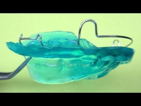
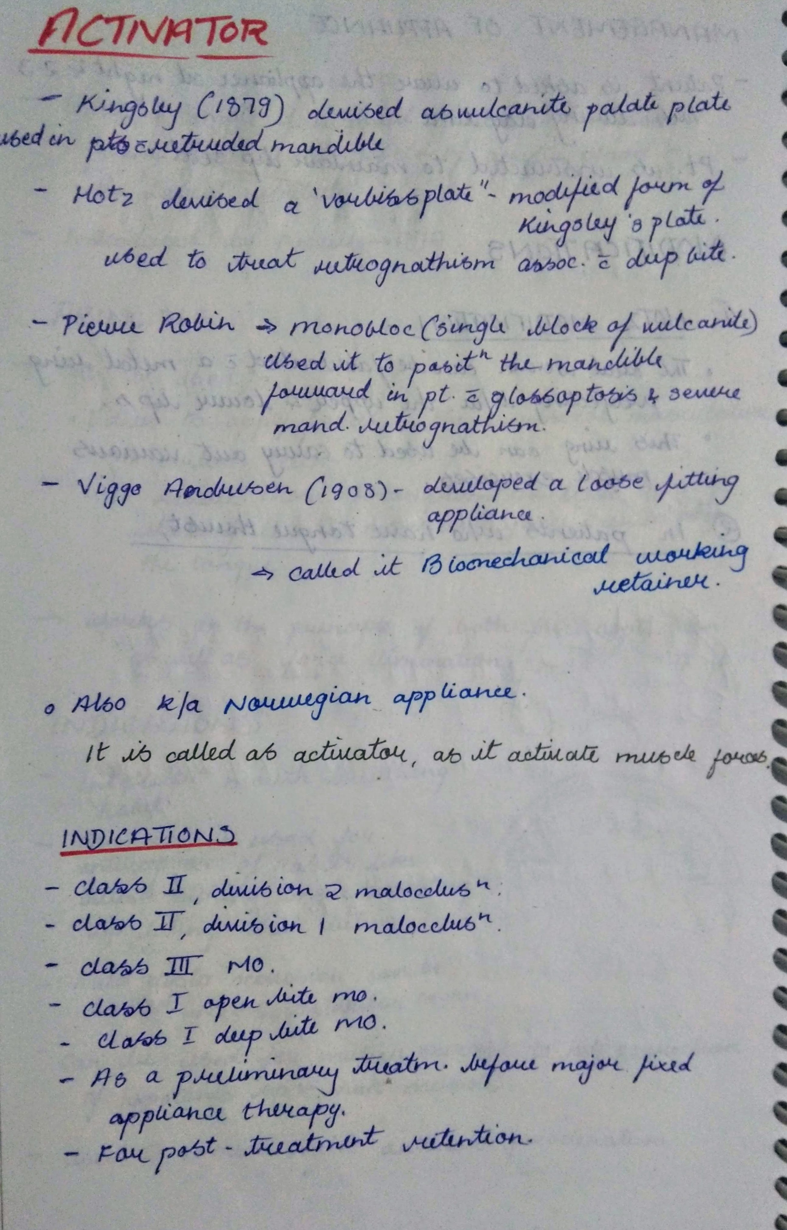


TRIMMING OF ACTIVATOR:

REFERENCES:



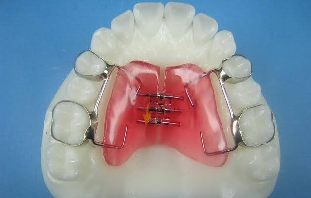
TYPES OF RME APPLIANCES:



REFERENCES:






Sources – Anil Ghoms textbook of oral medicine , osmosis.org
Image source – researchgate.net , Google images

