Sources – Anil Ghoms textbook of oral medicine , osmosis.org
Image sources – Google images , researchgate.net

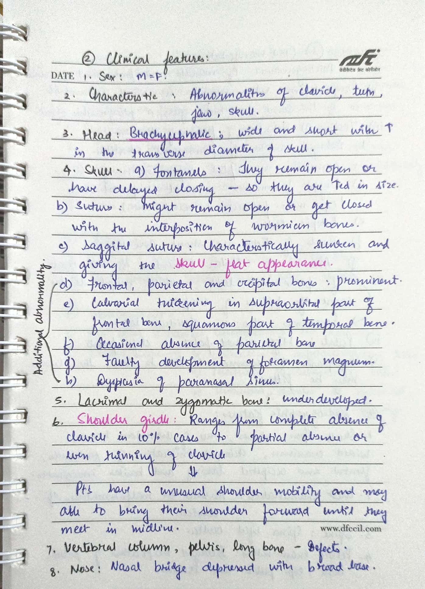



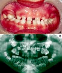
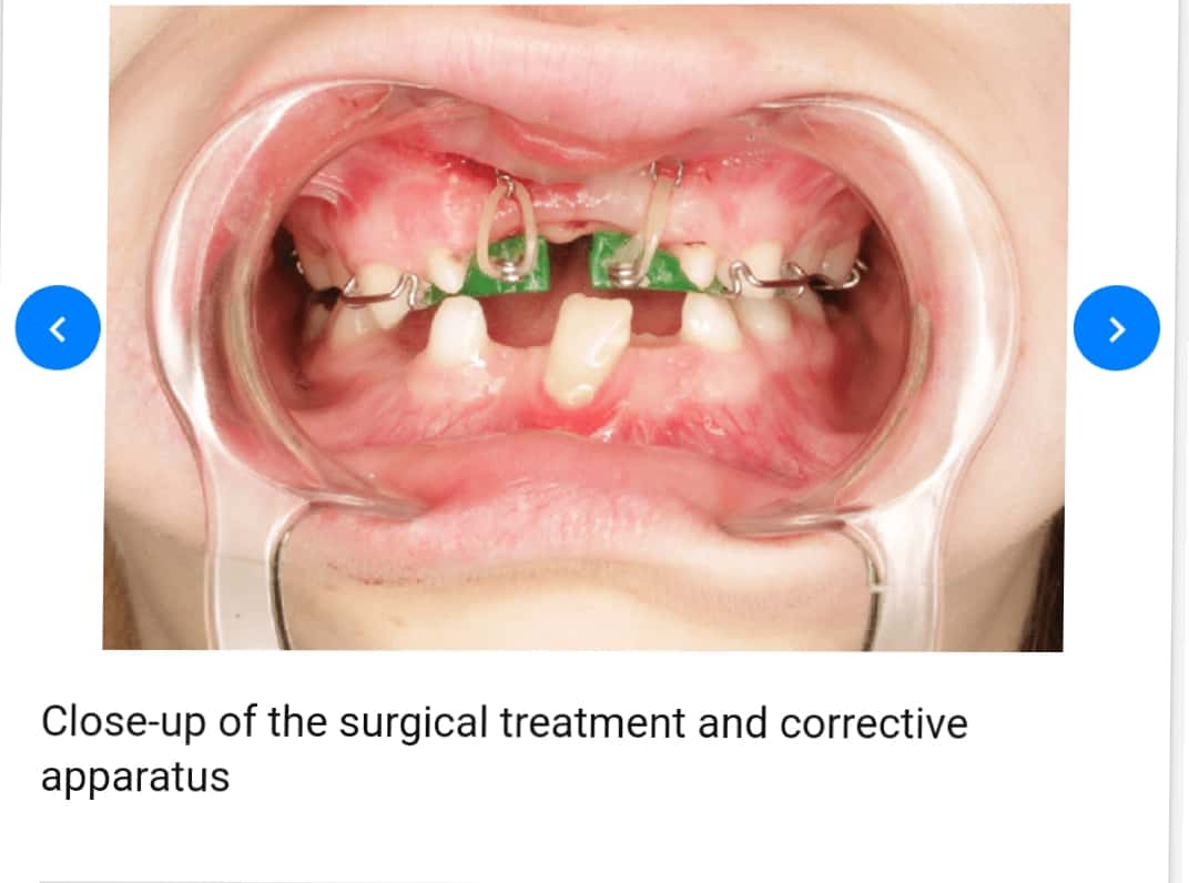
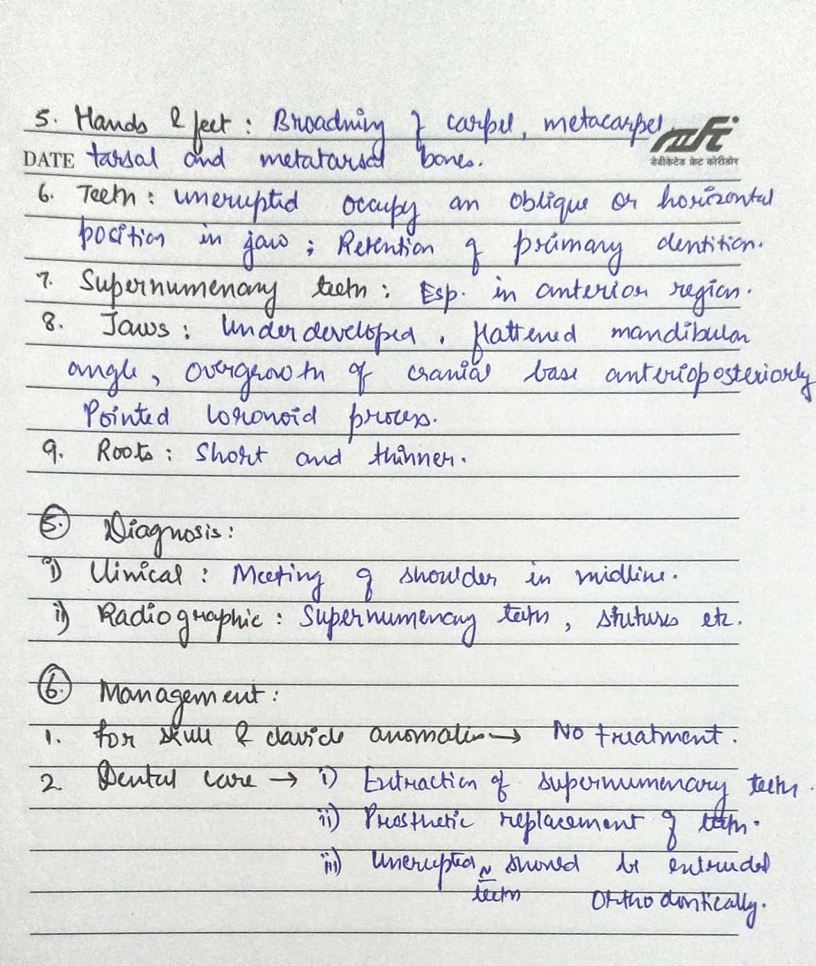


Sources – Anil Ghoms textbook of oral medicine , osmosis.org
Image sources – Google images , researchgate.net
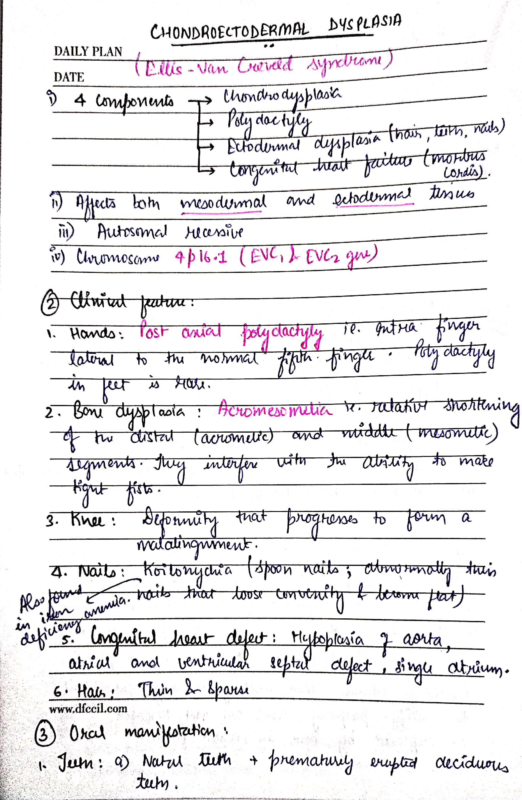
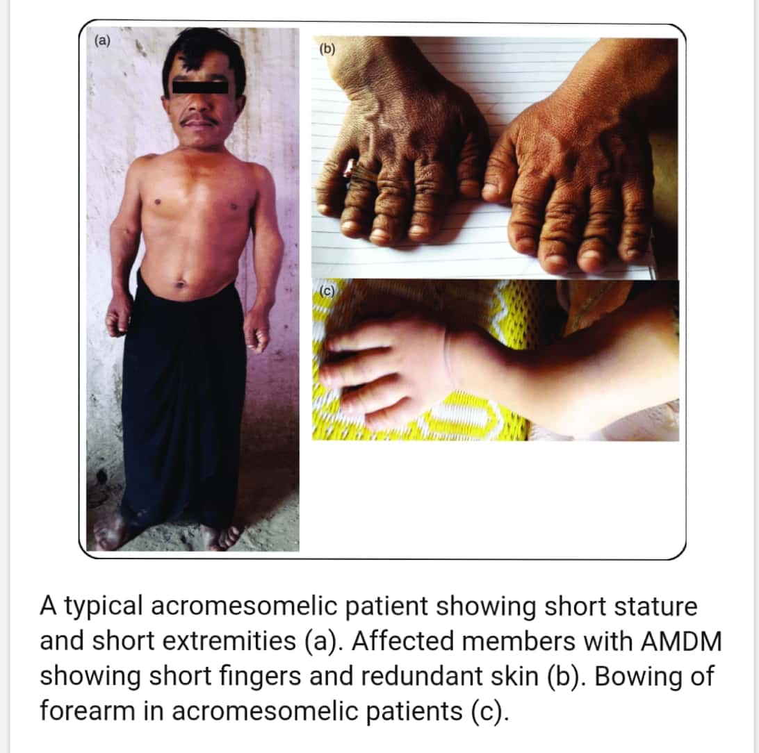


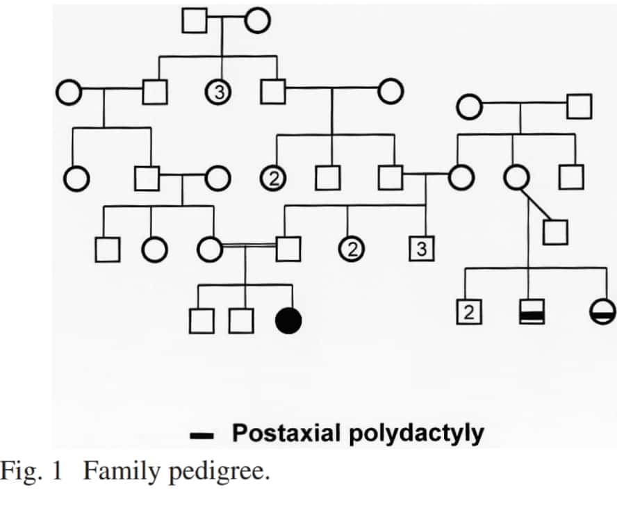
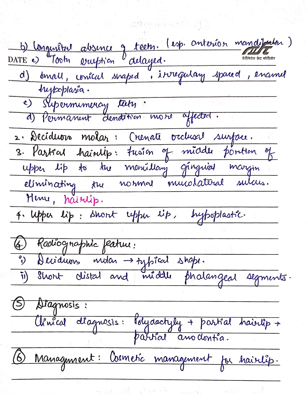
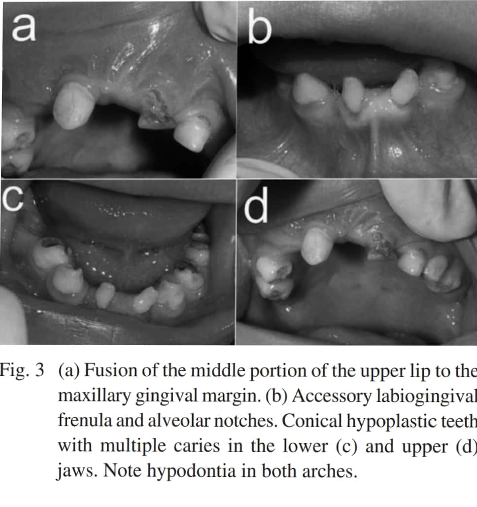


Ps. This is a good case report on Chondroectodermal dysplasia – https://api.semanticscholar.org/CorpusID:5721166
Sources – Shafer’s textbook of oral pathology
Image reference – Shafer’s textbook of oral pathology , semanticscholar.org
Written by : Dr. Urusa I Inamdar

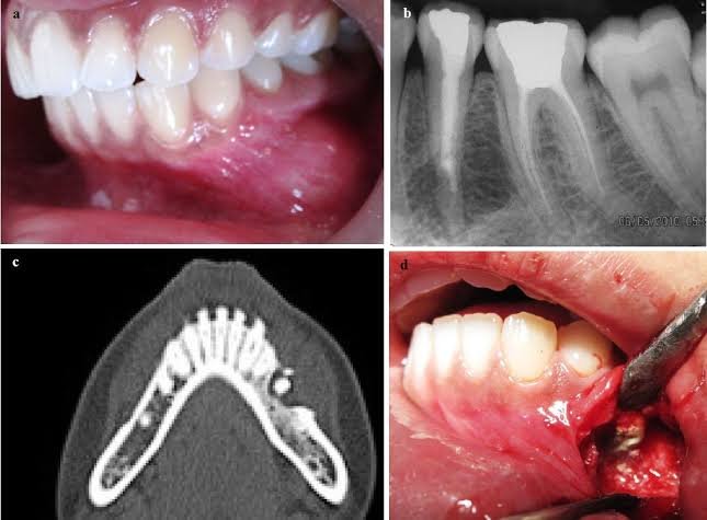

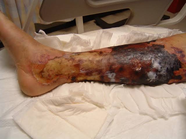


SINGLE STAGE V/S 2-STAGE SURGICAL PROCEDURES:
SURGICAL PROCEDURES:
There are 3 types:
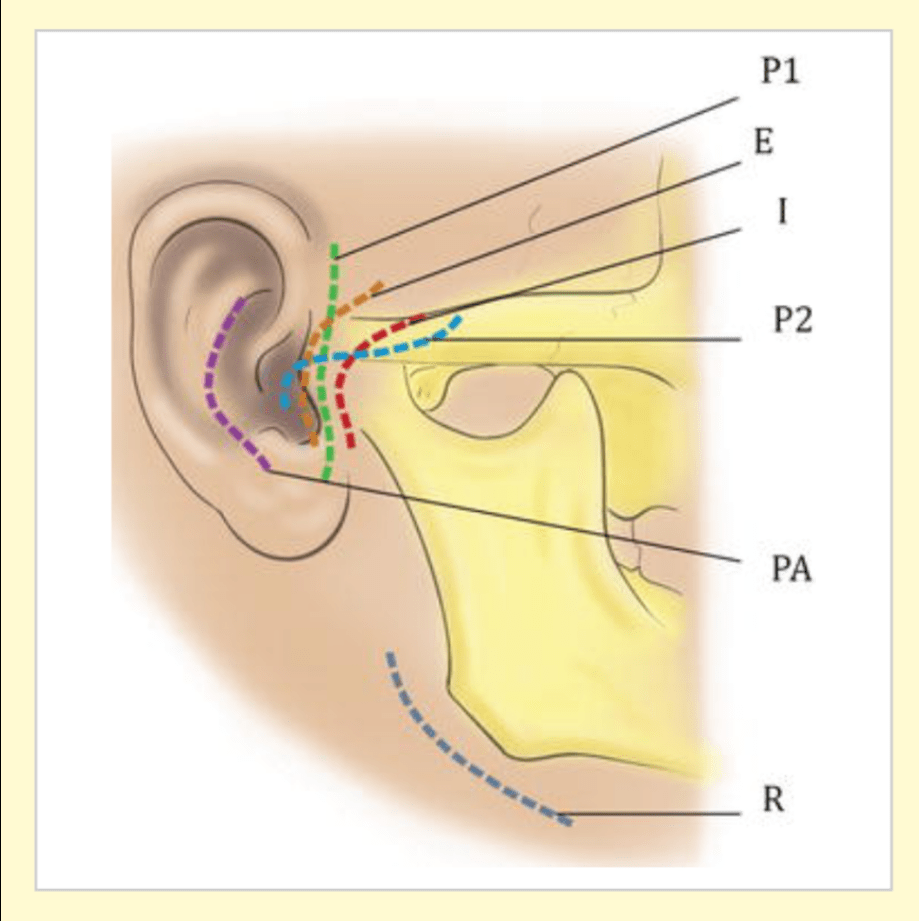
TMJ APPROACHES:
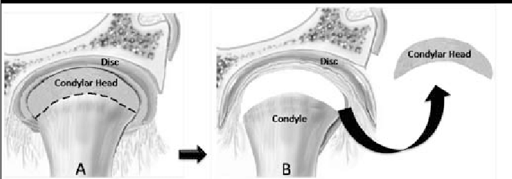
1. CONDYLECTOMY:
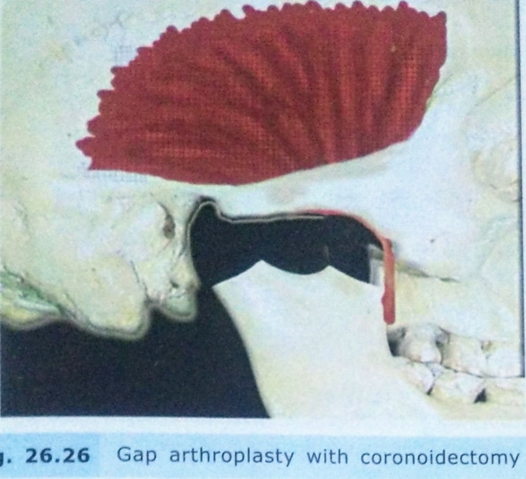
2. GAP ARTHROPLASTY:

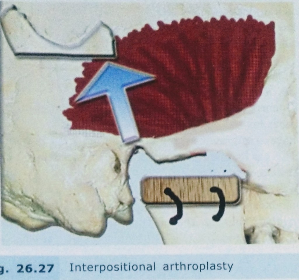
3. INTERPOSITIONAL ARTHROPLASTY:

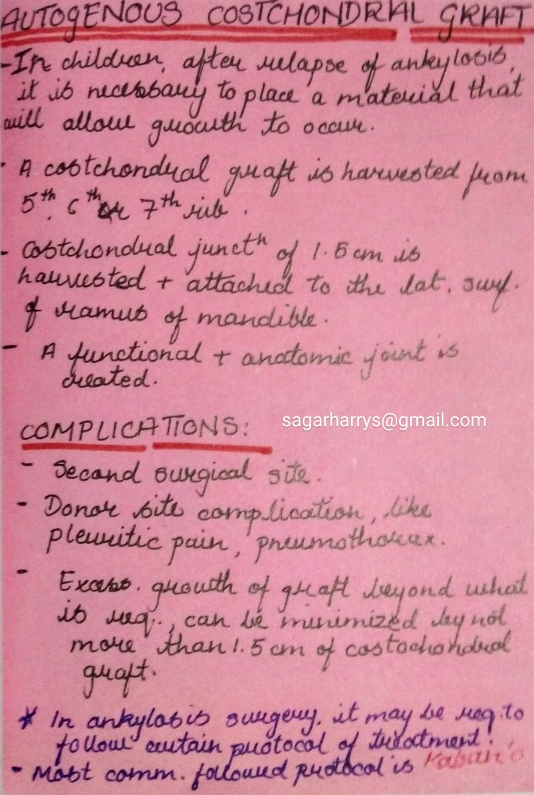
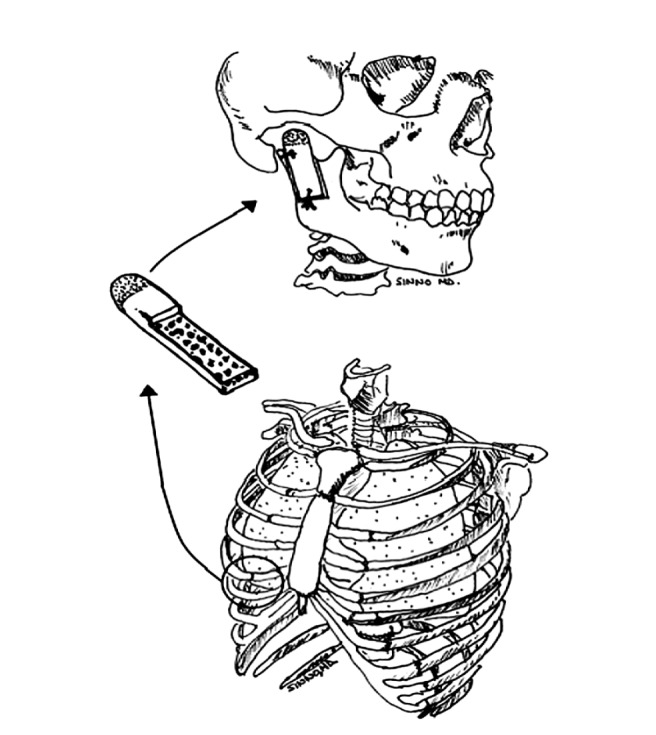
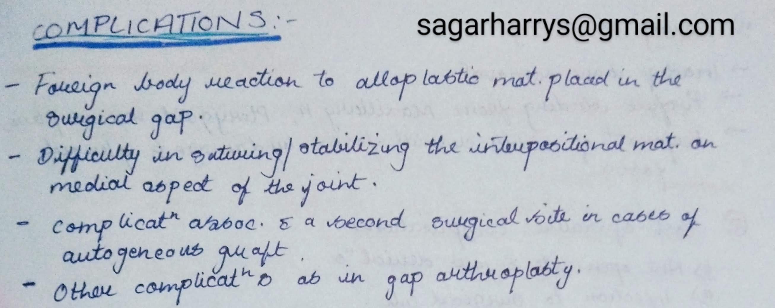
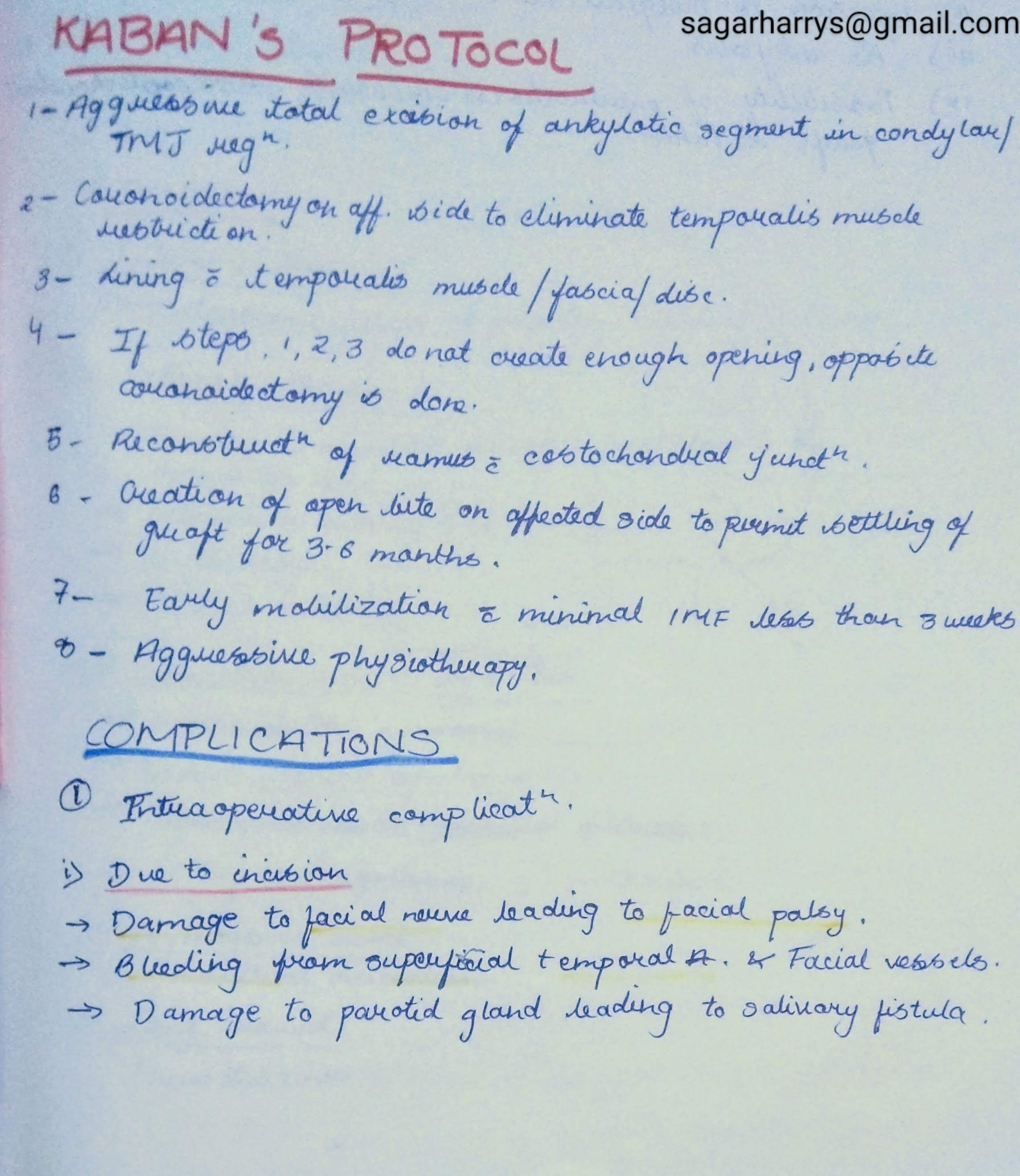
KABAN’S PROTOCOL:


REFERENCES:

CLASSIFICATION:
1. Based on type of tissue causing Ankylosis:


2. Based on the Side Involved :


3. Based on Severity of Ankylosis:
4. Based on Etiology of Trismus:
SAWHNEY’S CLASSIFICATION:

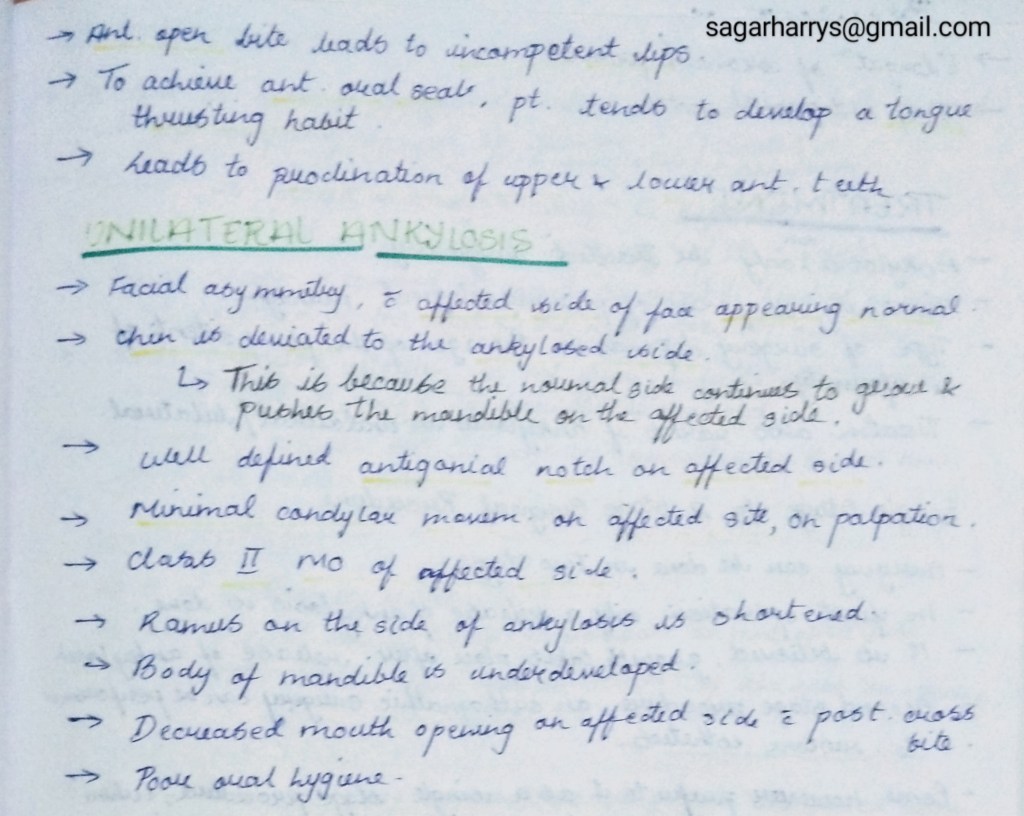
CLINICAL FEATURES:

INVESTIGATIONS:
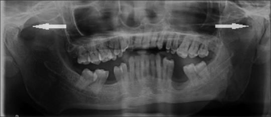
1. Radiographs:
– OPG
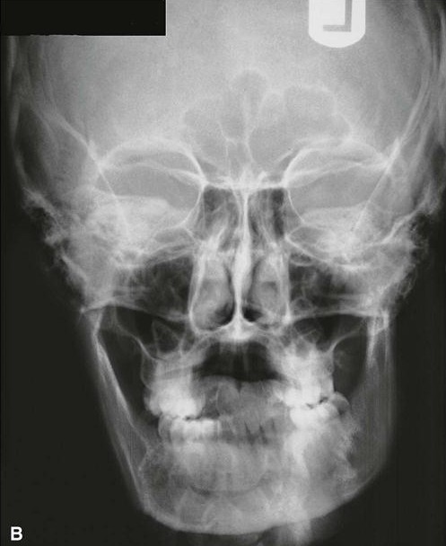
-PA VIEW:
– Lateral Oblique View of Mandible:
Ankylotic mass can seen in the Anteroposterior direction.
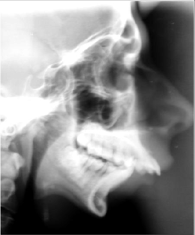
2. Lateral Cephalogram:
3. CT Scan:
Helpful as it gives an accurate picture of proximity of ankylotic mass to important structure,that cannot be seen in a radiograph.

RADIOGRAPHIC FEATURES:
REFERENCES:
Written by : Dr. Urusa I Inamdar
Diagnostic radiograph of tooth used to estimate the working length by measuring the tooth from a stable occlusal reference point till radiographic apex
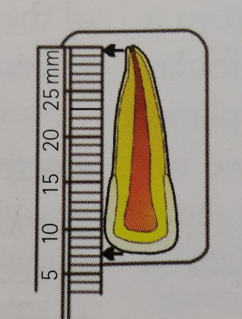
Subtract atleast 1 mm from this length as minor constriction is present short of the anatomic apex and compensation for radiographic image distortion.
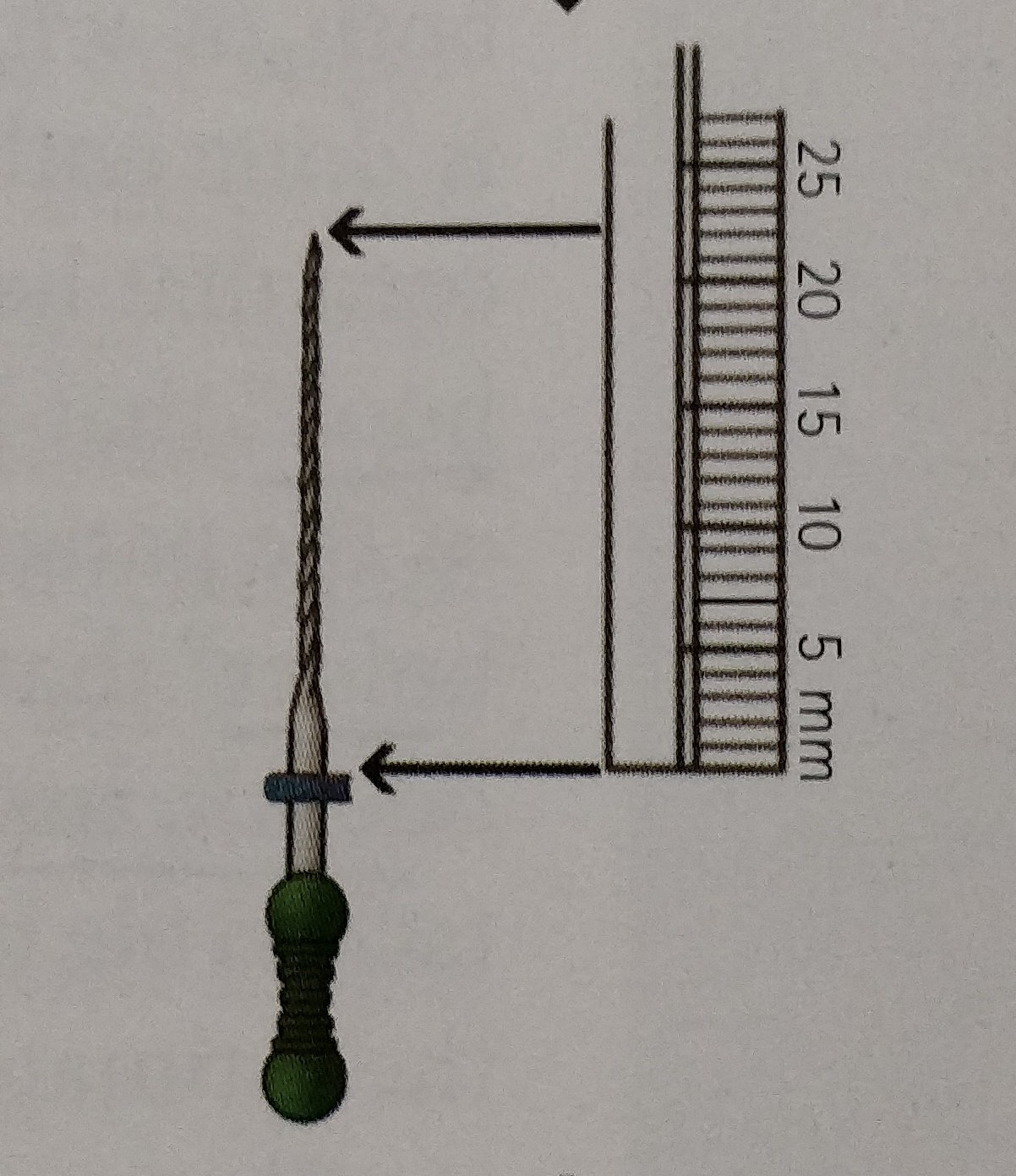
This measurement is transferred to a diagnostic instrument with a silicon stop then placed in the root canal and working length radiograph is taken.

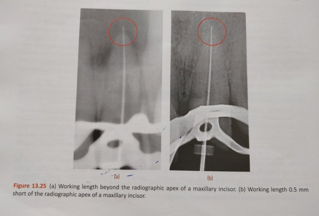
Now measure the difference between the end of the instrument and radiographic apex of the root , on the radiograph.
Tip of the instrument ends 0.5 mm – 1 mm from the radiographic root apex – working length established .
Retake the working length radiograph .

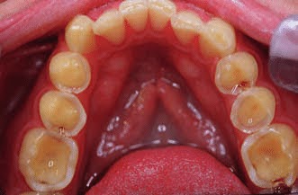


2. ABRASION:


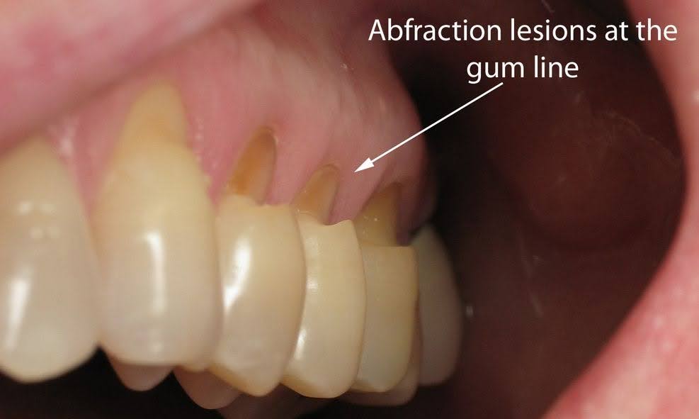
3. ABFRACTION:

MANAGEMENT:

REFERENCES:
Written by : Dr. Urusa I Inamdar
Krasner and Rankow’s Laws of Access Opening


• The Adams clasp was first described by Professor Phillip Adams.
• It is also known as Liverpool clasp, universal clasp and modified arrowhead clasp.
• When properly constructed this clasp offers maximum retention.
• The clasp is constructed using 0.7mm hard round stainless steel wire.
The Adams clasp is made of the following parts:
a) Two arrowheads
b) Bridge
c) Two retentive arms.
The two arrowheads engage the mesial and the distal proximal undercuts.
The arrow head are connected to each other by a bridge that is at 45° to the long axis of the tooth.
ADVANTAGES:
• It is rigid and offers excellent retention.
• It can be fabricated on deciduous as well as permanent teeth.
• TheyThey can be used on partially or fully erupted teeth.
• It can be used on molars, premolars and on incision.
• No specialized instrument is needed to fabricate the clasp.
• Young’s universal pliers that are used routinely for most wire bending can be used.
MODIFICATIONS:
• The Adams clasp can be modified in a number of ways.
• These modification is permit additional uses or enhanced retention.
• The following are some of the modifications of Adams clasp:
Adams with single arrowhead:

• The Adams clasp cam be modified to have a single arrowhead.
• This type of clasp is indicated in a partially erupted tooth, which usually is the last erupted molar.
• The single arrowhead is made to engage the mesio – proximal undercut of the list erupted molar.
• The bridge is modified to encircle the tooth distally and ends on the palatal aspect as a retentive arrowhead.
Adams with J Hook:

• A Hook can be soldered on to the bridge of the Adams clasp.
• These hooks are useful in engaging elastics.
• Adams with incorporated helix: A helix can be inc porated into the bridge of the Adams clasp This helps in engaging elastics.
Adams with additional arrowhead:
• Adams clasp can be constructed with an additional arrowhead.
• The additional arrowhead engages the proximal undercut of the adjacent tooth and is soldered on to the bridge of the Adams.
• This type of clasp offers additional retention.
Adams with additional arrowhead:

Adams With Incorporated Helix:

Adams with soldered buccal tube:
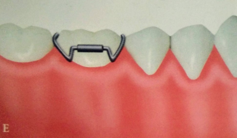
• A buccal tube can be soldered on to the bridge of the Adams clasp.
• This modification permits use of extra-oral anchorage using face bow- headgear assembly .
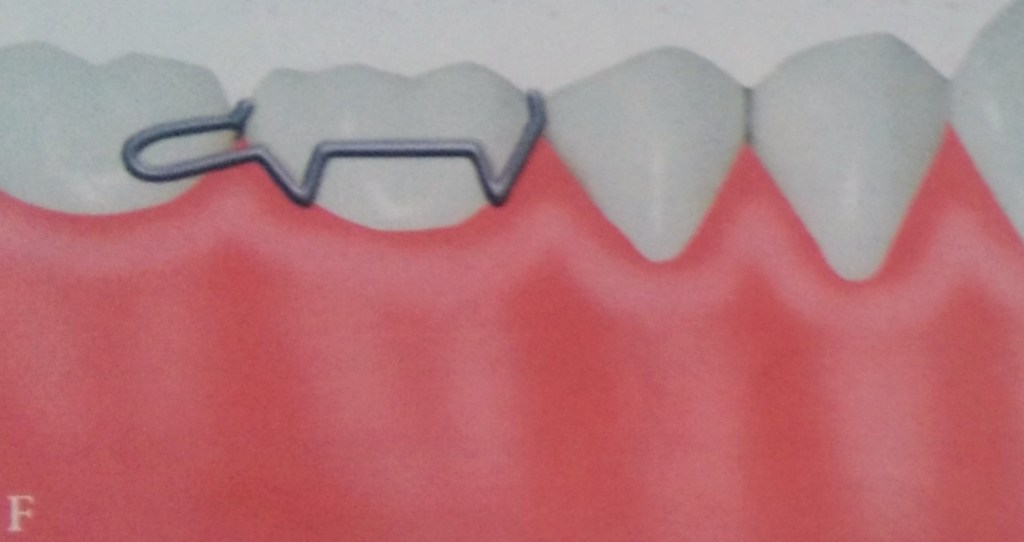
Adams with distal extension:
• The Adams clasp can be modified so that the distal arrowhead has a small extension incorporated distally.
• This distal extension helps in engaging elastics.
Adams With Distal Extension:
Adams on incisors and premolars:

• Adams clasp can be fabricated on the incisors and premolars when retention in those areas is required.
• They can be constructed to span a single tooth or two teeth.
DISADVANTAGES:
• Needs special arrowhead forming pliers the o fabricate.
• Occupies a large amount of space on buccal side.
• Arrowheads can injure interdental soft tissues.
• Difficult & time consuming to fabricate.
REFERENCES:
• Orthodontics: The Art & Science,SI Balajhi (7th Edition).
• SlideShare
Reference:Dental Anatomy B.D.Chaurasia.
Image:google

