ANTIBIOTIC GROUPS ( Part 2 )

Written by : Dr. Urusa I Inamdar

  1. Macrolides : inhibits bacterial protein synthesis by reversible binding to the 50s ribosomal subunit . Example : azithromycin, clarithromycin, erythromycin
  2. Monobactams : inhibit synthesis of peptidoglycan causing osmotic lysis , resistant to beta lactamases and active against gram negative rods. Example: aztreonam
  3. Nitrofurantoin : block aerobic energy production and synthesis of proteins , DNA ,RNA and cell walls . Example : nitrofurantoin
  4. Oxacephalosporins : bind to penicillin – binding proteins (PBP) of bacteria , inhibit bacterial cell wall peptidoglycan synthesis and activate bacterial cell wall autolytic enzymes. Example : flomoxef , latamoxef
  5. Oxazolidinones : cause faulty bacterial protein synthesis by binding to the 50s ribosomal subunit . Example : linezolid , tedizolid
  6. Penicillins : inhibit synthesis of peptidoglycan causing osmotic lysis . Example : Amoxicillin, ampicillin,dicloxacillin
  7. Penicillins with beta lactamase inhibitors : bind to penicillin binding proteins (PBP) of bacteria , inhibit bacterial cell wall peptidoglycan synthesis and activate bacterial cell wall autolytic enzymes. Example : Amoxicillin + clavulanic acid
  8. Quinolones : inhibit topoisomerases that are essential for bacterial DNA replication and transcription, inhibit DNA gyrase . Example : Ciprofloxacin, ofloxacin
  9. Sulfonamides : competitive inhibition of folic acid synthesis by acting as structural analogue of para-aminobenzoic acid (PABA) . Example : sulfabenzamide , sulfathiazole
  10. Tetracyclines : bind reversibly to receptors on the 30s subunit of the bacterial ribosome inhibiting protein synthesis. Example : minocycline , doxycycline
  11. Tyrocidins : alter cytoplasmic membrane causing cellular leakage . Example : bacitracin , tyrothricin

Reference:

  • Essentials of medical pharmacology – K D Tripathi
  • CIMS – prescribers handbook
  • Dental notes
ANTIBIOTIC GROUPS ( Part 1 )

Written by : Dr. Urusa I Inamdar

  1. Aminoglycosides : irreversible inhibition of protein synthesis by binding to receptors on the 30s subunit of bacterial ribosome. Example : amikacin , gentamicin , kanamycin
  2. Carbacephems : inhibit synthesis of peptidoglycan causing osmotic lysis. Example : loracarbef
  3. Cephalosporin : bind to penicillin – binding proteins (PBP) of bacteria , inhibit bacterial cell wall peptidoglycan synthesis and activate bacterial cell wall autolytic enzymes. Example : cefaclor , cefadroxil , cefalexin , cefazolin , cefepine
  4. Cephalosporins with Beta lactamase inhibitors : bind to penicillin – binding proteins (PBP) of bacteria , inhibit bacterial cell wall synthesis. Example : cefoperazone + sulbactam , cefuroxime + clavulanic acid
  5. Chloramphenicol : bind reversibly to a receptor site on the 50s subunit of bacterial ribosome . Example : chloramphenicol
  6. Cyclic Lipopeptides : bind to bacterial membranes and cause a rapid depolarisation of membrane potential . Hence , inhibit the protein , DNA and RNA synthesis which result in bacterial cell death . Example : daptomycin
  7. Diaminopyridines : inhibits dihydrofolic acid reductase of bacteria and blocks metabolic sequences in DNA synthesis . Example : trimethoprim
  8. Glycopeptides : prevent further elongation and cross linking of bacterial peptidoglycan synthesis , active against gram positive bacteria including methicillin-resistant Staphylococci. Example : oritavancin
  9. Glycylcyclines : bind reversibly to receptors on the 30s subunit of the bacterial ribosome inhibiting protein synthesis . Example : tigecycline
  10. Ketolides : inhibits bacterial protein synthesis by reversible binding to the 50s ribosomal subunit. Example : telithromycin
  11. Lincosamides : inhibit protein synthesis by interfering w/ initiation complexes and translocation reactions on the bacterial 50s subunit . Example : clindamycin , lincomycin
  12. Macrocyclic antibiotics : inhibit bacterial protein synthesis by inhibiting RNA polymerase sigma subunit. Example : fidaxomicin

References

  • Essentials of medical pharmacology – K D Tripathi
  • CIMS – prescribers handbook
  • Dental notes

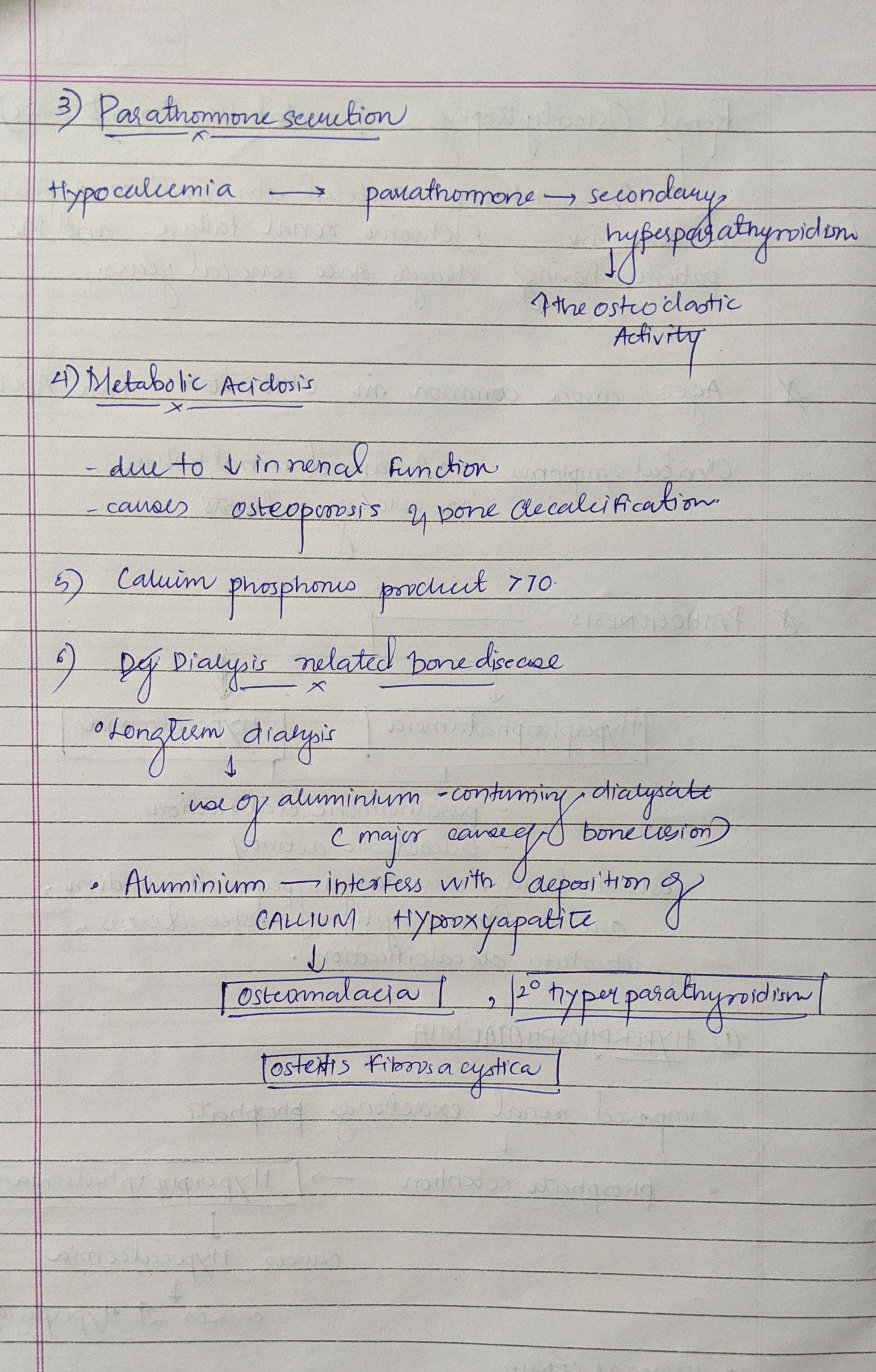
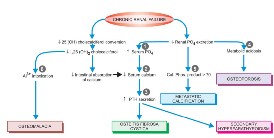
Renal osteodystrophy ( metabolic bone disease)

So what is renal osteodystrophy?

Renal Osteodystrophy: Definition, Treatment, Causes, Symptoms ...
Renal Osteodystrophy

MORPHOLOGIC FEATURES.

The following skeletal lesions can be identified in renal osteodystrophy:

  1. Mixed osteomalacia-osteitis fibrosa is the most common manifestation of renal osteodystrophy resulting from
    disordered vitamin D metabolism and secondary hyperparathyroidism.
  2. Pure osteitis fibrosa results from metabolic complications of secondary hyperparathyroidism.
  3. Pure osteomalacia of renal osteodystrophy is
    attributed to aluminium toxicity.
  4. Renal rickets resembling the changes seen in children
    with nutritional rickets with widened osteoid seams
    may occur .
  5. Osteosclerosis is characterised by enhanced bone
    density in the upper and lower margins of vertebrae.
  6. Metastatic calcification is seen at extraosseous sites
    such as in medium-sized blood vessels, periarticular
    tissues, myocardium, eyes, lungs and gastric mucosa.
12DaysinMarch, Renal Osteodystrophy for the USMLE Step One - YouTube

source -textbook of pathology for dental students c p baveja and image source- Google

MANAGEMENT:OAF

Buccal Advancement Flap

PALATAL ADVANCEMENT FLAP

  • This technique is very similar to buccal advancement technique where the gingiva is close to the oroantral defect is mildly advanced to approximate it.
  • May be done for small sized defects.
  • It is not possible to get too much of advancement, as the palatal tissue is very rigid and thick.
  • In this,the palatal tissue surrounding the OAC, is reflected +may be advanced downwards and sutured to the buccal tissue to cover the detect.
  • Not a very successful procedure.

ROTATIONAL ADVANCEMENT FLAP or ASHLEY’S FLAP OPERATION:


• It is posteriorly based flap which gets its supply from the greater palatine artery.
• Local anesthesia is infiltrated on the palatal aspect. Infilteration is also given on the buccal side of the oroantral defect.
• Removal of fistula tract and clearing any signs pathology.
• The flap is outlined with surgical marking an excess of what appears to be required to rotate the flap.
• Incision is placed to the bone making sure the palatal pedicle is kept intact.
• A full thickness mucoperiosteal flap is reflected with care taken to visualize the greater palatine vessels.
• A small amount of tissue along the margin of the defect on the buccal aspect is also reflected to enable suturing.
• The flap is reflected and rotated to fit the defect. There are chances of ending of the tissue when it is turned to cover the oroantral opening.
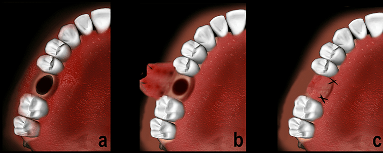
• The flap is sutured to the buccal flap ensuring a good water tight seal.
• The raw surface of the palate is left to granulate only.
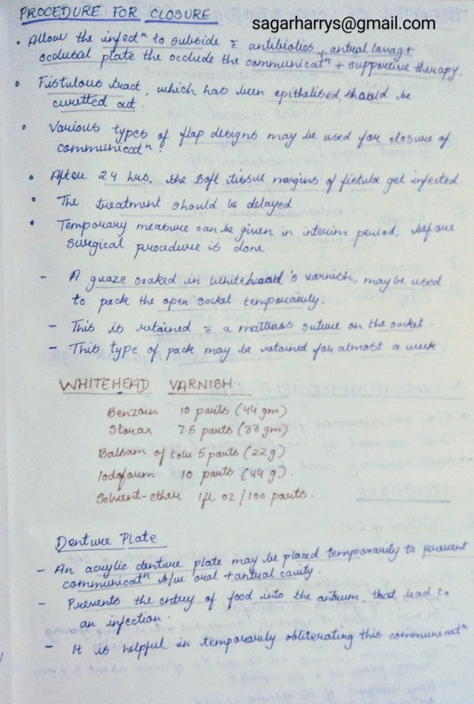
• A small gauze soaked in Whitehead’s varnish may be placed on that raw surface for a few days.

REFERENCES:

  • Textbook of Oral & Maxillofacial Surgery, Chitra Chakravarthy (2nd Edition)
  • Slideshare
  • researchgate.net

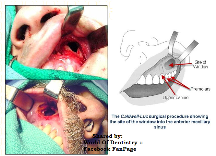
Caldwell-Luc Procedure

George Caldwell (New York)(1893)

Described a method of gaining space on maxillary sinus via canine fossa with nasal antrostomy.

Henric Luc (Paris)(1897)

Reported the same procedure as his own.

Thus a procedure by which the antrum is entered intraorally through the anterior wall & all irreversible disease is removed.

It is followed by antrostomy to promote permanent cure.

INDICATIONS:

  • Chronic Maxillary sinusitis.
  • Removal of foreign bodies in the antrum,such as root apices.
  • Treatment of benign dental cyst & tumours.
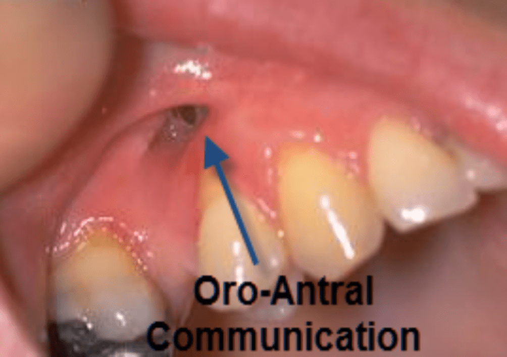
  • Treatment of Oro-antral fistula,that fails to heal.
  • Biopsy procedure for a suspected malignancy in the antrum.
  • Recurrent antrochoanal polyp.
  • Approach to Pterygopalatine fossa, Sphenoid sinus, Ethmoidal sinus.

CONTRAINDICATIONS:

  • Age: Not performed in patients below 17yrs as developing tooth buds in that region, may be damaged.
  • Acute infection.
  • Other systemic causes contraindicating surgery.

PROCEDURE:

1. Incision:

A semilunar incision is placed on the muvobuccal fold.

2. Exposure:

  • A Full thickness mucoperiosteal flap is reflect extending upto the intraorbital nerve.
  • Care is to be taken to protect the intraorbital nerve.

3. Approach to Antrum:

  • A micromotor with a large round bur is used to create a window about 1.5 to 2 cm in the anterior wall of antrum.
  • Sinus mucosa is seen below the bone.

4. Antral Lesion:

  • The lesion may be excised.
  • A biopsy may be done/sinus mucosa may be removed with the help of a currette if the case demands for it.

5. Antrostomy:

  • An opening is made in the medial wall in the lowermost and anterior aspect of the inferior meatus.

6. Packing:

  • The sinus cavity may be packed in with ribbon guaze impregnated with vaseline .
  • Guaze is packed in layers & the free end is brought out through created antrostomy opening.

7. Sutures:

  • Bone margins are smoothened.
  • The flap is replaced.
  • Flao may be sutured using resorbable suture material.

POST OPERATIVE CARE:

  • Ice packs are placed over the cheek for the first few hours after the surgery.
  • Sinus pack to be removed after 24-48 hours.
  • Avoid blowing nose for the at least 2-4 weeks after surgery.

COMPLICATIONS:

1. Intra-Operative:

  • Bleeding
  • Damage to Infraorbital Nerve.
  • Damage to Orbital content.

2. Post-Operative:

  • Reactionary Hemorrhage
  • Infection
  • Recurrence of lesion.
  • Antrostomy opening may get occluded.

REFERENCES:

  • Textbook of Oral & Maxillofacial Surgery, Chitra Chakravarthy (2nd Edition)
  • WorldofDentistry.com

DERMATOPHYTES

(1) Dermatophytes are a group of fungi that infect only superficial keratinised tissue (skin, hair and nails) without involving the living tissue.

(ii) They break down and utilise keratin.

(iii) They are incapable of penetrating subcutaneous tissue.

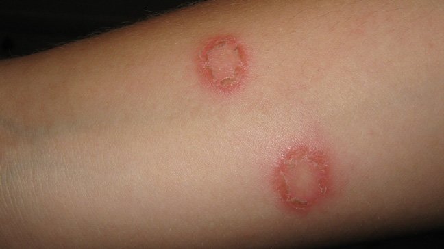
(iv) They cause dermatophytosis, also known as tinea or ringworm

Ring worm

Classification-

Dermatophytes are classified into three genera as follows:

Genus. Infection of

  1. Trichophyton- hair,skin,nail
  2. Microsporum- hair ,skin
  3. Epidermophyton- skin, nail

Clinical types

Clinically, ringworm can be classified depending on the site involved. These include

1.Tinea capitis (scalp)

2.Tinea corporis (non-hairy skin of the body)

3. Tinea cruris (groin)

4.Tinea pedis (foot) or athlete’s foot

5. Tinea barbae or barber’s itch (bearded areas of the face and neck).

🔶Favus- is a chronic type of ringworm involving the hair follicles. It leads to alopecia and scarring.

• In favus, there is sparse hyphal growth and formation of air spaces within the hair shaft.

Two types of hair infection may be present, ectothrix and endothrix.

🛑Ectothrix–

In ectothrix, a sheath of arthrospores is present on the surface of hair shaft.

🛑Endothrix

In endothrix the arthrospore formation occurs entirely within the hair shaft.

Source – slide share

Lab diagnosis-

🔻Specimens

• Skin scrapings

• Hair clippings

• Nail

🔻Direct microscopy

• Direct 10% KOH mount may show fungal hyphae.

🔻Culture

• SDA and SDA with antibiotics are used.

• Culture media are incubated at 25-30°C for three weeks.

• Identification of dermatophytes is based on

🔶colony morphology

🔶pigment production

🔶microconidia and macroconidia

🔻Colony characters

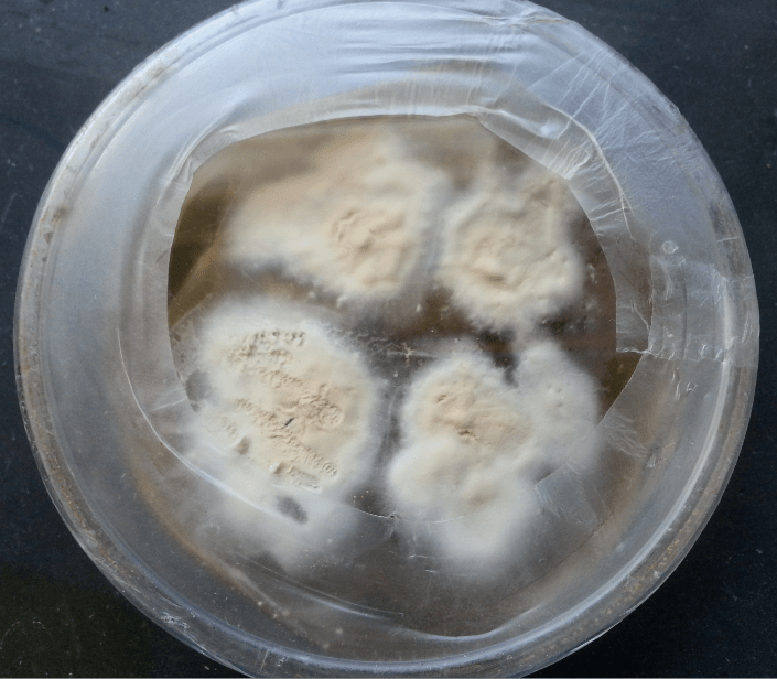
🔶Reverse of media is red in T. rubrum.

🔶White to creamy, cottony growth

Dermatophytes On SDA media

🔻Microscopy

🔶 Lactophenol cotton blue preparation from colony reveals microconidia, macroconidia or both. The following are the characteristics of three genera:

🔶Genus Trichophyton –More microconidia, very few macroconidia

🔶Genus Microsporum- Predominant macroconidia

🔶Genus Epidermophyton-macroconidia

🔻Treatment of dermatophytoses

🔶Topical antifungal agents are generally used for treatment.

🔶Oral griseofulvin is the drug of choice.

Source – textbook of microbiology for dental students c p baveja