It is also called as ‘stress lesion’. It is the loss of tooth structure that results from flexure which is caused by occlusal stresses. The magnitude of tooth tissue loss depends on the size, duration, direction, frequency and location of the forces.
Causes and mechanism
• Occlusal restoration—some suggested that occlusal restoration may lead to weakening of tooth ability to resist the stresses of occlusion leading to abfraction.
• Predisposing factors—factors, such as erosion and abrasion may play a significant role in tooth tissue loss.
Clinical features
Location—itusuallyaffectsbuccal/labialcervicalareas of teeth. Commonly affects single teeth with excursive interferences or eccentric occlusal loads.
Appearance—itappearsasdeep,narrowV-shapednotch. The lesion is typically wedge shaped with sharp line angles, but occlusal abfraction may present as circular invaginations.
REFERENCE- ANIL GHOM TEXTBOOK OF ORAL MEDICINE [2nd ed]
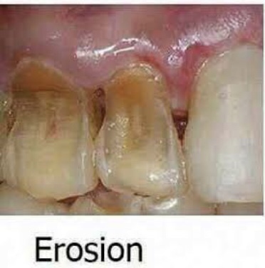
It is the loss of tooth substance by chemical process that does not involve known bacterial action. Dissolution of mineralized tooth structure occurs due to contact with acids. Erosion is a chemical process in which the tooth surface is removed in the absence of plaque.
• Local acidosis—it is seen in periodontal tissue from damage due to traumatogenic occlusion.
• Chronic vomiting—complete loss of enamel on lingual surfaces of teeth through dissolution by gastric hydrochloric acid. Vomiting can also occur in alcoholics, peptic ulcer, gastritis, pregnancy and drug side effect.
• Acidic foods and beverages—Large quantities of highly acidic carbonated beverages or lemon juice can produce erosion. Most of the fruits and fruits juices have a low pH and can cause erosion. Frequent consumption of carbonated drinks, which are acidic in nature, may result in the erosion of teeth.
• Anorexia nervosa—it induces chronic vomiting often after bouts of uncontrolled eating that is interspersed between periods of starvation, because of inner rejection of food.
• Occupational—workersinvolvinginmanufacturingof lead batteries, sanitary cleaners or soft drinks can develop erosion.
• Poorly monitored pH swimming pool—in cases of poorly monitored pH swimming can also cause erosion of the teeth.
• Medication—medication like chewable vitamin C and aspirin tablet may lead to erosion of teeth.
Clinical features
• Sites—It occurs most frequently on labial and buccal surfaces of teeth; some times, may occur on proximal surfaces of teeth. Usually confined to gingival thirds of labial surface of anterior teeth. Erosion may involve several teeth of dentition. From extrinsic source, it causes erosion on labial and buccal surface and from intrinsic source, it causes erosion on lingual or palatal source.
Appearance—it is usually a smooth lesion which exhibits no chalkiness.
Symptoms—loss of enamel often causes hypersensitivity in teeth and may also trigger secondary dentin formation.
Signs—lossoftoothsubstanceismanifestedbyshallow, broad, smooth, highly polished and scooped out depression on enamel surface adjacent to cementoenamel junction. When erosion affects the palatal surfaces of upper maxillary teeth, there is often a central area of exposed dentine surrounded by a border of unaffected enamel. In most cases, it results in little more than a loss of normal enamel contour, but in severe cases, dentin or pulp may be damaged.
Pink spot—there may be pink spot on tooth which is attributable to the reduced thickness of enamel and dentin making the pink hue of pulp visible.
Cupping—erosive lesions cause ‘cupping’ in dentin.
Radiographic features
• It appears as radiolucent defect in the crown margins may be well defined or diffuse.
Management
Dietcontrol—inapatientwherelossoftoothsurfaceis essentially caused by erosive fluids, advise regarding diet and use of sugar free chewing gum.
Fluoride mouthwash—prescription of a fluoride mouthwash is certainly indicated here.
Brushinghabits—brushinghabitsshouldbemodified.
Restoration—restoration of the defect, usually by glassinomer cement.
Systemic management—for systemic management ofvomiting, patient should be referred to the physician.
REFERENCE- ANIL GHOM TEXTBOOK OF ORAL MEDICINE [2nd ed]
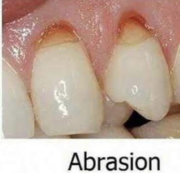
Abrasion is the pathological wearing away of tooth substance through some abnormal mechanical process. Abrasion usually occurs on the facial surface of the crown and the exposed root surfaces of teeth, but under certain circumstances it may be seen elsewhere such as on incisal or on proximal surfaces.
Etiology
Abrasivedentifrices—useofabrasivedentifricescanleadto abrasion of the incisal surface.
Habitual—Habitualpipesmokermaydevelopabrasion on the incisal edges of lower and upper anterior teeth. In some cases habitual opening of bobby pins may lead to abrasion.
Horizontal tooth brushing—horizontal tooth brushing may lead to abrasion of the cervical area of teeth.
Occupational—itoccurswhenobjectsandinstrumentare habitually held between the teeth by people during working. Holding nails or pins between teeth e.g. in carpenters, shoemakers or tailors.
Dentalflossortoothpicksinjury—improperuseofdental floss and tooth picks.
Ritual abrasion—it is mainly seen in Africa.Clinical featuresTooth brush injury • Sites—itusuallyoccursonexposedsurfacesofrootsofteeth. It is more commonly seen on left side of right handed persons and vice versa.
• Mechanism—it occurs due to back and forth movement of brush with heavy pressure causing bristles to assume wedge shaped arrangement between crown and root.
• Appearance—in horizontal brushing there is usually a ‘V’ shaped or ‘wedge’ shaped ditch on the root at cementoenamel junction . It is limited coronally by enamel.
• Symptoms—patient develops sensitivity as dentin becomes exposed.
• Signs—the angle formed in the depth of the lesion as well as that of enamel edge is a sharp one. Cervical lesions caused purely by abrasion have sharply defined margins and a smooth, hard surface. The lesion may become more rounded and shallow, if there is an element of erosion present.
• Dentinal features—exposed dentin appears highly polished Exposure of dentinal tubules and consequent irritation of the odontoblastic processes stimulates secondary dentin formation which is sufficient to protect the pulp from clinical exposure.
Dental floss or tooth pick injury • Site—Cervical portion of proximal surfaces ,just above the gingival margin, is affected. Grooves on distal surface are deeper than on mesial surface
Radiographic features
Tooth brush injury
Location—radiolucent defect at the cervical level of teeth.
Shape—well defined semilunar shape ,with borders of increasing density.
Pulp—pulp chamber may be partially or fully sclerosed in severely affected teeth.Dental floss injury • Appearance—narrow semilunar grooves in theinterproximal surfaces of teeth near cervical area.
Management
• Modified teeth cleaning habits—modification of teeth cleaning habits will be indicated.
• Removal of cause—elimination of causative agent should be carried out.
• Restoration—restoration should be done for esthetics purpose and to prevent further tooth wear.
REFERENCE- ANIL GHOM TEXTBOOK OF ORAL MEDICINE [2nd ed]
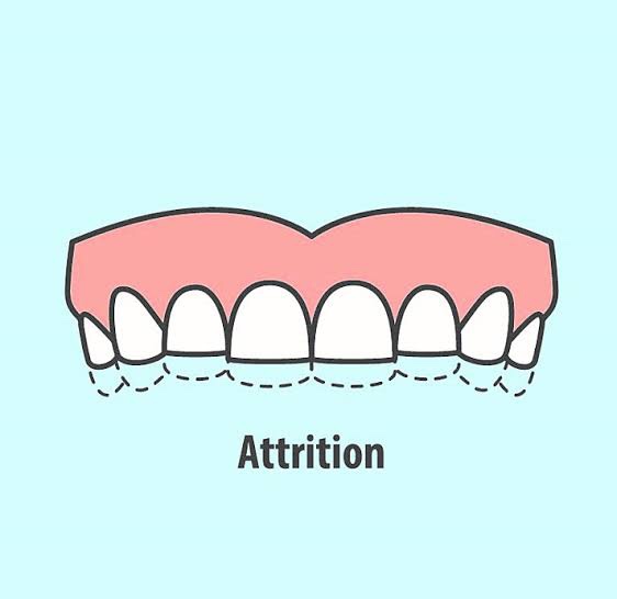
It is the physiologic wearing away of teeth because of tooth- to-tooth contact, as in mastication. It plays an important physiological role as it helps to maintain an advantageous crown-root ratio and gains intercoronal space of 1 cm, which facilitates third molar eruption. Attrition can be considered pathological when it cause functional, esthetics and dental sensitivity problems.
Types
• Physiological attrition—attrition which occurs due to normal aging process, due to mastication.
• Pathological attrition—it occurs due to certain abnor- malities in occlusion, chewing pattern or due to some structural defects in teeth.
Etiological factors for pathological attrition
• Abnormal occlusion • Developmental—malocclusion and crowning of teeth, may lead to traumatic contact during chewing, which may lead to more tooth wear. • Acquired—due to extraction of teeth. Extraction causes increased occlusal load on the remaining teeth, as the chewing force for the individual remains constant.
• Premature contact in case of edge-to-edge contact,pathological attrition can also occur. • Abnormal chewing habits parafunctional chewing habit like bruxism and chronic persistent chewing of coarse and abrasive food or other substances like tobacco. • Occupation in certain occupations, workers are exposed to an atmosphere of abrasive dust and cannot avoid it getting into mouth. • Structural defect in defects like amelogenesis imperfecta and dentinogenesis imperfecta, hardness of enamel and dentin is reduced and such teeth become more prone to attrition.
Clinical features
• Sex—men usually exhibit more severe attrition than women due to greater masticatory forces.
• Sites—it may be seen in deciduous as well as permanent dentition. It occurs only on occlusal, incisal and proximal surfaces of teeth. Severe attrition is seldomly seen in primary teeth, as they are not retained for any great period. Palabal cusps of maxillary teeth and buccal cusps of mandibular posterior teeth show most wear.
• Appearance – the first clinical manifestation of attrition is the appearance of small polished facet on a cusp tip or ridge and slight flattening of an incisal edge.
Physiologic attrition
Physiological tooth surface loss results in a reduction, in both vertical tooth height and horizontal tooth width .Physiological attrition showing wearing of the occlusal surface of the molar teeth.
Contact points—due to slight mobility of teeth in their socket (which is a manifestation of resiliency of periodontal ligament) similar facets occur at contact points.
Color of teeth when the dentin gets exposed, it generally becomes discolored i.e. brown in color.
Signs—there is gradual reduction in cusp height and consequent flattening of occlusal inclined plane. There is shortening of the length of dental arch, due to reduction in the mesiodistal diameter of teeth. Secondary dentin deposition occurs.
• Pathologicalattrition
Severe tooth loss—in pathological attrition severe tooth loss is seen .
Dentoalveolar compensation—if attrition affecting the occlusal surfaces of teeth has occurred, then reduction in occlusal face height (vertical dimension of occlusion) and increase in the freeway space could be anticipated. This may be further complicated by forward posturing of mandible. It is often observed, however, that despite overall tooth surface loss, the freeway space and the resting facial height appear to remain unaltered primarily because of dentoalveolar compensation. This is important with respect to patient assessment. If restoration of worn teeth is being planned then the extent of dentoalveolar compen- sation would appear to determine the dentist’s strategy; defining the need to carry out measures such as crown lengthening, to ensure the same vertical dimension of occlusion and freeway space.
Radiographic features
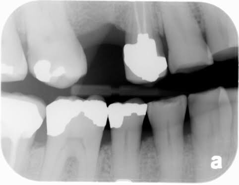
• Crown—smoothwearingofincisalandocclusalsurfaces of involved teeth is evident by shortened crown image
• Pulp—sclerosisofpulpchamberandcanalsisseendue to deposition of secondary dentin which narrows the pulp canals.
• Periodontal ligament—widening of periodontal ligament space and hypercementosis.
• Alveolar bone—some loss of alveolar bone.
Management
Modifying factors—treatment of patient depends upon degree of wear relative to the age of patient, etiology, symptoms and patient’s desire.
Habit breaking appliance—the provision of one of three different sorts of splints could be considered. A soft bite guard can help in breaking a bruxist habit or simply will protect the teeth during the bruxist habit. A localized occlusal interference splint is designed to break the bruxist habit and can be worn easily during the day. A stabilization splint reduces bruxism by providing an ideal occlusion: it also enables the clinician to locate and record centric relation. In case of bruxism, use of night guards may be effective in reducing attrition.
Correctivemethod—correctionofmalocclusion,stoppage of tobacco chewing habit and restriction of diet to non coarse food are useful in avoiding attrition.
Managementofsensitivityandesthetics—non-cariousloss of tooth tissue may require treatment for sensitivity, esthetics, function and space loss in the vertical dimension.
REFERENCE- ANIL GHOM TEXTBOOK OF ORAL MEDICINE [2nd ed]
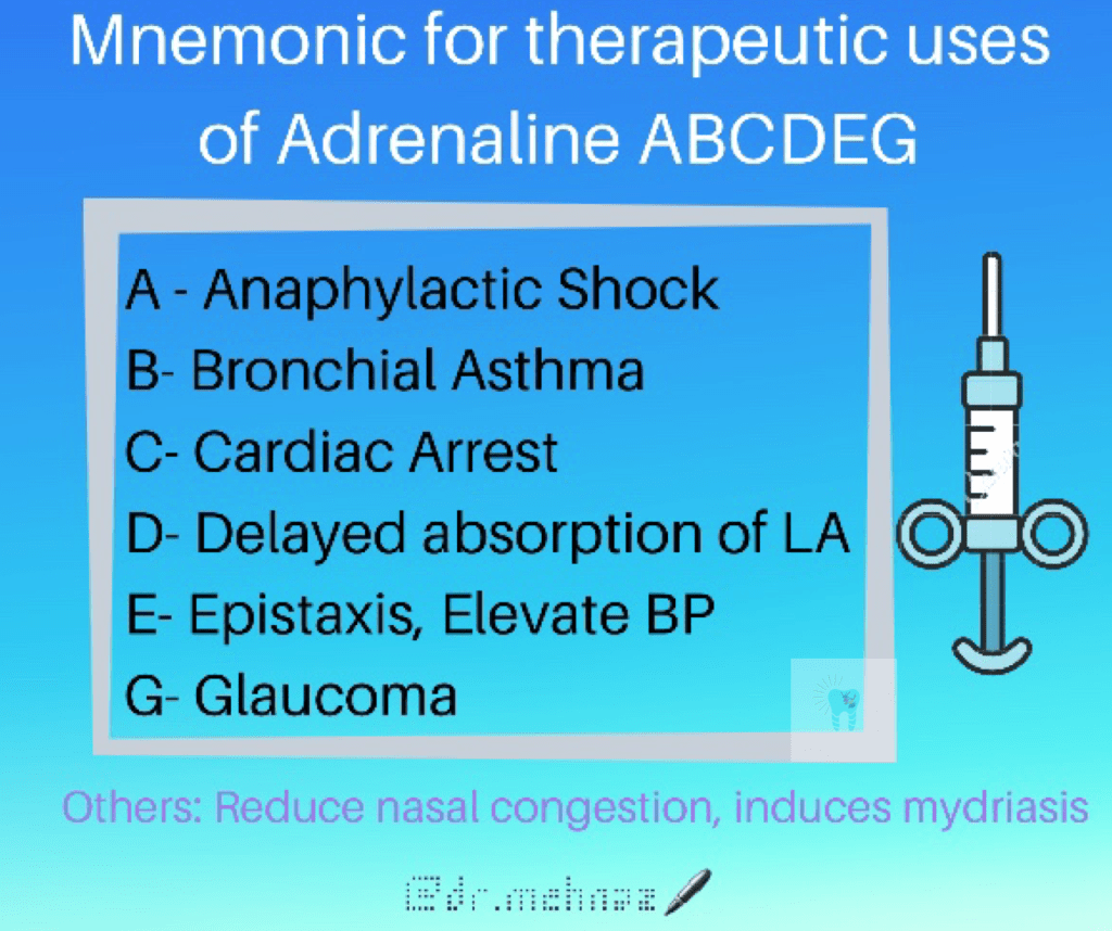
➡️ In case of anaphylactic shock or angioedema of Larynx or for bronchospasm attending drug hypersensitivity (Adrenaline + sub-class of gluco-corticoids) is recommended.
Put the patient in reclining position, administer oxygen at high flow rate
Inject adrenaline 0.5 mg (0.5 ml of 1 in 1000 solution for adult, 0.3 ml for child (6-12 years) & 0.15 ml for child (upto 6 years) i.m
Repeat every 5-10 min. in case patient does not improve.