INTRINSIC AND EXTRINSIC STAINS

Intrinsic Stains

Pre-eruptive Causes

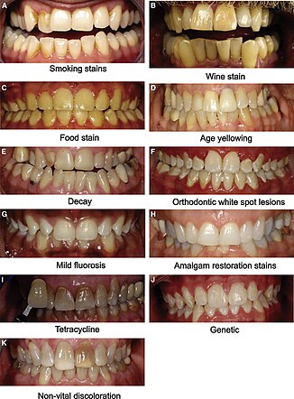
These are incorporated into the deeper layers of enamel and dentin during odontogenesis and alter the development and appearance of the enamel and dentin

.Alkaptonuria: Dark brown pigmentation of primary teeth is commonly seen in alkaptonuria. It is an autosomal recessive disorder resulting into complete oxidation of tyrosine and phenylalanine causing increased level of homogentisic acid.

Hematological disorders

• Erythroblastosis fetalis: It is a blood disorder of neonates due to Rh incompatibility. In this, stain does not involve teeth or portions of teeth developing after cessation of hemolysis shortly after birth. Stain is usually green, brown or bluish in color.
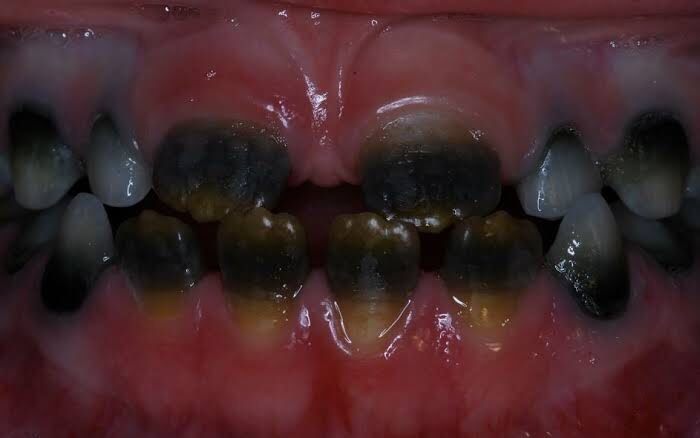
• Congenital porphyria: It is an inborn error of por- phyrin metabolism, characterized by overproduction of uroporphyrin. Deciduous and permanent teeth may show a red or brownish discoloration. Under ultraviolet light, teeth show red fluorescence.
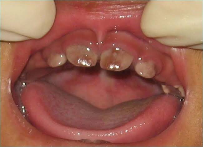
• Sickle cell anemia: It is inherited blood dyscrasia characterized by increased hemolysis of red blood cells. In sickle cell anemia infrequently the stains of the teeth are similar to those of erythroblastosis fetalis, but discoloration is more severe, involves both dentitions and does not resolve with time.

Amelogenesis imperfecta: It comprises of a group of conditions, that demonstrate developmental alteration in the structure of the enamel in the absence of a systemic disorders. Amelogenesis imperfecta (AI) has been classified mainly into hypoplastic, hypocalcified and hypomaturation type.

Fluorosis: In fluorosis, staining is due to excessive fluoride uptake during development of enamel. Excess fluoride induces a metabolic change in ameloblast and the resultant enamel has a defective matrix and an irregular, hypomineralized structure 

  • Vitamin D deficiency results in characteristic white patch hypoplasia in teeth.
  • Vitamin C deficiency together with vitamin A deficiency during formative periods of dentition resulting in pitting type appearance of teeth.
  • Childhood illnesses during odontogenesis, such as exanthematous fevers, malnutrition, metabolic disorder, etc. also affect teeth.
  1. Dentinogenesis imperfecta : It is an autosomal dominant development disturbance of the dentin which occurs along or in conjunction with amelogenesis imperfecta. Color of teeth in dentinogenesis imperfecta (DI) varies from gray to brownish violet to yellowish brown with a characteristic usual translucent or opalescent hue.
  2. Tetracycline and minocycline: Unsightly dis- coloration of both dentitions results from excessive intake of tetracycline and minocycline during the development of teeth. Chelation of tetracycline molecule with calcium in hydroxyapatite crystals forms tetracycline orthophosphate which is responsible for discolored teeth.

Posteruptive Causes

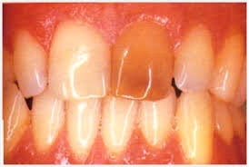
  • Pulpal changes: Pulp necrosis usually results from bacterial, mechanical or chemical irritation to pulp. In this disintegration products enter dentinal tubules and cause discoloration.
  • Trauma: Accidental injury to tooth can cause pulpal and enamel degenerative changes that may alter color of teeth.Pulpal hemorrhage leads to grayish discoloration and nonvital appearance. Injury causes hemorrhage which results in lysis of RBCs and liberation of iron sulfide which enter dentinal tubules and discolor surrounding tooth.
  • Dentin hypercalcification: Dentin hypercalcification results when there are excessive irregular elements in the pulp chamber and canal walls. It causes decrease in translucency and yellowish or yellow brown discoloration of the teeth.
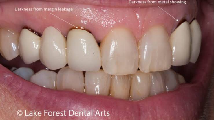
  • Dental caries: In general, teeth present a discolored appearance around areas of bacterial stagnation and leaking restorations.
  • Restorative materials and dental procedures: Discoloration can also result from the use of endodontic sealers and restorative materials.
  • Aging: Color changes in teeth with age result from surface and subsurface changes. Age related discoloration are because of:– Enamel changes: Both thinning and texture changes occur in enamel.
– Dentin deposition: Secondary and tertiary dentin deposits, pulp stones cause changes in the color of teeth.

• Functional and parafunctional changes: Tooth wear may give a darker appearance to the teeth because of loss of tooth surface and exposure of dentin which is yellower and is susceptible to color changes by absorption of oral fluids and deposition of reparative dentin.

Extrinsic Stains

Daily Acquired Stains

Plaque: Pellicle and plaque on tooth surface gives rise to yellowish appearance of teeth.

Food and beverages: Tea, coffee, red wine, curry and colas if taken in excess cause discoloration.

Tobacco use results in brown to black appearance of teeth.

Poor oral hygiene manifests as:

  • –  Green stain
  • –  Brown stain
  • –  Orange stain.

Swimmer’s calculus:
– It is yellow to dark brown stain present on facial andlingual surfaces of anterior teeth. It occurs due toprolonged exposure to pool water.

Gingival hemorrhage.

Chemicals

• Chlorhexidine stain: The stains produced by use of chlorhexidine are yellowish brown to brownish in nature.

Metallic stains: These are caused by metals and metallic salts introduced into oral cavity in metal containing dust inhaled by industry workers or through orally administered drugs.

Stains caused by different metals

• Copper dust—green stain
• Iron dust—brown stain
• Mercury—greenish black stain • Nickel—green stain
• Silver—black stain.

Reference- Nisha garg textbook of endosontics and Anil Ghom textbook of oral medicine

CLASSIFICATION OF TOOTH DISCOLORATION

Classification of discoloration

  • Intrinsic discoloration
  • Extrinsic discoloration
  • Combination of both

1.Based on the Etiology of tooth discoloration

Intrinsic stains

  • • Pre-eruptive causes– Disease
    i. Alkaptonuria
  • iiHematological disorders
    iii. Disease of enamel and dentin
  • iv. Liver diseases.– Medications
    i. Tetracycline stains and other antibiotic use
  • ii. Fluorosis stain.
  • • Posteruptive causes of discoloration– Pulpal changes
    – Trauma
    – Dentin hypercalcification
    – Dental caries
    – Restorative materials and operative procedures – Aging
    – Functional and parafunctional changes.

Extrinsic stains
• Daily acquired stains

– Plaque
– Food and beverages – Tobacco use
– Poor oral hygiene
– Swimmer’s calculus
– Gingival hemorrhage.  

  • Chemicals
    – Chlorhexidine
  • – Metallic stains.

2.Classification of extrinsic stains (Nathoo in 1997)

  • •  N1 type dental stain (direct dental stain): Here colored materials bind to the tooth surface to cause discoloration. Tooth has same color, as that of chromogen.
  • •  N 2 type dental stain (direct dental stain): Here chromogen changes color after binding to the tooth.
  • •   N3 type dental stain (indirect dental stain): In this type prechromogen (colorless) binds to the tooth and undergoes a chemical reaction to cause a stain.

3.Different Types of Stains according to Color:

BLACK COLOR-

It usually results due to contact with certain metallic elements such as silver, iron and lead.It may appear as thin line running approximately 1 mm or so above the gingival margin, it may occur on both the facial and lingual surfaces of teeth.Stain is firmly attached to the surface but remains extrinsic, thus it may be removed by brush and abrasives. But, it recurs later on.

Green stain

It usually occurs as thick deposit involving the cervical one-third of facial surface of maxillary incisors of young children. It affects boys more frequently than girls.

It is associated with poor oral hygiene and decalcification is sometimes present in enamel, underlying the deposit. It occurs due to chromogenic bacteria or fungi or it may be caused by bacterial action on remnants of enamel cuticle. It is extrinsic and may be removed by simple brushing and abrasive.

Orange stain

It occurs infrequently and usually involve both facial and lingual surfaces of the incisors. It is easily removed than green stain and its cause is unknown but it is believed to be the result of chromogenic bacteria. It is associated with poor oral hygiene and removed with the help of brush and abrasives.

Brown stain

It can be seen in non-smokers and is usually lighter brown than that of tobacco and form a tenacious, but delicate film on surface of the teeth. It is usually seen more commonly on lingual surface of lower incisors and buccal surface of maxillary molar teeth. It is formed due to altered salivary mucins which have undergone change through the action of bacterial enzymes.

REFERENCE- NISHA GARG TEXTBOOK OF ENDODONTICS AND ANIL GHOM TEXTBOOK OF ORAL MEDICINE

NATAL AND NEONATAL TEETH

NATAL AND NEONATAL TEETH

It is also called as ‘congenital teeth’, ‘fetal deciduous teeth’, ‘dentition proceox’ and ‘natal and neonatal teeth’. There is premature eruption of teeth or teeth like structures that are present at birth.

Natal teeth are the teeth which are present at the time of birth and neonatal teeth are the teeth which are present within 30 days after the birth.

Etiology

Hereditary—superficial position of tooth germ.

Hormonal influence—eruption accelerated by febrileincident or hormonal stimulation.

Classification

  • Mature—they are fully developed in shape and comparable in morphology to the primary teeth. Prognosis is relatively good.
  • Immature—their structure and development is incomplete. Poor prognosis of teeth.

Clinical features

  • Appearance—teeth may be conical or may be normal in size and shape and opaque yellow-brownish in color.
  • Signs—they are hypermobile because of their limited root development. Within relatively short time, premature erupted tooth will become stabilized and other teeth of the arch are erupted. Teeth appear to be attached to a small mass of soft tissue.
  • Significance—some teeth are so much mobile that there is danger of displacement and possible aspiration and in this case, removal is indicated.
  • Riga fede ulcer—there is ulceration of the ventral surface of the tongue caused by the sharp incisal edges. It leads to interference with proper suckling and feeding and thus the neonate is at risk of nutritional deficiency.
  • Associated syndromes—it may associate with syndromes like Ellis-van Creveld syndrome and cleft palate.

Management

  • Extraction—extraction of the teeth should be carried out if it is causing inconvenience during suckling, interference with breastfeeding and causing traumatic injury.
  • NOTE- Extraction should be done after 10th postpartum day due to the inability of the clotting but nowadays, it is no longer considered because of prophylactic administration of vitamin K as a standard procedure in most of the hospitals.
  • Rounding of sharp angle—the other option that may be used is rounding of the sharp angle of incisal edges of teeth.
  • Retaining the tooth—if not necessary, tooth should not be removed.The preferable approach, however, is to leave the tooth in place and to explain to the parents the desirability of maintaining this tooth in the mouth because of its importance in the growth and uncomplicated eruption of the adjacent teeth.
  • A retained natal or neonatal tooth may cause difficulty for a mother who wishes to breast-feed her infant. If breast-feeding is too painful for the mother initially, the use of a breast pump and bottling of the milk are recommended. However, the infant may be conditioned not to “bite” during suckling in a relatively short time if the mother persists with breast-feeding. It seems that the infant senses the mother’s discomfort and learns to avoid causing it.

REFERENCE- MC DONALD TEXTBOOK OF PEDODONTICS AND ANIL GHOM TEXTBOOK OF ORAL MEDICINE

RADIOLOGICAL APPEARANCES & FEATURES(PART II)

Many lesions that occur on the jaw present with similar radiographic appearances making it difficult to differentiate among them . Despite development of various cross sectional imaging modalities ,the radiographs are still remain the first and most important investigations.

So some of the common features that we as dentist might come across are listed below.I believe this would come handy for your radiodiagnosis😊.

  • Ghost teeth appearance-regional odontodysplasia
  • Ground glass appearance-monostotic fibrous dysplasia, hyperparathyroidism, middle stage of paget’s disease & ossifying fibroma,sickle cell anemia
  • Garrington sign-osteosarcoma (localised symmetrical widening of PDL
  • Hanging drop appearance-orbital blow out fracture
  • Heart shaped radiolucency-nasopalatine /incisive canal cyst
  • Honey comb appearance-anerysmal bone cyst,odontogenic myeloma,central giant cell granuloma,central hemangioma
  • Lincoln sign-paget’s disease (increased uptake in the mandible mimicking Lincoln’s beard.
  • Mass of color/ cauliflower like appearance-calcified lymph nodes
  • Moth eaten appearance-chronic osteomyelitis, early stage of ostesarcoma,radiolucent stage of fibrous dysplasia,squamous cell carcinoma,leukemia,malignant lymphoma
  • Mottled appearance-ossifying fibroma,fibrous dysplasia
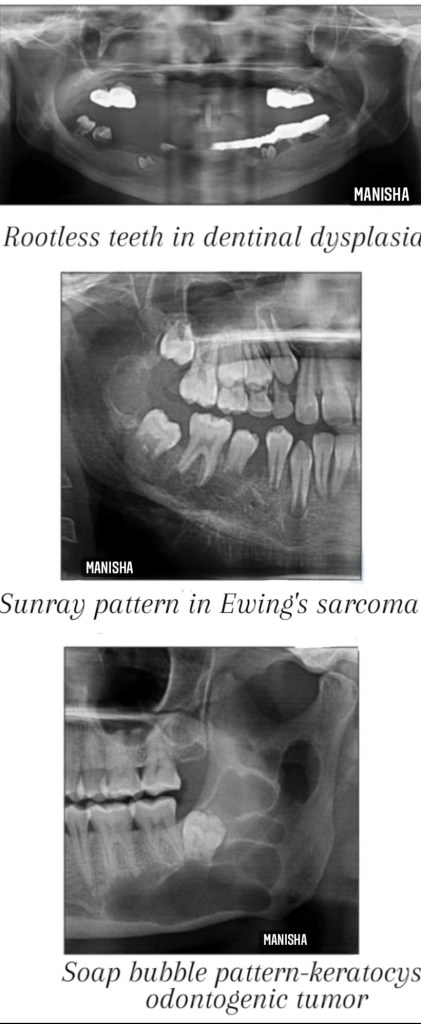
  • Onion peel /skin appearance-Garre’s osteomyelitis, Ewing’s sarcoma,eosinophilic granuloma,caffey’s disease.
  • Orange peel appearance-mixed stage of fibrous dysplasia (peud-de-orange)
  • Pear shaped appearance-globulomaxillary cyst
  • Pepper pot appearance-hyperparathyroidism
  • Peripheral cuffing of bone-peripheral giant cell granuloma
  • Pressure type appearance-squamous cell carcinoma of gingiva
  • Punched out appearance-multiple myeloma
  • Rootless teeth appearance-dentinal dysplasia
  • Salt and pepper appearance-hyperparathyroidism, thalassaemia
  • Sausage like appearance-sialographic appearance of sialodochitis
  • Sialectasis-sialographic appearance of sialadenitis
  • Sharpened pencil appearance-osteoarthritis /rheumatoid arthritis of TMJ
  • Shell teeth appearance-type III dentinogenesis imperfecta
  • Soap bubble appearance-ameloblastoma,aneurysmal bone cyst,central hemangioma,central giant cell granuloma, odontogrnic keratocyst
  • Step ladder appearance-sickle cell anemia
  • Sunburst/sunray appearance-central hemangioma,osteosarcoma,Ewing’s sarcoma
  • Target sign-(rounded radiopacity with a central radiolucency)-impacted tooth in bucco lingual direction(lower 3rd molar)
  • Tennis racket appearance-odontogenic myxoma
  • Thistle tube appearance-type II dentinal dysplasia
  • Thumb print /fingerprint appearance-mixed stage of fibrous dysplasia
  • Tram track sign/calcifications-Sturge Weber syndrome
  • Trap door appearance-orbital blow out fracture
  • Tree in winter appearance-normal sialographic appearance of parotid gland

Some of the radiographic features are pathogonomic to a specific disease, thus can be helpful in narrowing down of differential diagnosis.

🖋Manisha M.A

Sources:White and Pharoah’s -Oral radiology textbook,Shafer’s textbook of oral pathology (8th edition) , Websites-www.ijournalhs.org, Article-Phore S,Panchal RS,Bhagla P, Nabi N.Dental radiographic signs.Indian j health sci 2015;8:85-90.

RADIOLOGICAL APPEARANCES & FEATURES(PART I)

The real importance of learning radiographic signs associated with specific disease is of relevance to clinical examination of the head & neck ; at the same time aiding in differentiating – what is normal from abnormal & hence appropriate treatment can be instituted for such conditions /abnormalities.

Here are the list of few radiographic appearances .

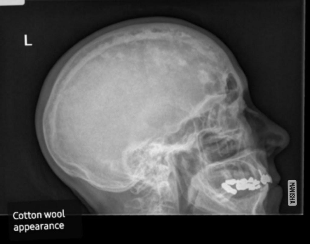
Cotton wool appearance seen on the lateral skull in Paget’s disease
  • Antral halo appearance-acute sinusitis
  • Ball in hand appearance-sialographic appearance of intrinsic benign tumor
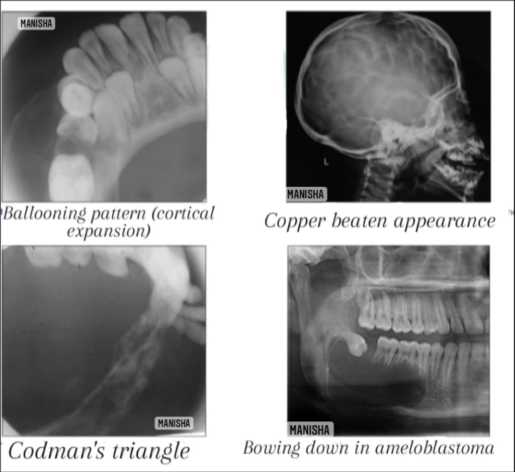
  • Balloon like appearance-follicular cyst
  • Beaten silver/Copper beaten appearance-Crouzen syndrome,hypophosphatasia,craniofacial dysostosis ,obstructive hydrocephalus
  • Bush in winter appearance-normal sialographic appearance of submandibular gland
  • Candlestick appearance-Progressive systemic sclerosis,pycnodysostosis
  • Cherry blossom/branchless fruit laden tree/snowstorm/punctate sialectasis-sialographic appearance of Sjogren’s syndrome
  • Codman’s triangle-osteogenic sarcoma,Ewing sarcoma,carcinoma of alveolar ridge
  • Cotton wool appearance-paget’s disease,fibrous dysplasia(thick,amorphous radiopaque stage),cemmento-osseous dysplasia,chronic diffuse sclerosis osteomyelitis
  • Crew cut/Hair-on-end appearance-sickle cell anemia,thalassemia
  • Driven snow appearance-calcifying epithelial odontogenic tumor /cyst(Pindborg tumor)
  • Downward bowing appearance-cemento-osseous fibroma,ameloblastoma
  • Ely’s cyst-osteoarthritis
  • Filling defect appearance-salivary gland tumor
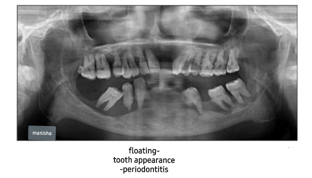
  • Floating tooth appearance-squamous cell carcinoma,malignant lymphoma,periodontitis ,eosinophilic granuloma,osteomyelitis

🖋Manisha M.A

Sources:White and Pharoah’s -Oral radiology textbook,Shafer’s textbook of oral pathology (8th edition).Websites-www.dreamstime.com, www.ijournalhs.org,Article-Phore S,Panchal RS,Bhagla P, Nabi N.Dental radiographic signs.Indian j health sci 2015;8:85-90.

SUPERNUMERARY TEETH

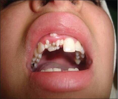
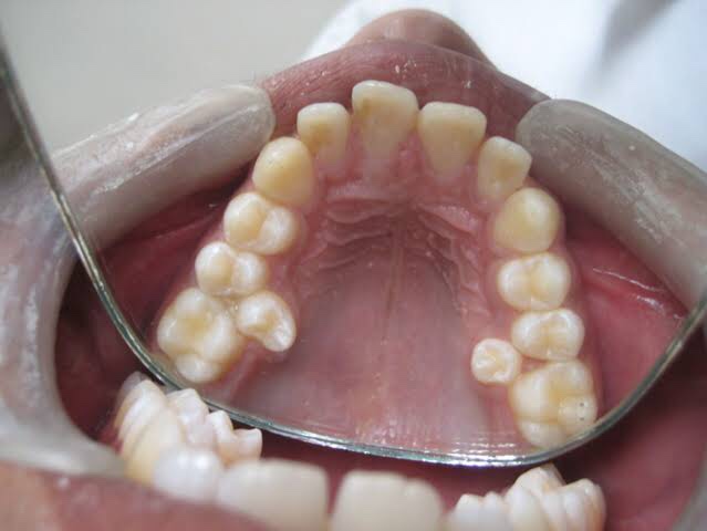
A supernumerary tooth is an additional tooth to the normal set of teeth. It may closely resemble the teeth of the group to which it belongs, i.e, molars, premolars or anterior teeth, or it may bear little resemblance in size or shape to the teeth with which it is associated.

It is also called as ‘hyperdontia’. It is defined as any tooth or tooth substance in the excess of the usual configuration of twenty deciduous or thirty two permanent teeth.

Pathogenesis and etiology

  • The various factors which are responsible for formation of supernumerary teeth as follows:
  • Phylogenetic reversion theory—this theory is nowadaysdiscarded.
  • Dichotomy theory—this theory states that tooth bud issplit to form two different tooth,
  • Hyperactivity of dental lamina—a supernumerary toothdevelops from 3rd tooth bud arising from dental lamina near the permanent tooth bud.
  • Hereditary—it is inherited as an autosomal dominant trait, when associated with syndromes. It is inherited as an autosomal recessive trait when associated with only supernumerary teeth. Supernumerary teeth are most commonly found in relative of affected children than in general population.

Four different morphological types of supernumerary teeth have been described:

• Conical
• Tuberculate
• Supplemental
• Odontome

Conical. This small peg-shaped conical supernumerary tooth is most commonly found in the permanent dentition. It develops with root formation ahead of or at an equivalent stage to that of permanent incisors and usually presents as a mesiodens. The conical supernumerary can result in rotation or displacement of the permanent incisor, but rarely delays eruption.

Tuberculate. The tuberculate type of supernumerary possesses have more than one cusp or tubercle. It is frequently described as barrel-shaped and may be invaginated. Root formation is delayed compared to that of the permanent incisors. Tuberculate supernu- meraries are often paired and are commonly located on the palatal aspect of the central incisors. They rarely erupt and are frequently associated with delayed eruption of the incisors.

Supplemental. The supplemental supernumerary refers to a duplication of teeth in the normal series and is found at the end of a tooth series. The most common supplemental tooth is the permanent maxillary lateral incisor, but supplemental premolars and molars also occur. The majority of supernumeraries found in the primary dentition are of the supplemental type and seldom remain impacted.

Odontome. The term ‘odontoma’ refers to any tumour of odontogenic origin. Odontoma represents a hamartomatous malformation rather than a neoplasm. The lesion is composed of more than one type of tissue and consequently has been called a composite odontoma. Two separate types have been described, the diffuse mass of dental tissue which is totally disorganized is known as a complex composite odontoma; whereas, the malformation which bears some superficial anatomical similarity to a normal tooth is referred to as a compound composite odontoma.

According to location
• Mesiodens—it is located at or near the midline in the incisal region of maxilla between central incisors . It is a small tooth with cone shaped crown and short root. It may cause retarded eruption, displacement or resorption of adjacent root. It frequently causes improper alignment.

• Distomolar—it is found in molar region frequently located distal to 3rd molar. Generally, these teeth are smaller than normal 2nd and 3rd molar, crown morphology is highly abnormal.

• Paramolar—it is supernumerary molar, usually small and rudimentary and is situated buccally or lingually to one of the maxillary molars or interproximally between 1st, 2nd and 3rd maxillary molars.

• Peridens—supernumerary teeth that erupt ectopically, either buccally or lingually to the normal arch are referred as peridens.

Radiographic features

When it is needed—if abnormal clinical signs are present you can go for OPG examination, IOPA, occlusal radiographic examination.

Significance—radiograph will aid in determining the location and number of unerupted teeth. It can also used to see if there is any cyst formation.

Appearance—theirradiographicpictureischaracteristic of teeth.

Management

• Surgical extraction—it depends on potential effect on normal dentition, their position, number and complications that may result from surgical removal. If required, they should be extracted.

REFERENCE- SHAFER’S TEXTBOOK OF ORAL PATHOLOGY 8TH ED AND ANIL GHOM TEXTBOOK OF ORAL MEDICINE TEXTBOOK OF ORAL MEDICINE