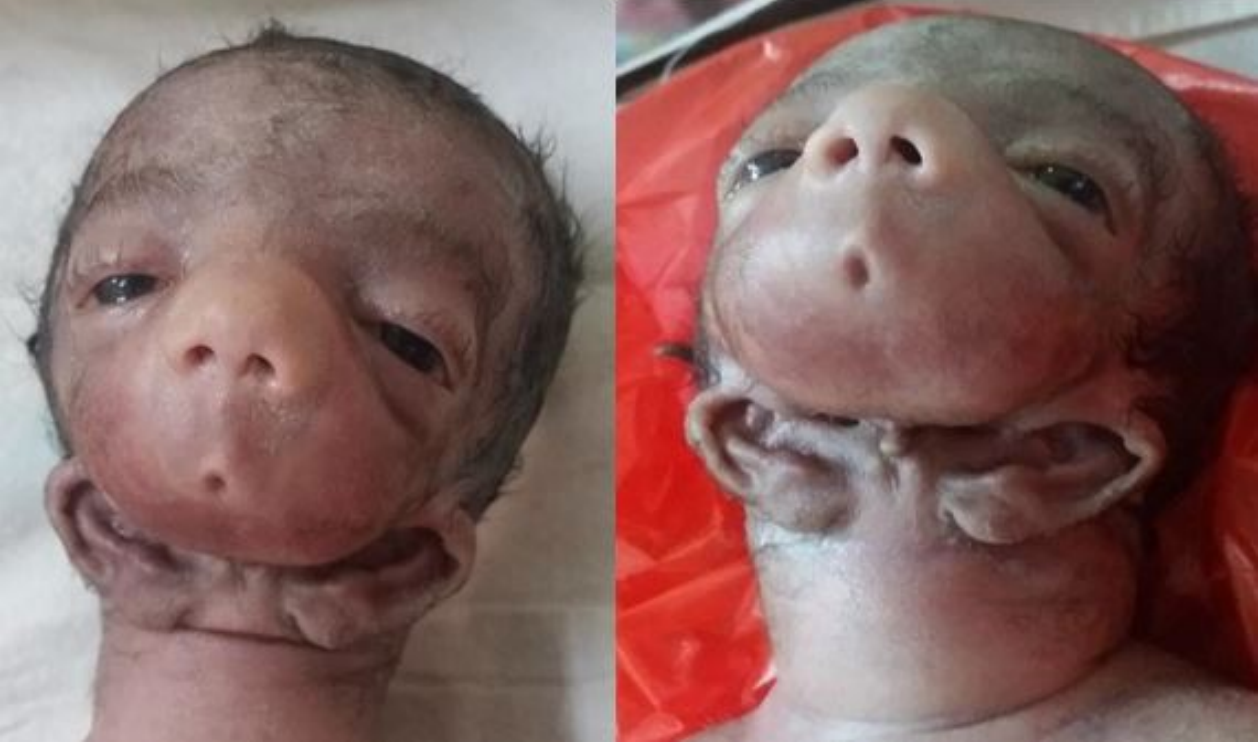
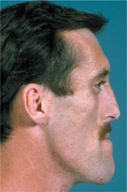
Macrognathia

General factors which would influence and tend to favour mandibular prognathism are as follows:

• Increased height of the ramus

• Increased mandibular body length

• Increased gonial angle

• Anterior positioning of the glenoid fossa

•Decreased maxillary length

• Posterior positioning of the maxilla in relation to the cranium

• Prominent chin button

•Varying soft-tissue contours

Source- textbook of oral pathology Shafers and Google images

FORENSIC ODONTOLOGY PART-2

LIP PRINTS

The wrinkles and grooves visible on the lips have been named by Tsuchihashi as ‘sulci labiorum rubrorum’. The imprint produced by these grooves is termed as ‘lip print’, the examination of which is referred to as ‘cheiloscopy’.

Tsuchihashi proposed a classification dividing the pattern of grooves into six types:
Type I: Clear-cut vertical grooves that run across the entire lip
Type I’: Similar to type I, but do not cover the entire lip Type II: Branched grooves
Type III: Intersected grooves
Type IV: Reticular grooves
Type V: Grooves that cannot be morphologically differentiated.


  • A combination of these grooves may be found in any given set of lips.
  • To simplify recording, the lips are divided into quadrants similar to the dentition- a horizontal line dividing the upper and lower lip and a vertical line and a vertical dividing right and left sides. By noting the type of groove in each quadrants, the individual’s lip print pattern may be recorded. Thus classification, thus, enabled differentiation of lip print patterns between any two individuals.
  • Lip prints are usually left at crime scenes, and can provide a direct link to the suspect. 
  • Although invisible, these prints can be lifted using materials such as aluminium powder and magnetic powder. 
  • Ball states that the vermillion border has minor salivary glands, and the edges of the lips has sebaceous glands, with sweat glands in between. 
  •  One may therefore, assume that secretions of oils and moisture from these enable development of ‘latent’ lip prints, analogous to latent fingerprints, in most crime scenes where close contact between the victims and culprit has occurred.

DISADVANTAGE

  • A major disadvantage of lip print investigation pertains to uncertainty about the permanence of lip patterns. While they are believed to remain unchanged throughout one’s life, 
  • Sivapathasundharam B and coworkers doubted this, as major trauma to the lips resulting in scarring, pathosis, surgical treatment  rendered to correct the pathosis, affect the size and shape of the lip, thereby may alter the pattern and morphology of the grooves.

REFERENCE-SHAFER’S TEXTBOOK OF ORAL PATHOLOGY 8TH EDITION

FORENSIC ODONTOLOGY PART 1


Forensic Odontology defined by the Federation Dentaire International (FDI) as ‘that branch of dentistry which, in the interest of justice, deals with the proper handling and examination of dental evidence, and with the proper evaluation and presentation of dental findings’.

Scope of Forensic Odontology

• Record preparation—the correct handling and exami- nation and the proper preparation and presentation of dental evidence in both civil and criminal legal procedures.

• Identification—personal identification, either individually or in context of mass disasters.

• Age assessment—to calculate the age of patient.
• Bite mark investigation—investigation of criminal cases where bite marks are involved and the interpretation of

• Humanabuse—recognitionofdomestic,andchildabuse.

• Lipprint—comparisonandidentificationoflipprint.

• Legalaspect—legalaspectofdentaltraumatology.

A vital role the forensic dentist plays is in criminal investigation.

  • Crime investigation includes the investigation of 
  • BITE MARKS
  • LIP PRINTS
  • CHILD ABUSE

BITE MARKS

McDonald (1974) has defined bitemark as “a mark caused by the teeth either alone or in combination with other mouth parts”.

Bite marks may be caused by humans or animals; they may be on tissue, food items, or other objects. 

CLASSIFICATION OF BITE MARKS

Cameron and Sims classification

  1. Depending on biting agent

a. Human : Children, Adults

b. Animals : Mammals, Reptiles, Fish

c. Mechanical : Full denture, belt marks etc

  1. Depending on material bitten 

a. Skin : Human, Animal

b.  Perishable items : Food items like cheese,  apple etc.

c. Non- perishable items : Unanimated objects such as pipes, pens, pencils.


WEBSTER’S CLASSIFICATION

Type I – food item fractures readily with limited depth of tooth penetration e.g. hard chocolate.

Type II – food item fractures with considerable penetration of teeth e.g. bite marks in apple & firm fruits.
Type III – Complete or near complete penetration of the food item with slide marks e.g cheese

FACTORS AFFECTING BITE MARK INJURIES

  • Inherent skin factor
  • Age
  • Sex
  • Time
  • Vascularity

CHARACTERISITCS OF HUMAN BITE MARK FOR IDENTIFICATION

  • Human bite mark characteristics include an elliptical or ovoid pattern containing tooth and arch marks.
  • Simplest form of a bite mark consists of tooth marks produced by antagonistic teeth.
  • An arch mark may indicate the presence of 4 to 5 teeth marks reflecting the shape of their incisal or occlusal surfaces.
  • Class features: differentiate between tooth type
  • Incisors- rectangular
  • Canines- triangular
  • Premolars + molars – spherical/point shaped
  • Depends on attrition
  • Other significant findings to identify a bite mark to give the identify of the suspect are :
  • Presence or absence of each tooth
  • Peculiar shape of each tooth
  • Mesiodistal dimensions
  • Arch form and size
  • Relationship between the upper and the lower jaws
  • Any unusual features, such as rotation, fractured teeth, supernumerary teeth, microdontia, diastema etc.  

BITE MARK INVESTIGATION

  • The guidelines for bite marks analysis are given by American Board of Forensic Odontology (ABFO) and careful use of these helps in enhancing the quality of the investigation and conclusions. The collection of evidence regarding the bite marks falls in following categories :

1. Description of the bite marks

  1. Demographic data
  2. Location of bite marks
  3. Shape Color
  4. Type of injury (Abrasion, Ecchymosis, Laceration, Petechial hemorrhage and Incision) 

2. Collection of evidence from victim

3. Collection of evidence from suspect

REFERENCE- SHAFER’S TEXTBOOK OF ORAL PATHOLOGY AND SLIDE SHARE

CHARACTERISTICS OF A DIGITAL IMAGE

Image Characteristics

  • Contrast Resolution
  • Spatial Resolution
  • Detector Latitude
  • Detector Sensitivity
  • Signal to noise ratio

CONTRAST RESOLUTION

  • Contrast resolution is the ability to distinguish different densities in the radiographic image. 
  • Current digital detectors capture data at 8-, 10-, 12-, or 16-bit depths. 
  • The bit depth is a power of 2. 

Spatial Resolution

  • Spatial resolution is the capacity for distinguishing fine detail. 
  • The theoretical limit of resolution is a function of picture element (pixel) size for digital imaging systems.

DSCN6702

Film based IOPA – 20 lp / mm.

Digital receptors 7 lp / mm.

Film  >  CCD  >  PSP 

Detector Latitude

It is the ability of the image receptor to capture a range of x-ray exposures as different densities.

  • The latitude ofCCD and CMOS detectors is similar to film

Photostimulable phosphor receptors have larger latitudes and have a linear response to five orders of magnitude of x-ray exposure.

Detector  Sensitivity

  • Sensitivity of a detector is its ability to respond to small amounts of radiation. Intraoral film sensitivity is classified according to speed.
  • High resolution CCD and CMOS systems achieve less dose reduction than lower resolution PSP systems. CCD and PSP systems or extraoral imaging require exposures similar to those needed for 200-speed screen-film systems.

REFERENCE-WHITE AND PHAROAH 5TH EDITION

Antimalarial drugs

What is malaria ?

•Malaria is a life-threatening disease.

• It’s typically transmitted through the bite of an infected Anopheles mosquito.
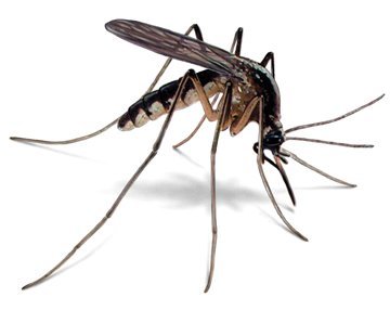
Female anopheles mosquito

•Infected mosquitoes carry the Plasmodium parasite.

•When this mosquito bites you, the parasite is released into your bloodstream.

•Once the parasites are inside your body, they travel to the liver, where they mature. After several days, the mature parasites enter the bloodstream and begin to infect red blood cells.

•Within 48 to 72 hours, the parasites inside the red blood cells multiply, causing the infected cells to burst open.

•The parasites continue to infect red blood cells, resulting in symptoms that occur in cycles that last two to three days at a time.

AREAS WHERE MALARIA IS FOUND –

Malaria is typically found in tropical and subtropical climates where the parasites can live.

Life cycle of malaria

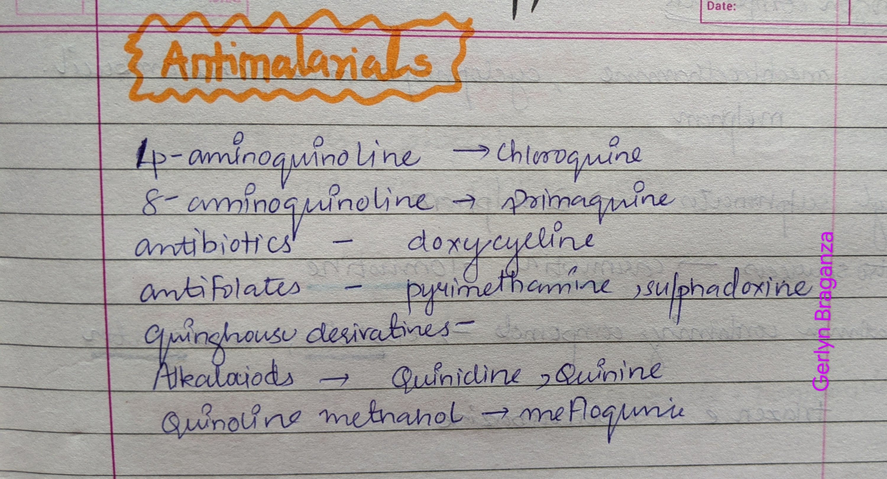
Drugs used in malaria

Source – 1.textbook of pharmacology for dental students tara shanbhag

2. Healthline

3 pinterest and Google images

Viral Childhood Exanthems

  • Viral rashes are often caused by immune reactions to the virus and cell damage caused by the virus.
  • A key bacterial cause of rash is Streptococcus pyogenes, which causes Scarlet Fever.

Helpful distinguishers between the viral exanthems:

  • We can categorize them by the initial location and pattern of the rash.
    – Three viral exanthems tend to initiate on the face:
    Measles, rubella, and erythema infectiosum.
    – Chickenpox arises on the face/scalp and trunk.
    – Roseola infantum typically first appears on the trunk
    – Hand, Foot, and Mouth disease produces rash on the hands and feet, in the mouth.

Be aware that these are meant to be helpful generalizations, and may not always hold true; for example, hand, foot, and mouth disease can also produce rash on the buttocks.

  • Rash types:
    – Multiple rash types can exist at once.
    – Macules are flat, colored spots on the skin.
    – Papules are solid, raised areas; larger papules are called nodules.
    – Vesicular rashes comprise raised “pockets” of fluid in the skin.
  • The timing of the rash and presence of other symptoms can also help distinguish among the exanthems.
    – For example, some infections are associated with fever, malaise, and respiratory symptoms.
    – Knowing the time lapse between virus introduction and symptom appearance can also help, although the incubation periods of the various viruses often overlap and may include a wide range.

VIRAL EXANTHEMA

Hand, foot, and mouth disease:

  • Usually caused by Coxsackievirus A.
  • Average incubation period of 3-6 days.
  • Most common in children younger than 5.
  • As its name suggests, hand, foot, and mouth disease is characterized by a rash that can be macular, maculopapular, or vesicular on the hands, feet, and in and around the mouth.

Erythema infectiousum, aka, Fifth Disease

  • Caused by Parvovirus B19.
  • Average incubation period of 7 days
  • Tends to affect children 5-15 years old.
  • Initial symptoms can include fever, runny nose, headache; diarrhea is also possible.
  • These flu-like symtpoms are followed by a malar facial rash that spreads to the trunk and extremities.
    – Facial rash takes on a characteristic “slapped cheeks” pattern, whereas the rash on the extremities often comprises maculopapular rash in a “lacy” pattern.

Roseola infantum, aka, exanthema subitum

  • Most commonly caused by Human Herpes Virus 6, and sometimes Human Herpes Virus 7.
  • Roseola infantum is also sometimes called “6th disease”, because it was the 6th exanthema identified (erythema infectiosum was the fifth).
  • Average incubation period is 9 days.
  • Although disease can occur in a wide range of ages, it most commonly affects children younger than 2 years old.
  • Initial symptoms include a very high fever (exceeding 104 degrees Fahrenheit/40 degrees Celsius).
    – The fever lasts approximately 3 days, which is why Roseola infantum is sometimes called “3-Day Fever”.
  • Macular or maculopapular “blanching rash” arises first on the trunk.
    – “Blanching rash” means that when the skin is pressed upon, often with a clear glass, the rash fades from red to pale.
  • Another common finding are red uvulopalatoglossal spots, aka, Nagayama spots,.
  • A range of other symptoms, including gastrointestinal, respiratory, ocular, and auditory problems, can occur.
  • Febrile seizures are a common complication in roseola infantum.

Chickenpox

  • Caused by Varicella-Zoster virus (aka, Human Herpes Virus-3)
  • Average incubation period is 16 days.
  • Often affects children younger than 5.
  • Prior to rash, patients may experience fever, malaise, sore throat, and low appetite.
  • Rash is characterized by crops of lesions that pass through macular, vesicular, and crusted phases.
    – Lesions usually first appear on the head/neck, and spread to the rest of the body.
  • Clinical correlation: Shingles is an illness that occurs in adults upon reactivation of the Varicella-Zoster Virus; the reactivated virus is called Herpes-Zoster Virus.
    – Whereas the chickenpox rash is often itchy, the shingles rash can be very painful. Vaccination against Varicella-Zoster virus also prevents shingles.

Measles (aka, rubeola)

  • Caused by the Measles virus.
  • Average incubation period is 14 days.
  • Prior to rash, patients often experience Fever and the “Three C’s”: Cough, Coryza (runny nose), and Conjunctivitis.
  • These symptoms are followed by a *maculopapular rash that begins on the face and neck and spreads.
  • Before the body rash, many patients also develop Koplik spots, which are spots along the palate and internal buccal surfaces (these spots are sometimes calked Koplik’s sign).
  • Serious complications from measles virus infection include potentially fatal pneumonia and encephalitis; vaccination helps to prevent these and other complications.

Rubella (aka, German measles)

  • Caused by Rubella virus.
  • Average incubation period is 14 days.
  • Rubella is characterized by the acute onset of a pink maculopapular rash that begins on the face and spreads.
    – The rash lasts about 3 days, so Rubella is sometimes called “3-Day measles” – careful not to confuse this with Roseola infantum, which is sometimes called “3-Day Fever.”
  • Some patients also have swollen lymph nodes in the neck area; systemic symptoms, such as headache, are mild if present.
  • Congenital rubella, which is contracted during fetal development, is associated with severe birth defects; this form of rubella can also be prevented by the rubella vaccine.