Powerful ways to remember what you study.

Muhad Noorman P – Final year -Team Dentowesome

Most often we get frustrated by studying for days before exams, often we fail to recollect or forget while writing exams. It’s a quite natural process for a human body to forget.
However there are tricks to master our hippocampus and remember for long. Excelling in exams are only possible based on how much you remember topics.

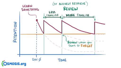
According to Ebbinghaus curve of forgetting information is lost from brain and we’re inable to recollect it.A typical graph of the forgetting curve purports to show that humans tend to halve their memory of newly learned knowledge in a matter of days or weeks unless they consciously review the learned material.

In oder to master long term memorization, we need to practice following methods

Revision : You still remember, A for apple and mitochondria is power house of cell. Constant and frequent revison makes your hippocampus to convert short term memory to long term memory.

Spaced repetition learning technique
Review Your Notes. Within 20-24 hours   of the initial intake of information, make sure the information is written down in notes and that you have reviewed them.
Recall the Information for the First Time. Recall the Materials Again.
Study It All Over Again
Difficult topics are checked regularly while easy topics could be reviewed occasionally

Take a break method
  Study for 20 minutes take a 5 minute break repeat pattern for 3 to 4 hours. It helps to gain more focus, At the end you’ll be happy for the productive hours. Without break in intervals your brains rejects input eventually your output becomes non productive. Mastering this techinque daily, your graph of productivity hits up.

Use body movements while learning,helps to Tigger muscle memory.


Make a story to memorize long topics. Pieces of information are always connected each other when a story link is given.

Organise your study table. Neat study table and fresh environment boost your intake . Bright light, fresh air, erect spine enhance brain functioning. Feel comfortable stay away from cluttered environment

Try to understand what you learn,  things you understand and studied are memorised 9 times.


Learn opposite things .

Switch your topics frequently. Similiar memory get’s intermixed (interference theory).

Things learned at the beginning and end are most memorized. Plan your topics accordingly.


Dicatate your topics and record in dictaphone you can download in your phone. Hear audios before you sleep, going to a beach or restaurant… Brain makes short term memory to long term memory while relaxed.

Visualise your topics. You still remembers the colour of precipitate and titration from your 12th chemistry lab practicals. Visualized memory is far beyond your imaginations.

Read first from books, 2 or 3 days later watch related topics videos from Youtube or any informative apps. Audio+ video learning brushes your previous stored information

Always make use of Sticky notes of alternating colours (prefer light colours- eye rejects dark colour for long time. Use sticky notes apps In your phone screen ( numericals, years etc.could be written in it).

  Last days before your exams should be used for rough reading or revison not for studying. Brain rejects things learned in stressed or a state of anxiety .( Your neurotransmitters makes it mess. Respect them 🤣)

Credits : 1) Forgetting curve definition:Wikipedia. Image : Internet. 2) Spaced repetition technique images from Internet and Osmosis.org website . Spaced repetition method content from Google.

How to study efficiently and score more marks in exams.

Muhad Noorman P, Final year Student – Team Dentowesome

Scoring good and better marks are always priority of any student irrespective of their level and class .  Hard work and smart work helps to score good marks.

Some tips to study smart and score more marks

1) Always organise yourself, Never procrastinate. Don’t think about wasted days ,look forward days ahead and make efficient planning.

2) Give importance to every subject equally, start with easy and end in hard nuts.


3) Identify your best time, and place to study. Ignore and never seek how you’re peer group works. Always your peer lies about studies, focus on yourself 🙂

4) Cut your social distraction, even though it sounds like a rocket science, regular practice helps to cut your Distraction. Utilise focus mode in android phones, Install Forest app. It Helps to prioritise your study hours.


5) Teach yourself as if you’re a teacher, trust me you’ll crack a million topics.


6) Teach you’re peer group , it’ll help to recollect and brush up your brain.


7) Regularly shift your studyplace , between a period of 1 hour or 2 hour later,  brain and mind always rejects learning from a same environment.


8) Get familiarise with the exam layout, use previous year question, understand nature of questions , prepare accordingly.


9) Always finish with previous questions first and if time allows study other topic left, mentioned in  University syllabus.


10) Reading a book not studying :  Revise topic after 1 or 2 hour, Prepare notes, put away books . Attempt topic as an exam question. Self realization is best methodology to improve yourself.


11) Never cut too many corners: Often we get devastated listening rumors , predictable questions . And the  truth is anything can come.


12) Practice mock exams during free times. Practice always make a man perfect.


13) Organize your answer while writing in exam papers. Never forget, Presentation matters. Include as much as figures, flowcharts, pie diagrams..etc.. Proove examiner you have an edge over topics. Underline important points with seperate ink.


14) Last but not least get an adequate sleep.(Ideally 6 to 7 hours)  Give some time for your brain and hippocampus to process your memory.

References: Image : Google

BURKITT LYMPHOMA

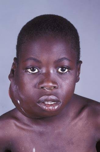
It is also called as ‘African jaw lymphoma’. It is a lymphoreticular cell malignancy. In the African form jaw involvement is 75% and in cases of the American form, abdomen involvement is more common. It is a B-cell neoplasm.

Etiology

• Epstein-Barrvirus(EBV)which also causes nasopharyn- geal carcinoma and infectious mononucleosis is considered to be the etiological factor. There are higher EBV antibody levels in patients of Burkitt’s lymphoma.

Clinical Features

  • Age and sex—peak incidence is in children between 6to 9 years. Males are affected more commonly than the females, with a ratio of 2:1.
  • Site distribution—more are found in maxilla than in mandible, where it may spread rapidly to the floor of the orbit. Almost always occurs in molar area. In the African form, more than one quadrant is involved while in the American form, only one quadrant is involved.
  • Onsetandprogress—the most important hall mark of this tumor is the fast growth with a tumor doubling time of less than 24 hours.
  • Symptoms—the most common presenting features are swelling of the jaws, abdomen and paraplegia. It is painless.
  • Sign—peripheral lymphadenopathy is common.
  • Prognosis—it is rapidly fatal in the absence of treatment,with death occurring within 6 months.

Oral Manifestations

  • • Onset and extent—it begins generally as a rapidly growing tumor mass of the jaws, destroying the bone with extension to involve maxillary, ethmoid and sphenoid sinus as well as orbit.
  • Symptoms—loosening or mobility of permanent teeth.There is gross distortion of the face due to swelling. Paresthesia and anesthesia of inferior alveolar canal or other sensory facial nerves are common.
  • Signs—gingiva and mucosa adjacent to the affected teeth become swollen, ulcerated and necrotic. As the tumor mass increases, the teeth are pushed out of their sockets. Swelling of the jaw occurs and it may cause facial asymmetry. They are capable of blocking nasal passages, displacing orbital contents and eroding through skin. There is derangement of arch and occlusion. There may be large quantity of mass protruding into the mouth, on the surface of which may be seen rootless, developing permanent teeth.
  • Spread—once the tumor perforate the bone, it is initially confined by the periosteum, but subsequently it spreads to the soft tissues of the oral cavity and face where rapid tumor growth soon obliterates the entire face and skin becomes tense and shiny.

Histology

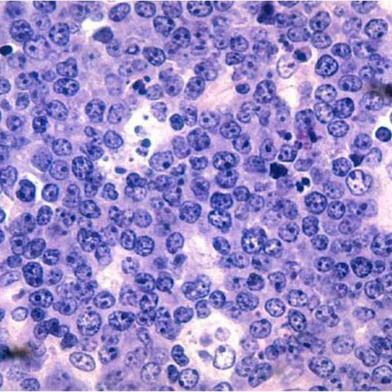
Shows characteristic starry sky appearance.

  1. Radiographic Features
    • Motheaten appearance—small radiolucent foci scattered throughout the affected area. These small foci coalesce and form a multilocular moth eaten appearance.
    • Sunray appearance—if periosteum is elevated, it will produce sunray appearance.
    • Margins—margins are ill defined and non-corticated.
    • Shape—they expand rapidly and are ballooned shaped.
    • Teeth—Lesions are osteolytic with loss of lamina dura about the erupted teeth and crypts of developing teeth are enlarged.
    • Effect ons urrounding structures—they expand very rapidly and breach its outer cortical limits.
  1. Diagnosis
  2. • Clinical diagnosis—swelling of the jaw and abdomen with peripheral lymphadenopathy can give clue to the diagnosis.

• Radiological diagnosis—moth eaten appearance is seen with loss of lamina dura around the teeth.

• Laboratorydiagnosis—monotonous sea of un differentiated monomorphic lymphoreticular cells, usually showing abundant mitotic activity. There is also hyperchro- matosis and loss of cohesiveness. Characteristic ‘starry sky’ appearance is seen.

Management

• Cytotoxicdrugs—cytotoxicdrugs like cyclophosphamide 40 mg/kg in single IV administration and repeated about 2 weeks later. Vincristine and methotrexate have been successful in some cases.

• Multiagent chemotherapy—combination of drugs such as cyclophosphamide, vincristine and methotrexate give better results than any single drug. Majority of patients show dramatic response to the therapy. The swelling regresses and the displaced teeth return to their normal position within 1 to 2 weeks.

REFERENCE- SHAFER’S TEXTBOOK OF ORAL PATHOLOGY AND ANIL GHOM TEXTBOOK OF ORAL MEDICINE

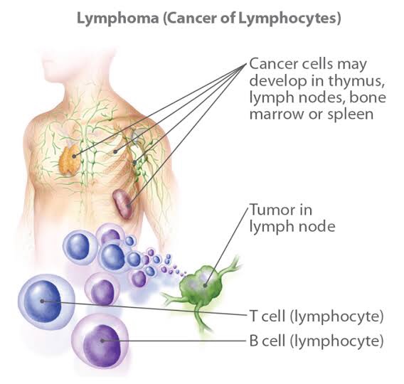
HODGKIN’S AND NON-HODGKIN’S LYMPHOMA

  • Primary tumors of the lymphoid system
  • Cancers involving lymphocytes during maturation or storage in the bone marrow
  1. Hodgkin’s lymphoma
  2. Non-Hodgkin’s lymphoma
  3. Burkitt’s lymphoma

Hodgkin’s Lymphoma

Hodgkin’s disease (HD) is a lymphoid malignancy characterized by the presence of Reed-Sternberg cells of B cell origin. The disease arises usually in single nodal area and successively spreads to contiguous lymph node areas. Extranodal involvement is rare.

Types (Histological)

• Lymphocyte predominant—abundant lymphocytes, few plasma cells, occasional Reed-Sternberg cell, localized involvement of one side of diaphragm and most favorable prognosis.

• Mixed cellularity—lymphocytes, plasma cells, eosino- phils, easily identified Reed-Sternberg cell.

• Nodular sclerosis—sparse lymphocytes, stromal cells, fibrosis and numerous but bizarre Reed-Sternberg cells. It has poor prognosis.

• Lymphocyte depletion—lymphocytes, plasma cells, eosinophils with localized involvement.

ETIOLOGY

  • Unkonown
  • However both genetic and environmental factors including EBV ( Ebstein Barr Virus) play role in pathogenesis.

CLINICAL FEATURES

  • • Age and sex—it is characterized by a bimodal age incidence, peak one in young adults and the second in the 5th decade of life with equal distribution between sexes.
  • First sign – asymptomatic enlargement of a supradiaphragmatic lymph node often in the neck
  • .Symptoms—theinvolvednodesarepainless.Generalized weakness, loss of weight, cough, dyspnea and anorexia are seen. Pain in back and abdomen owing to splenic enlargement, due to pressure of enlarged nodes or involvement of vertebrae.
  • Pel Ebstein fever—characteristic features of this disease are Pel-Ebstein fever, a cyclic spiking of high fever and generalized severe pruritis of unknown etiology.
  • Mediastinal lymph node involvement is common
  • Night sweats, fever, weight loss
  • Rarely produces oral manifestations

Hodgkin’s lymphoma. This high-power photomicrograph shows the characteristic Reed-Sternberg cell (arrow) of Hodgkin ‘s lymphoma, identifiedby its “owl-eye” nucleus.

  1. Radiographic Features
    • Site—it is rarely seen in jaws. The common regions are the posterior maxilla and mandible.
    • Appearance—malignant lymphoma arising in the oral cavity spreads to bone and cause irregular bone loss to the area of the lesion. There are radiolucent areas sepa- rated from each other by normal appearing bone which later become confluent, unless treatment is carried out.
    • Margins—typically,theradiolucentlesionshavediffuse ill defined margins which suggest infiltration of bone.
    • Osteoblastic type—osteoblastic type is uncommon in jaws, but it is seen in the vertebrae and pelvis. In it, there is frank sclerosis with filling of the marrow spaces by bone. It presents as grayness or whiteness which is abnormal. The margins may be well defined and sharp or irregular and trailing off gradually into the normal bone.

Diagnosis

  • Clinicaldiagnosis—discreteenlargementoflymphnode which is rubbery in consistency with some systemic signs.
  • Radiologicalfeatures—thereisfociofradiolucencyseen in the jaw.
  • Laboratorydiagnosis—itischaracterizedbyreplacement of normal lymph node architecture by an admixture of malignant lymphoid cells and non-neoplastic inflammatory cells. Characteristic Reed Sternberg cells are present. Multinucleated giant cells are also present. There is also presence of anemia which is normocytic and normochromic. ESR is raised. There is also raised level of LDH.

MANAGEMENT

Localized disease: The patients with localized disease (IA, IIA) are treated with 3 cycles of chemotherapy (ABVD-doxorubicin, bleomycin, vinblastine and dacarbazine) followed by radiotherapy of involved nodal areas. 

Extensive disease: Patients with B symptoms or extensive disease receive complete course (6-8 cycles) of ABVD chemotherapy.

Long-term cure can be achieved in >90% patients with localized disease and in 50-75% patients with extensive disease.

Non-Hodgkin’s Lymphoma

It is also called as ‘lymphosarcoma’. In this group, there is neoplastic proliferation of lymphoid cells, usually affecting the B-lymphocytes. Unlike Hodgkin’s lymphoma, the disease is frequently widespread at the time of diagnosis, often involving not only the lymph nodes but also bone marrow, spleen and other tissue. Early involvement of bone marrow is typical of this lymphoma.

Types (Histological)

  • Nodular—neoplastic cells tend to aggregate in such a way that large clusters of cells are seen.
  • Diffuse—thereismonotonousdistributionofcellswith no evidence of nodularity or germinal center pattern.
  • Etiology
  • Viral—the etiology is unclear but herpes virus and Epstein barr etiology has been suggested.
  • Immunological—there may be induced immunologic effect permitting a malignant clone to proliferate.
  1. Clinical Features
    • Age and sex—it affects persons of all ages from infants to the elderly. But is commonest in middle age group. Males are affected more commonly than the females.
    • Onset—the onset of symptoms may be insidious. Painless lymph node enlargement of abdominal and mediastinal region are the most common finding. Very often the first group of lymph nodes affected may be cervical, axillary or inguinal.
    • Symptoms—the patient complains of tiredness, loss of weight, fever and sweating. Pain is the main symptom of bone involvement which may present as a patho- logical fracture. Patient may complain of abdominal pain, nausea, vomiting, diarrhea or intestinal obstruc- tion which may occur due to involvement of gastro- intestinal tract. Pressure effect of lymphoma may cause dysphagia, breathlessness, vomiting, intestinal obstruction or ascites and paraplegia.
    • Signs—if liver and spleen are involved, hepato- splenomegaly is present. The growth is fleshy and is prone to ulceration.

The matted, nontender lymph node enlargement in the lateral cervical region represents a common presentation of lymphoma.

Oral manifestations

  • Gingival or mucosal tissue swelling or masses
  • Rapid growth of tumour from non-healing tooth extraction site.
  • Paresthesia or anaesthesia due to nerve invasion

Radiographic Features

• Appearance—asthediseaseprogressessmallradiolucent foci scattered throughout the area may be seen. Subsequent radiographs of the expanding lesion will show that these small foci have coalesced to form large multilocular moth eaten radiolucency with poorly defined margins.

• Margins—lesion blends imperceptibly with adjacent normal bone, in most of the cases.

• Effect on surrounding structures—lesions may cause marked expansion of bone. Erosion and perforation of cortex may occur.

• Maxillary sinus involvement—if the lesion involves maxillary sinus, possible opacification with breached cortical walls and associated paracentral or intracentral mass.

• Teeth—cortices of unerupted tooth buds and lamina dura of adjacent teeth are lost. Teeth may be resorbed.

Diagnosis

  • Clinical diagnosis—bluish color mass of the palate with multiple lymph node involvement may suspect the diagnosis of non-Hodgkin’s lymphoma.
  • Radiological features—expansion of bone with radio- lucency is present.
  • Laboratory diagnosis—blood count usually shows hypersplenism or hemolytic anemia. The reduced WBC and RBC counts are seen along with reduced hemo- globin levels and reticulocytosis. In some cases, there may be slight increase in lymphocytes and thrombo- cytopenia. Moderate degree of anemia will also present when there is considerable bone marrow involvement. Some very aggressive high grade non-Hodgkin’s lymphomas are associated with very high urate levels which can precipitate renal failure.

Treatment

The disease stage is determined after clinical evaluation and investigations.
Treatment of low grade lymphoma: It depends on the stage of the disease and clinical status.

  • Asymptomatic patients generally do not require therapy.
  • Symptomatic localized (stage I) disease can be treatedwith radiotherapy.
  • Symptomatic patients with extensive disease (stage III,IV) are treated with chlorambucil or combination chemotherapy (CVP; cyclophosphamide, vincristine, and prednisolone or CHOP; cyclophosphamide, doxorubicin, vincristine and prednisolone).
  • Other newer effective agents are fludarabine, monoclonal antibody (anti-CD20) with or without radionuclides, and lymphoma vaccine.
  • The relapse following therapy is common. Hence, low grade NHL is generally not curable. However, the cure is now possible in some cases with the use of radio- immunotherapy.
  • Treatment of high grade lymphoma:
  • Localized high grade NHL (stage I and non-bulky stage II) is treated with three cycles of chemotherapy (CHOP with monoclonal antibody, anti-CD20) followed by radiotherapy.
  • Extensive disease (bulky stage II, stage III, and stage IV) is treated with six to eight cycles of chemotherapy.
  • Autologous bone marrow transplantation is indicatedin relapsed cases.
  • The cure rate is 60-90% in stage I and II and 30-40% instage III and IV.

REFERENCE- SHAFER’S TEXTBOOK OF ORAL PATHOLOGY, ANIL GHOM TEXTBOOK OF ORAL MEDICINE AND GOGLE SLIDE SHARE