ATTRITION

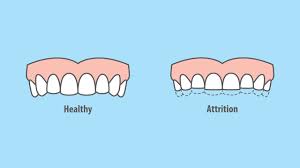
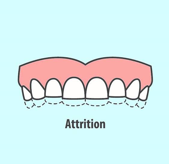
  1. It is the physiologic wearing away of teeth because of tooth- to-tooth contact, as in mastication. It plays an important physiological role as it helps to maintain an advantageous crown-root ratio and gains intercoronal space of 1 cm, which facilitates third molar eruption. Attrition can be considered pathological when it cause functional, esthetics and dental sensitivity problems.

Types

• Physiological attrition—attrition which occurs due to normal aging process, due to mastication.

• Pathological attrition—it occurs due to certain abnor- malities in occlusion, chewing pattern or due to some structural defects in teeth.

Etiological factors for pathological attrition

• Abnormal occlusion
• Developmental—malocclusion and crowning of teeth, may lead to traumatic contact during chewing, which may lead to more tooth wear.
• Acquired—due to extraction of teeth. Extraction causes increased occlusal load on the remaining teeth, as the chewing force for the individual remains constant.

• Premature contact in case of edge-to-edge contact,pathological attrition can also occur.
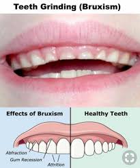
• Abnormal chewing habits parafunctional chewing habit like bruxism and chronic persistent chewing of coarse and abrasive food or other substances like tobacco.
• Occupation in certain occupations, workers are exposed to an atmosphere of abrasive dust and cannot avoid it getting into mouth.
• Structural defect in defects like amelogenesis imperfecta and dentinogenesis imperfecta, hardness of enamel and dentin is reduced and such teeth become more prone to attrition.

Clinical features

• Sex—men usually exhibit more severe attrition than women due to greater masticatory forces.

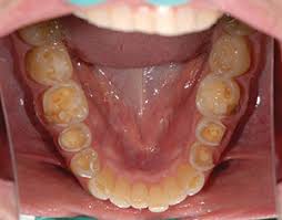
• Sites—it may be seen in deciduous as well as permanent dentition. It occurs only on occlusal, incisal and proximal surfaces of teeth. Severe attrition is seldomly seen in primary teeth, as they are not retained for any great period. Palabal cusps of maxillary teeth and buccal cusps of mandibular posterior teeth show most wear.

• Appearance – the first clinical manifestation of attrition is the appearance of small polished facet on a cusp tip or ridge and slight flattening of an incisal edge.

Physiologic attrition

  • Physiological tooth surface loss results in a reduction, in both vertical tooth height and horizontal tooth width .Physiological attrition showing wearing of the occlusal surface of the molar teeth.
  • Contact points—due to slight mobility of teeth in their socket (which is a manifestation of resiliency of periodontal ligament) similar facets occur at contact points.
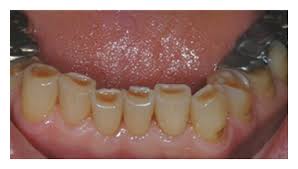
  • Color of teeth when the dentin gets exposed, it generally becomes discolored i.e. brown in color.
  • Signs—there is gradual reduction in cusp height and consequent flattening of occlusal inclined plane. There is shortening of the length of dental arch, due to reduction in the mesiodistal diameter of teeth. Secondary dentin deposition occurs.

• Pathologicalattrition

Severe tooth loss—in pathological attrition severe tooth loss is seen .

Dentoalveolar compensation—if attrition affecting the occlusal surfaces of teeth has occurred, then reduction in occlusal face height (vertical dimension of occlusion) and increase in the freeway space could be anticipated. This may be further complicated by forward posturing of mandible. It is often observed, however, that despite overall tooth surface loss, the freeway space and the resting facial height appear to remain unaltered primarily because of dentoalveolar compensation. This is important with respect to patient assessment. If restoration of worn teeth is being planned then the extent of dentoalveolar compen- sation would appear to determine the dentist’s strategy; defining the need to carry out measures such as crown lengthening, to ensure the same vertical dimension of occlusion and freeway space.

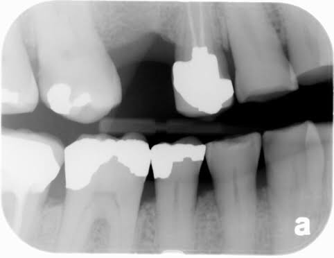
Radiographic features

• Crown—smoothwearingofincisalandocclusalsurfaces of involved teeth is evident by shortened crown image

• Pulp—sclerosisofpulpchamberandcanalsisseendue to deposition of secondary dentin which narrows the pulp canals.

• Periodontal ligament—widening of periodontal ligament space and hypercementosis.

• Alveolar bone—some loss of alveolar bone.

Management

  • Modifying factors—treatment of patient depends upon degree of wear relative to the age of patient, etiology, symptoms and patient’s desire.
  • Habit breaking appliance—the provision of one of three different sorts of splints could be considered. A soft bite guard can help in breaking a bruxist habit or simply will protect the teeth during the bruxist habit. A localized occlusal interference splint is designed to break the bruxist habit and can be worn easily during the day. A stabilization splint reduces bruxism by providing an ideal occlusion: it also enables the clinician to locate and record centric relation. In case of bruxism, use of night guards may be effective in reducing attrition.
  • Correctivemethod—correctionofmalocclusion,stoppage of tobacco chewing habit and restriction of diet to non coarse food are useful in avoiding attrition.
  • Managementofsensitivityandesthetics—non-cariousloss of tooth tissue may require treatment for sensitivity, esthetics, function and space loss in the vertical dimension.

REFERENCE- ANIL GHOM TEXTBOOK OF ORAL MEDICINE [2nd ed]

Internal Vs. External Resorption

What are internal and external resorption and the causes?

🔅The American Association of Endodontics defines resorption as, “a condition associated with either physiologic or a pathologic process resulting in a loss of dentin, cementum, and/or bone. Vital tissue is necessary for either external or internal resorption to occur.”

🔅By this definition, internal resorption is “a defect of the internal aspect of the root following necrosis of odontoblasts as a result of chronic inflammation and bacterial invasion of the pulp tissue.”

🔅External resorption is “resorption initiated in the periodontium and initially affecting the external surfaces of the tooth—may be further classified as surface, inflammatory, or replacement, or by location as cervical, lateral, or apical; may or may not invade the dental pulpal space.”

Difference between Internal Resorption Vs. External Resorption

Histology:

Clinical Case:

It may present initially as a pink-hued area on the crown of the tooth; the hyperplastic, vascular pulp tissue filling in the resorbed areas. This condition is referred to as a pink tooth of Mummery.
External Inflammatory lesion

Dr. Mehnaz Memon🖊


References:

  1. https://www.byendo-cupertino.com
  2. https://www.dentistryiq.com
  3. Image Source: Google

NRT DRUGS

Written by : Dr. Urusa I Inamdar

Nicotine gum

Nicotine polacrilex .

  • Buccal absorption
  • 4 mg of nicotine ( India )
  • Two varieties in India : gutkha and mint flavour
  • Duration of treatment is 4-6 weeks ; start weaning after 2-3 months .
  • Brand name : nicorette , nulife , nicotex

Nicotine patch ( transdermal )

  • Ready absorption of nicotine across the skin .
  • 3 of the patches are for 24 hr use and one is for 16 hr ( waking ) use .
  • Starting doses are 21-22 mg/ 24 hr patch and 15 mg/ 16 hr patch .
  • The recommended total duration of treatment is usually 6-12 weeks .
  • Brand name : habitrol , nicodern – cq , nicotrol

Patient instruction for nicotine patch:

  • Do not smoke while using the patch .
  • Rotate the patch site to minimize skin irritation .
  • If insomnia occurs , remove patch before going to bed or use 16 hr patch .
  • Apply a new patch every day ( remove old patch ) in a location between the neck and waist that is relatively hairless and where the skin is not broken . Apply to a different location each day .

Nicotine nasal spray

  • 8-40 dosage per day .
  • Nasal irritation may occur .
  • Treatment time : 3-6 months .
  • Brand name : nicotrol nasal spray

Nicotine inhaler

  • 6-16 cartridges /day
  • Mouth and throat irritation may occur .
  • Treatment time : upto 6 months .
  • Brand name : nicotrol inhaler

References

  • Dental notes
  • Google.com

Fordyce granules( fordyce disease)

Pathology Outlines - Fordyce granules

What are Fordyce granules? How does it occur ?

  • Firstly it isn’t a disease as the name says it.
  • Rather this can be called a developmental anomaly.
  • It is characterised by heterotopic collection of sebaceous glands in various sites of the oral cavity.
  • It is said that the occurance of sebaceous glands in the mouth may be by inclusion in the oral cavity of the ectoderm.
  • This has some of the potentialities of skin during the development of the maxillary and the mandibular processes during the embryonic life.

Clinical features-

1.Appearance-

  • as yellow spots, seperated
  • or forming large plaques
  • project slightly above the suface of tissue
Sebaceous glands (Fordyce spots or Tyson glands)

2. Site of appearance- found frequently in a bilateral symmetical pattern

  • mucosa of cheeks (opposite the molar teeth)
  • inner surface of lips
  • retromolar region
  • tongue
  • gingiva
  • palate
  • frenum

Besides the oral cavity they also appear in the oesophagus ,the female genital tract ,cervix uteri, male genitilia ,nipples, palms ,soles ,parotid ,larynx and the orbit .

3. Usually seen more in adults than children . This is due to the better development of sebaceous glands and hair system is not seen until puberty.

Histology

  • These are heterotopic collection of sebaceous glands and they are identical with those that are seen in the skin.
  • But they are unassociated with hair follicles and hair shaft from the gingiva. (this may be a very rare occurance )
  • Glands are located superficially.
  • There may be few or many lobules.
  • They are grouped around one duct or more ducts and they open at the surface of the mucosa .
  • The ducts may show keratin plugging.

Treatment-

It requires no treatment.

source – textbook of oral pathology shafers and google images .

Tooth Attrition

7 Common Reasons You Could Be at Risk for Teeth Grinding

Defined as physiological wearing away of tooth structure.

Biologically Based Restorative Management of Tooth Wear

This phenomenon is more physiological than pathological

🛑SITES OF OCCURRENCE-

  • occlusal surface
  • incisal surface
  • proximal surface

It is also associated with the aging process. More the older the person gets more it regresses

🛑MAIN CAUSE- Tooth to Tooth contact during Mastication

  • It is seen in deciduous as well as in permanent dentition.

🛑Types of attrition

🔸Physiological attrition: Attrition which occurs progressively during normal aging process as a result of masticatory occlusion. Usually a slow process through out life .

🔸Pathological attrition: This is a severe form of attrition that occurs as a result of abnormalities in occlusion ,chewing pattern and structural defects in teeth .

Examples are bruxism or clenching

Kids and Grinding

🛑AETIOLOGY

  • Abnormal chewing habits: Parafunctional chewing habits like Bruxism and chronic persistent chewing of coarse foods or other substances like tobacco.
  • In other occupations workers are exposed to an atmosphere of abrasive dusts e.g. silica
  • Amelogenesis imperfecta and dentinogenesis imperfecta in which the hardness of enamel and dentine is reduced and such teeth become more prone to attrition and is seen in children

🛑Clinical presentation –

  • Appearance of small polished facet on the cusp tip /ridge or slight flattening of the incisal edge
  • Sensitivity and pain : attrition may be entirely asymptomatic or there may be dentine hypersensitivity.
  • Tooth discoloration : attrition and erosion of the enamel exposes inner and darker dentine giving a yellower appearance
  • Compromised periodontal support leading to drifting of teeth
  • Altered occlusion due to decreasing occlusal vertical dimension

Men usually have more attrition than women as a result of greater masticatory force .

Also variations are seen with the coarseness in diet and chewing tobacoo

Bio-Rejuvenation Dentistry: Utilizing Nanohybrid Flowable ...
advanced attrition

Sources -textbook of oral pathology – shafers, slide share , pictures – google photos.

SYNDROMES🤯-Made easy(Part-1)

Syndromes are defined as combination of medical signs & symptoms that together represent a disease process .

As mentioned above, being a set of features ,most of us might have a tough time trying to mug up all of the characteristics pertaining to a syndrome which are often confusing too.

So here’s a humble attempt to make it easy .How??…..Short forms/mnemonics ofcourse!

Some of these are already familiar to you & for the rest -the author of this post holds patent 🙂 .Only some of the important syndromes pertaining to dentistry have been discussed here.Hope you would find it helpful.

Sources :Shafers textbook of oral pathology,Instagram -_dentistars_,dental_exams,www.cartoonstock.com

Cleidocranial dysplasia

Sources – Anil Ghoms textbook of oral medicine , osmosis.org

Image sources – Google images , researchgate.net