The Closed mandibular nerve block 💉

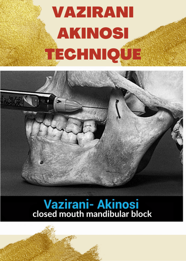
❇️ The Vazirani-Akinosi technique:

🔆“It is a specific method of nerve block in the mandibular region, carried out with the mouth closed.

❇️ The area of distribution of the anaesthesia includes

a) the corresponding dental arch,

b) the body of the jaw and the inferior ramus,

c) the gingiva/mucosa and vestibular periosteum, anterior to the mental foramen

d) the area of distribution of the lingual nerve: 2/3 of the anterior of the tongue and floor of the mouth, the gingiva/ mucosa and lingual periosteum.

🔆» The main indication, already anticipated, is trismus: classically this is contraction of the masticatory musculature which prevents the performance of effective inferior alveolar anaesthesia, for example, in cases of pulpitis or an abscess of a lower molar.

– By Dr. Sneha poeghal , Mallareddy institute of dental sciences , hyd .

References – Malamed

ODONTOTOMY

The extraction of a tooth requires the separation of its attachment to the alveolar bone via the crestal and principal fibers of the periodontal ligament. Also involved is a process of expanding the alveolar socket.

After the tooth has been removed, the form of alveolar process is restored by finger pressure. Bleeding is arrested by means of a pressure pack placed between the jaws, and the wound is allowed to fill with a blood clot.

EXODONTIC PROCEDURES:

The following techniques may be used for tooth removal:

  1. the forceps technique
  2. the elevator technique (https://dentowesome.wordpress.com/2020/06/18/dental-elevators/)
  3. the open view technique

Odontotomy can be used to facilitate any of these three procedures.

ODONTOTOMY

  • In some instances the extraction may be simplified by cutting a tooth apart. This is especially desirable in the case of multi-rooted deciduous or permanent teeth with severely divergent roots.
  • It is also useful in cases where the crown is so decayed that only a shell remains. (As shown in the figure👇🏻)
  • If the roots are divergent, the bur cut, instead of being vertical, may be made parallel with one of the roots, and this part of the tooth then is extracted first.
  • The other roots may be removed either with forceps or an elevator.

Dentowesome|@drmehnaz🖊


References: Textbook of Oral Surgery – Daniel M Laskin

Swellings in the angle of Mandible, Floor of Mouth & Palate

Swellings at the angle of Mandible include: ✍🏻👇🏻

🔅Congenital disease

• Branchial Cleft Cyst

🔅Neoplasm

(i) Benign

  • Hemangioma
  • Lymphangioma, Cystic hygroma
  • Pleomorphic adenoma (mixed tumor)
  • Warthin tumor
  • Neurofibroma
  • Angiolipoma
  • Adenoma
  • Hamartoma
  • Lipoma
  • Oncocytoma

(ii) Malignant

  • Mucoepidermoid carcinoma
  • Squamous cell carcinoma
  • Adenoid cystic carcinoma
  • Acinic cell carcinoma
  • Adenocarcinoma
  • Rhabdomyosarcoma
  • Lymphoma, leukemia
  • Metastatic adenopathy

🔅Inflammation/Infection

  1. Parotitis
  2. Parotid Abscess
  3. Tuberculosis
  4. Sarcoidosis
  5. Sjögren disease
  6. HIV

Detailed View🔍

1) Branchial Cleft Cyst:

  • Failure of involution of clefts and pouches lead to cysts, fistulas or sinus tracts.
  • Its a painless fluctuant swelling
  • First branchial cleft cysts are rare usually located at parotid gland or periparotid region.
  • Second branchial cleft cyst – Type II are the most common
  • Typically, second branchial cleft cysts present as a rounded swelling just below the angle of mandible, anterior to the sternocleidomastoid

2) Hemangiomas:

They are the most common benign salivary gland mass. Capillary hemangiomas involve parotids

3) Lymphangiomas

They are congenital malformations of the lymphatic system that may involve the parotid gland (Soft asymptomatic neck mass associated with facial asymmetry)

4) Pleomorphic Adenoma:

Hard painless slow growing mass

5) Warthin Tumor:

Incorporation of heterotopic salivary gland ductal epithelium within intraparotid & periparotid nodes

6) Parotitis & Parotid Abscess:

  • Most common in children
  • Mumps is the most common viral cause of parotitis
  • The condition manifests tender swelling at the angle of Mandible
  • Sialadenitis is most commonly due to bacterial infections caused by Staphylococcus aureus.
  • Premature neonates and immunosuppressed individuals are affected.

Swellings in the floor of Mouth: 👇🏻✍🏻

Ranula presents as a translucent blue, dome-shaped fluctuant swelling & contains viscid, glairy jelly like fluid
  1. Ranula – a type of mucocele found on the floor of the mouth. Present as a swelling of connective tissue consisting of collected Mucin from a ruptured salivary gland by local trauma.
  2. Swellings in the floor of the mouth are more likely to arise from structures above the Mylohyoid muscle. The commonest swellings in the floor of the mouth are denture induced hyperplasia & salivary calculus.
  3. Swellings in the floor of the mouth may inhibit swallowing & speech.
  4. Mandibular tori produce bony hard swelling lingual to the lower premolars.

Differential diagnosis of swellings of the floor of the mouth or neck (Jham et al., 2007): https://www.researchgate.net/figure/Differential-diagnosis-of-swellings-of-the-floor-of-the-mouth-or-neck-Jham-et-al-2007_tbl1_287206404


Swellings on the Palate: 👇🏻✍🏻

  1. Torus palatinus is an intrinsic bone lesion whereas a dental abscess pointing on the palate (usually from the palatal roots of the 1st & 2nd maxillary molars or from upper lateral incisors) is extrinsic.
  2. Salivary neoplasms
  3. Invasive carcinoma from the maxillary sinus may produce a palatal swelling.
  4. Kaposi’s sarcoma, typical of HIV/AIDS may also present as lump on palate.
  5. Paget’s disease.

Differential diagnosis of palatal swellings: https://www.researchgate.net/figure/Differential-diagnosis-of-palatal-swellings_tbl1_221967546

Dentowesome|@drmehnaz🖊


Image source: Google.com