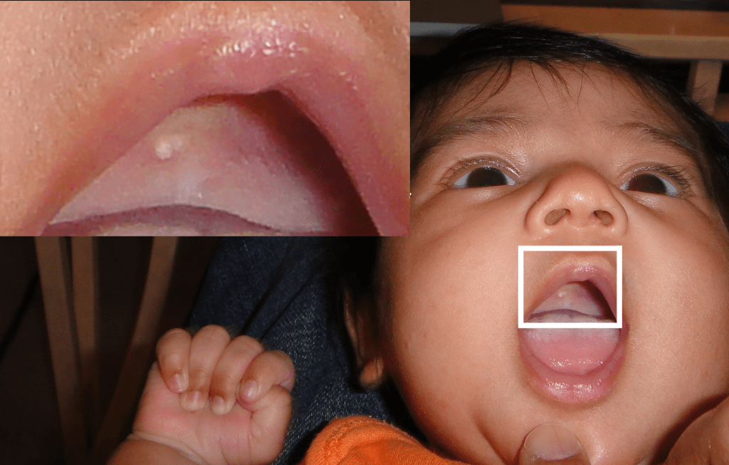
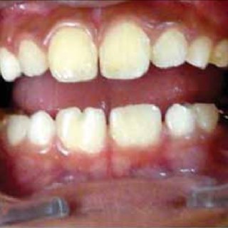
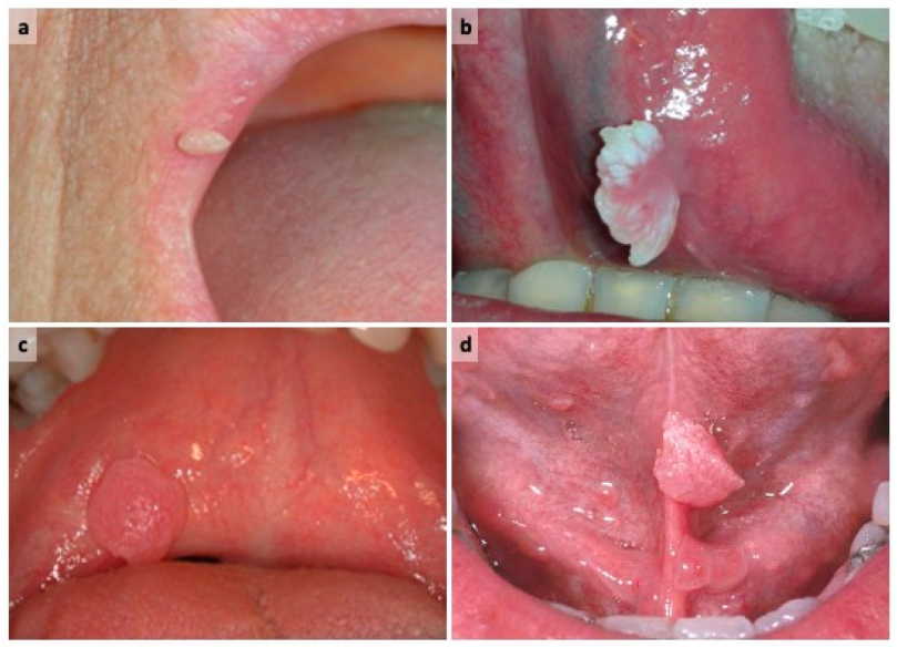
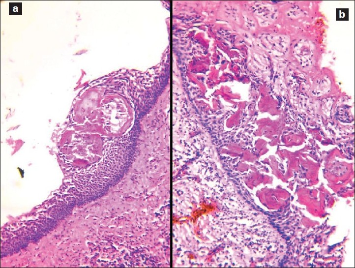
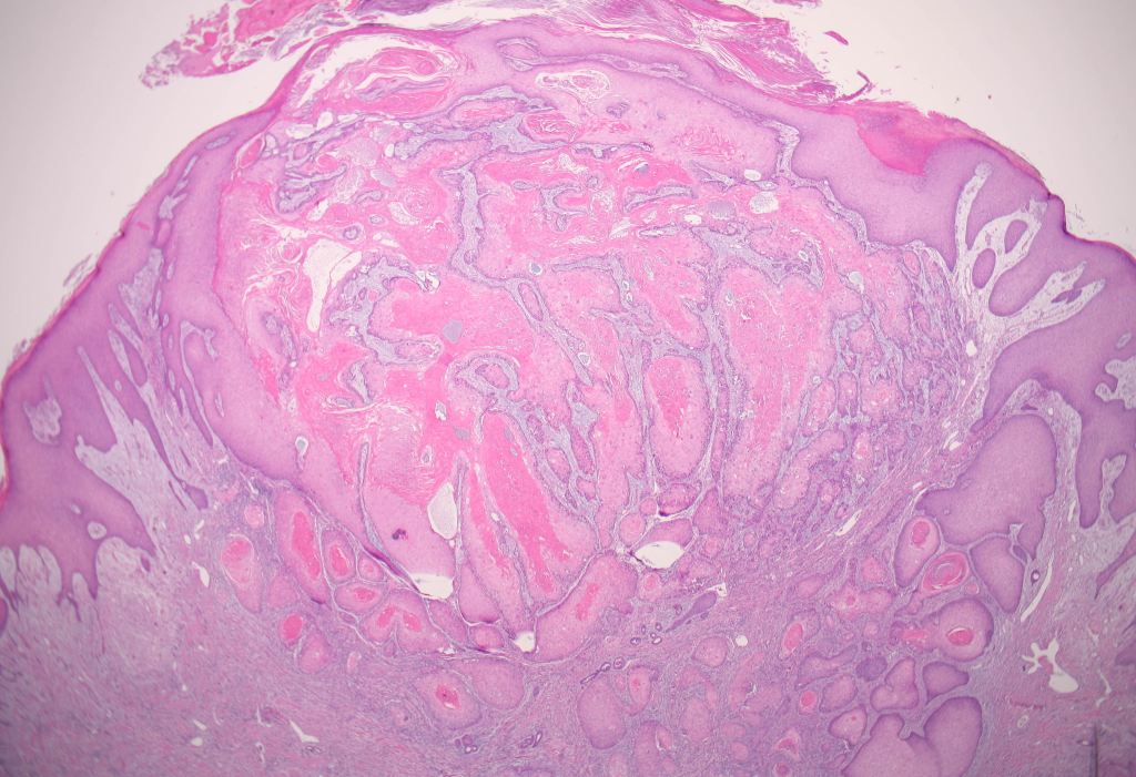
A 1-week-old girl was referred by the paediatrician because of white nodules on her mandibular alveolar ridge.
As a dental student, it’s important to learn about various oral conditions that can affect patients of different age groups. Gingival cysts are one such condition that can occur in both neonates (newborns) and adults, although they are more commonly associated with newborns.
In neonates, gingival cysts are often referred to as Epstein’s pearls. These cysts are small, white or yellowish bumps that appear along the gum line or on the roof of the mouth. They are considered a normal finding in newborns and are typically harmless. Epstein’s pearls are thought to result from the accumulation of epithelial cells (the cells that make up the outer layer of the skin) during the development of the gums. They usually resolve on their own within a few weeks or months after birth without requiring any treatment.
On the other hand, gingival cysts can also occur in adults. In adults, these cysts are usually referred to as adult gingival cysts or adult Epstein’s pearls. Adult gingival cysts are similar in appearance to neonatal Epstein’s pearls and can occur anywhere along the gum line. However, unlike neonatal Epstein’s pearls, adult gingival cysts are considered pathological and may require treatment. They are thought to develop due to various factors such as trauma, chronic irritation, infection, or the entrapment of epithelial remnants within the gum tissue.
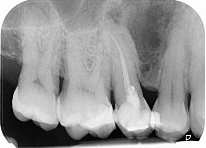
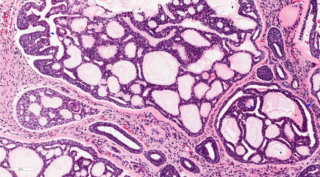
A multilocular radiolucent lesion was found in the interdental bone between the upper first and second premolars. The teeth were vital and after enucleation, the pathologist reported that the cyst had features of a developmental periodontal cyst lined by squamous epithelium with focal thickened areas.
As a dental student, it’s essential to learn about various dental conditions. One specific condition you may come across is called a botryoid cyst or a lateral periodontal cyst.
Botryoid cysts develop from small pieces of tissue called odontogenic epithelial remnants, which are found in the periodontal ligament. These remnants are leftover tissue from tooth development. The term “botryoid” refers to the appearance of these cysts under a microscope, which resembles a cluster of grapes.
Unlike some other cysts caused by inflammation or infection, botryoid cysts are not primarily driven by inflammation. Instead, they are considered a developmental abnormality originating from these remaining epithelial tissues.
When examining a botryoid cyst under a microscope, you may observe focal areas where the cyst lining appears thicker or denser compared to the surrounding epithelium. These areas are known as focally thickened epithelial plaques. The presence of these plaques is a characteristic feature that helps identify botryoid cysts microscopically.
As a dental student, it’s important to keep in mind that this information is based on knowledge available until September 2021. Stay engaged in your studies, seek guidance from your professors, and consult trusted dental resources for the most up-to-date and comprehensive information on dental conditions like botryoid cysts.
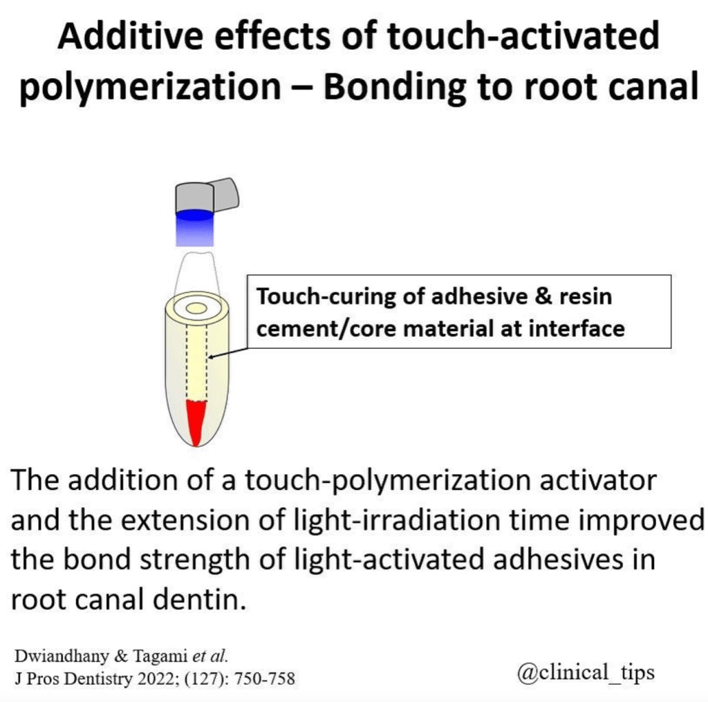
Additive effects of touch activated polymerization have been studied in the context of bonding to root canal dentin. Studies have shown that stresses from polymerization shrinkage and problems with adequate access to the root canal complicate the formation of high-strength bonds when cementing endodontic posts with resin cements [1].
The effects of different bonding systems with various polymerization modes and root canal regions on the bond strength of core build-up resin composite to dentin have also been investigated [2][4].
The interaction of geometrically related factors predicts that bonding of adhesive root-filling materials to root canals is highly unfavorable when compared with indirect intracoronal restorations with a similar resin film thickness [3].
Resin cement polymerization results in shrinkage, creating micro gaps between the fiber-reinforced composite (FRC) posts and the root canal wall [7].
The BS of fiber posts to root canals can be improved by soft-started polymerization [6].
The effect of composite post placement and/or light-irradiation of dual-cure resin composite on adhesion to root canal dentin using the chemical activation mode of a 1-step self-etch dual-cure adhesive has also been evaluated [8].
The activity of matrix metalloproteinases (MMPs) following endodontic irrigation protocols and correlation to the push-out bond strength (PBS) and nanoleakage expression (NL) in the root dentin have been studied [9].
Finally, the mechanical properties of the bonding agent, which could be enhanced by adequate polymerization, have been shown to affect the bond strength to root canal dentin [10].
[2] (2021). Effects of different bonding systems with various polymerization modes and root canal region on the bond strength of core build-up resin composite. J Prosthodont Res, 4(65), 521-527. https://doi.org/10.2186/jpr.jpr_d_20_00251
[4] (2021). Effects of different bonding systems with various polymerization modes and root canal region on the bond strength of core build-up resin composite. J Prosthodont Res, 4(65), 521-527. https://doi.org/10.2186/jpr.jpr-d-20-00251
[5] (2020). Novel Bioactive and Therapeutic Root Canal Sealers with Antibacterial and Remineralization Properties. Materials, 5(13), 1096. https://doi.org/10.3390/ma13051096
[6] (2017). Is the bonding of self-adhesive cement sensitive to root region and curing mode?. J. Appl. Oral Sci., 1(25), 2-9. https://doi.org/10.1590/1678-77572015-0430
[7] (2020). The Effects of Differences in Post Canal Widths on Microleakage in Prefabricated Fiber Reinforced Composites. sod, 2(5), 1-8. https://doi.org/10.28932/sod.v5i2.2432
[8] (2010). Effect of composite post placement on bonding to root canal dentin using 1-step self-etch dual-cure adhesive with chemical activation mode. Dent. Mater. J., 6(29), 642-648. https://doi.org/10.4012/dmj.2010-006
[9] (2022). In Situ Zymography Analysis of Matrix Metalloproteinases Activity Following Endodontic Irrigation Protocols and Correlation to Root Dentine Bond Strength. Polymers, 17(14), 3567. https://doi.org/10.3390/polym14173567
[10] (2005). Regional Bond Strength of Four Self-etching Primer/Adhesive Systems to Root Canal Dentin. Dental Materials Journal, 2(24), 261-267. https://doi.org/10.4012/dmj.24.261
A cyst was removed along with an unerupted third molar tooth. The pathologist reported a fibrous capsule that was myxoid in places and that was lined by a thin layer of squamous and cuboidal epithelium. The cyst originated from the amelocemental junction.
As a dental student, it’s important to have a good understanding of dentigerous cysts. Dentigerous cysts are a type of cyst that form around unerupted or impacted teeth. They develop from a structure called the dental follicle, which surrounds the tooth germ or the developing tooth.
When a tooth fails to erupt properly, sometimes a cyst can form around it. This cyst is called a dentigerous cyst. It is characterized by a fibrous capsule, which is like a protective layer, and it is lined by stratified squamous epithelium, which is a type of tissue that forms the outer layer of the cyst.
Histologically, dentigerous cysts may show various features such as the thickness of the epithelial lining or the presence of inflammation or keratinization. However, these features are not unique to dentigerous cysts and can also be seen in other types of cysts.
In terms of diagnosis, clinical features are important. Dentigerous cysts are usually discovered during routine dental exams or through radiographic evaluations. One of the key clinical features is that the cyst originates from the amelocemental junction, which is the junction between the enamel (the hard outer layer of the tooth) and the cementum (the specialized tissue covering the root of the tooth).
On an X-ray, the dentigerous cyst appears as a well-defined radiolucent area around the crown of the impacted tooth. It’s important to note that dentigerous cysts often don’t cause any symptoms and are found incidentally. However, if they grow in size or become infected, they can lead to swelling, pain, or displacement of nearby teeth.
Treatment for dentigerous cysts usually involves surgical removal or marsupialization, which is a procedure that creates a surgical opening to allow drainage and shrinkage of the cyst. This is done to prevent complications and facilitate the eruption of the impacted tooth.
Overall, understanding the clinical and histological features of dentigerous cysts is crucial for diagnosing and managing these conditions effectively as a dental student.
First permanent molar is about to erupt, so the age = is 5 years
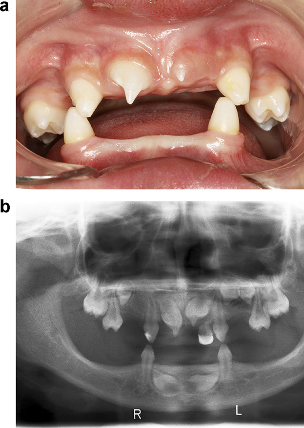
Number of teeth = 19, FUSION
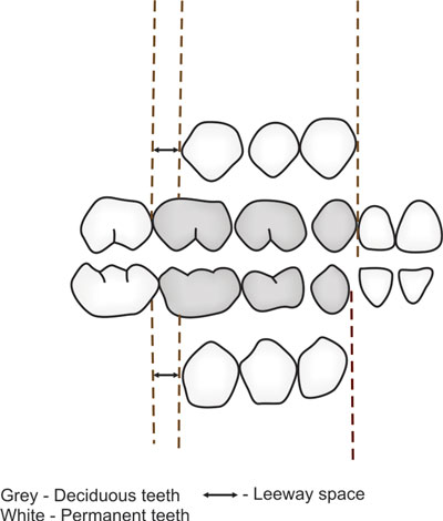
LEEWAY SPACE – Maxillary = 0.9 mm and Mandibular = 1.7 mm
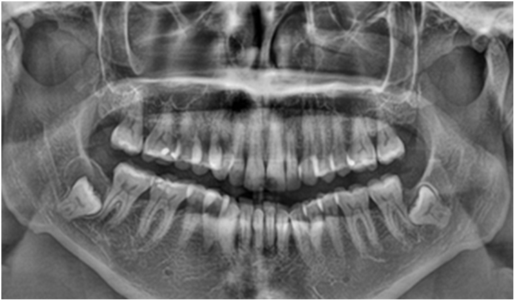
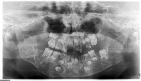
OPG of 10 year old child – permanent CI, LI and first molar has erupted. The primary canine, first molar and second molar are also present.
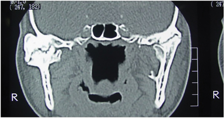
8 year old male patient comes to your clinic with bilateral swelling in the jaw, upturned eyes with excessive sclera visible beneath the iris = CHERUBISM
A cyst was enucleated from the posterior mandible of a 38-year-old man. The pathologist reported that the lining was composed of stratified squamous epithelium that showed parakeratosis and basal-cell palisading. Some areas were inflamed and cholesterol nodules were noted.
As a dental student, it’s important to understand the histological features of odontogenic keratocyst (OKCs) in order to recognize and diagnose them accurately. Here’s a breakdown of the key features:
Basal-cell palisading: Odontogenic keratocysts have a specific arrangement of cells in their lining. The cells at the base of the epithelium align in a palisade-like fashion, with their nuclei positioned away from the central cavity of the cyst. This is a distinctive characteristic often seen in OKCs.
Keratinisation: Odontogenic keratocysts exhibit a high degree of keratinization. This means that the cells within the lining of the cyst produce a protein called keratin, resulting in the formation of a layer of keratinized cells. This keratinization gives the lining a unique appearance when observed under a microscope.
Uniform thickness: The epithelial lining of an odontogenic keratocyst tends to have a relatively consistent thickness throughout. This means that when examining a tissue sample of an OKC, you will notice that the lining is of similar thickness in different areas. However, it’s important to remember that this feature alone is not sufficient for making a definitive diagnosis.
Lumen filled with keratinous material: The central space or lumen within an odontogenic keratocyst is typically filled with keratinous material. This material is composed of dead cells that have undergone keratinization. Essentially, the lining of the cyst sheds these keratinized cells, and over time, they accumulate within the cystic space, filling it with this keratin debris.
Remember, these histological features are indicative of an odontogenic keratocyst, but a comprehensive diagnosis also requires consideration of other clinical and radiographic findings. It’s crucial to consult with an experienced oral and maxillofacial pathologist for a definitive diagnosis and to determine the appropriate treatment plan for the patient.