Diagnosis Demystified – Case 12

A 25-year-old man presented with an anterior midline cystic swelling in the neck. When he protruded his tongue the cyst elevated.
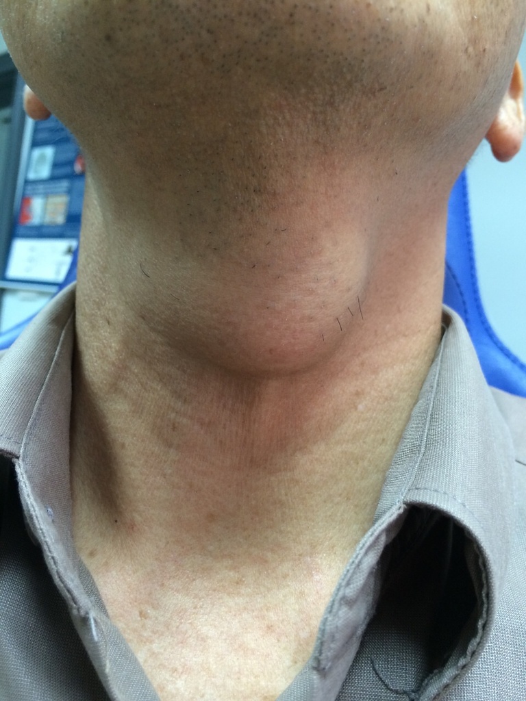
Thyroglossal tract cysts develop at any point from the foramen caecum to the thyroid along the line of the vestigal thyroglossal duct. Most occur below the level of the hyoid bone and in people under 30 years.

Thyroglossal tract cysts are abnormal fluid-filled sacs that develop along the path of a structure called the thyroglossal duct. To understand this, let’s break it down step by step.

  1. Thyroglossal tract: During the early stages of fetal development, the thyroid gland (a butterfly-shaped gland located in the neck) forms at the base of the tongue in an area called the foramen caecum. As the fetus grows, the thyroid gland descends down the neck to its final position in front of the trachea (windpipe). The path it takes while descending is called the thyroglossal tract.
  2. Vestigial thyroglossal duct: The thyroglossal tract is initially a duct-like structure that connects the thyroid gland to the base of the tongue. However, as the thyroid gland descends, the duct usually disappears, leaving only a remnant called the vestigial thyroglossal duct.
  3. Development of cysts: In some cases, the vestigial thyroglossal duct may not completely disappear, and remnants of it can persist. These remnants can potentially develop into cysts. Thyroglossal tract cysts usually form anywhere along the path of the vestigial thyroglossal duct, from the foramen caecum (at the base of the tongue) to the final location of the thyroid gland in front of the trachea.
  4. Location and age group: Most thyroglossal tract cysts occur below the level of the hyoid bone, which is a small U-shaped bone in the neck. They are commonly found in people who are under 30 years old. However, it’s important to note that thyroglossal tract cysts can occur at any age, including in adults.

In summary, thyroglossal tract cysts are fluid-filled sacs that develop along the vestigial thyroglossal duct, a remnant of the developmental path of the thyroid gland. They can occur anywhere from the base of the tongue to the final location of the thyroid gland. Most of these cysts are found below the level of the hyoid bone and are typically observed in individuals under 30 years of age.
Diagnosis Demystified- Case 11

A 26-year-old patient presented with a rapidly growing lesion that expanded the maxilla. There was bone destruction on the radiograph and therefore malignancy was suspected. On biopsy, the lesion was intraosseous and was cavitated. There was profuse bleeding and a small biopsy of the lining was taken. The pathologist reported osteoclast-like giant cells and granulation tissue with blood clots.

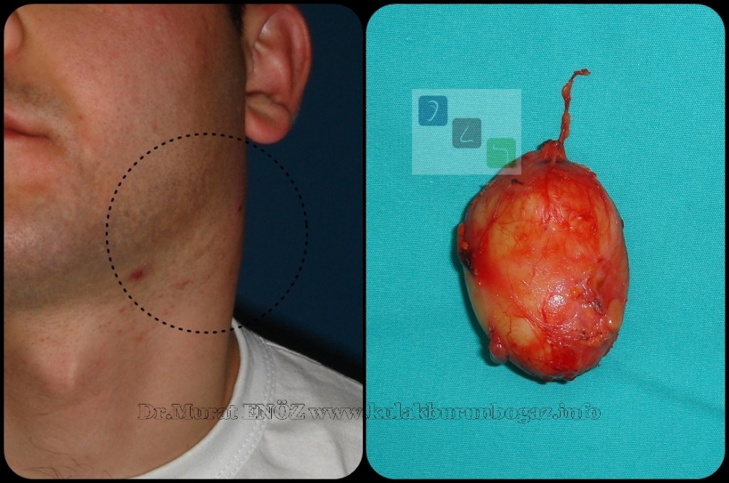
Branchial or lymphoepithelial cysts are typically present in the second and third decades after slow enlargement. They are lined by squamous epithelium and have lymphoid tissue with prominent follicles in the wall. In older patients, they may be confused with cystic metastatic squamous carcinoma in a lymph node. Metastatic thyroid cancer can also mimic branchial cysts. The cyst must be carefully examined by the pathologist.

RESEARCH PAPERS

  • Branchial or lymphoepithelial cysts are a type of congenital neck pathology that can occur due to the remnants of a branchial arch developing into a cyst, causing swelling [9].
  • These cysts are also known as branchial cleft cysts [1][3][6][7][9][10].
  • The prevalence of branchial cysts in children is reported to be around 24.2% [2].
  • They can occur in various locations, including the submandibular gland, parotid gland, and palatine tonsil [1][4][8][10].
  • The etiopathogenesis of these cysts is still a subject of debate, and various postulations have been made regarding the matter [8].
  • Thyroid lymphoepithelial cysts are reported to originate from follicular cells and are unrelated to solid cell nests and lateral cervical cysts arising from branchial-derived remnants [5].
  • There is no specific reference available that discusses branchial cysts in the maxilla. However, branchial cysts are typically found in the lateral neck region, deep to the sternocleidomastoid muscle, and are derived from remnants of the branchial apparatus [13][15][16].
  • They are congenital anomalies that usually present as a cystic mass in the neck [17].
  • In rare cases, branchial cysts can arise in the posterior mediastinum [11].
  • The classification of branchial cysts depends on the branchial cleft they originated from, with locations ranging from the external auditory canal to the lower part of the neck [12].
  • Branchial cysts are typically diagnosed based on clinical examination and imaging studies, such as ultrasound or computed tomography [13].
  • Treatment usually involves surgical excision of the cyst [14].

REFERENCES

  • [1] Ahamed, Kannan, Velaven, Sathyanarayanan, Roshni, E (2014). Lymphoepithelial cyst of the submandibular gland. J Pharm Bioall Sci, 5(6), 185. https://doi.org/10.4103/0975-7406.137464
  • [2] Irani, Zerehpoush, Sabeti (2016). Prevalence of Pathological Entities in Neck Masses: A Study of 1208 Consecutive Cases. Avicenna J Dent Res, 1(8), 4-4. https://doi.org/10.17795/ajdr-25614
  • [3] Delantoni, Onder, Orhan (2022). B-mode and color Doppler imaging of different types of branchial cleft cysts in children. A multicenter study and review of the literature. J Ultrason, 90(22), 174-178. https://doi.org/10.15557/jou.2022.0028
  • [4] Som, Brandwein, Silvers (1995). Nodal inclusion cysts of the parotid gland and parapharyngeal space: A discussion of lymphoepithelial, aids-related parotid, and branchial cysts, cystic warthin’s tumors, and cysts in sjogren’s syndrome. Laryngoscope, 10(105), 1122-1128. https://doi.org/10.1288/00005537-199510000-00020
  • [5] Suzuki, Hirokawa, Ito, Takada, Higuchi, Hayashi, … & Miyauchi (2018). Derivation of thyroid lymphoepithelial cysts from follicular cells. Endocr J, 5(65), 579-586. https://doi.org/10.1507/endocrj.ej17-0402
  • [6] Najib, Berrada, Lahjaouj, Oukessou, Rouadi, Abada, … & Mahtar (2021). Cervical lymphoepithelial cyst: Case report and literature review. Annals of Medicine and Surgery, (61), 185-187. https://doi.org/10.1016/j.amsu.2020.12.041
  • [7] Gill, Chhabra, Singh, Mehrotra, Rawat (2020). Lymphoepithelial Cyst, A Diagnostic Dilemma: Case Report. Ann of Pathol and Lab Med, 10(7), C137-140. https://doi.org/10.21276/apalm.2824
  • [8] Gurka (2020). Case Report of a Parotid Benign Lymphoepithelial Cyst. ADOH, 3(12). https://doi.org/10.19080/adoh.2020.12.555836
  • [9] Park, Yoon, Bang, Ahn (2019). Branchial cleft cyst in the parotid gland in a human immunodeficiency virus-negative patient. Arch Craniofac Surg, 3(20), 191-194. https://doi.org/10.7181/acfs.2019.00031
  • [10] Bingöl, Balta, Bingöl, Mazlumoglu, Kilic (2016). Lymphoepithelial Cyst in the Palatine Tonsil. Case Reports in Otolaryngology, (2016), 1-3. https://doi.org/10.1155/2016/6296840
  • [11] Han, Shang (2023). Branchial cleft cyst arising in posterior mediastinum: A case report. Front. Surg., (9). https://doi.org/10.3389/fsurg.2022.1088142
  • [12] Park, Yoon, Bang, Ahn (2019). Branchial cleft cyst in the parotid gland in a human immunodeficiency virus-negative patient. Arch Craniofac Surg, 3(20), 191-194. https://doi.org/10.7181/acfs.2019.00031
  • [13] Reynolds, Yap, Marikar, Roland (2019). Fifteen-minute consultation: The infant with a neck lump. Arch Dis Child Educ Pract Ed, 5(105), 258-261. https://doi.org/10.1136/archdischild-2019-316827
  • [14] Dhoke, Khadakkar, Dhote, Choudhary, Harkare, Kamal (2012). Type III Second Branchial Cleft Cyst. International Journal of Head and Neck Surgery, 2(3), 112-114. https://doi.org/10.5005/jp-journals-10001-1107

Diagnosis Demystified – Case 10/170

A 44-year-old woman presented with a swelling in the midline floor of the mouth that elevated the floor of mouth and tongue. The mucosa over the cyst was yellowish in colour.

Dermoid cysts are cystic lesions that develop from abnormal inclusion of skin tissue during embryonic development. These cysts can occur along the midline of the body, including in the oral region. In the oral cavity, they can be found either above or below the mylohyoid muscle.

When a dermoid cyst is located above the mylohyoid muscle, it causes a bulge into the floor of the mouth. On the other hand, if it is located below the mylohyoid muscle, it expands into the submental soft tissue. The mylohyoid muscle is a muscle located in the floor of the mouth that separates the oral cavity from the submental region below the chin.

Histological examination refers to the microscopic analysis of the cyst’s tissue. When a dermoid cyst is examined under a microscope, the lining of the cyst resembles epidermis, which is the outermost layer of the skin. This means that the tissue inside the cyst looks similar to the surface of the skin.

Additionally, adnexal structures are often found within dermoid cysts. These structures include hair follicles, sebaceous glands (oil glands), and smooth muscle. Hair follicles are responsible for hair growth, while sebaceous glands produce oil to moisturize the skin and hair. Smooth muscle is a type of muscle tissue that is not under voluntary control and is typically found in the walls of organs.

The presence of these adnexal structures within the lining of the cyst helps to confirm the diagnosis of a dermoid cyst. The combination of the epidermis-like lining and the presence of hair follicles, sebaceous glands, and smooth muscle is characteristic of dermoid cysts.

It’s important for dental students to be aware of dermoid cysts because they can present in the oral region and may require surgical intervention for removal. Proper diagnosis and treatment planning are crucial to ensure optimal patient care.

Cleaning root canal sealer and contaminated dentin

Root canal sealer and contaminated dentin can be cleaned using various methods to achieve proper coronal seal and adhesion to dentin. The presence of remnants of root canal sealers affects the coronal seal and adhesion to dentin. The following are some methods that can be used to clean root canal sealer and contaminated dentin:

1. Cleaning with gutta-percha solvents: A study by Topçuoğlu et al. (2015) investigated the effect of different cleaning procedures using gutta-percha solvents on bond strength of adhesive resins to AH Plus contaminated dentin (APCD). The study found that cleaning with gutta-percha solvents can improve bond strength of adhesive resins to contaminated dentin.

2. Removal of smear layer: [2] found that the presence of smear layer affects negatively the adhesion of root canal sealers because it forms an interface between the sealing material and dentin, hindering or impeding sealer penetration into the dentinal tubules.

3. Cleaning with irrigation solutions: Çakır (2023) cleaned the dentin of primary teeth contaminated with root canal sealers (AH Plus, MTA Fillapex) with different irrigation solutions (saline, NaOCI, ethanol). 4. Cleaning with ultrasound or combined with acetone: [4] found that cleaning epoxy resin-based sealer-contaminated dentin surfaces using ultrasound or combined with acetone could preserve the bond strength.

5. Laser-EDTA cleansing effect phenomenon: Miletić et al. (2016) validated the laser‐EDTA cleansing effect phenomenon as an optimal cleansing method, which could also enhance the bond strength of sealers to root canal dentin.

6. Cleaning with sodium chlorite or ethanol: [6] divided specimens into four groups according to the cleaning method of dentin used: G1, no root canal sealer (control); G2, 0.9% sodium chlorite (NaCl); G3, ethanol; and G4, followed by diamond drill.

7. Sealer removal protocols: [7] compared the efficacy of different sealer removal protocols on the microtensile bond strengths (MTBS) of single step adhesives to a calcium silicate-based bioceramic root canal sealer contaminated dentin.

8. Adhesive properties of root canal sealers: [8]investigated the adhesive properties of eight root canal sealers, applied as a thin layer between a dentine and a gutta-percha surface.

9. Rubber dam isolation: [10] suggest performing rubber dam isolation to avoid contamination of the enamel bonding surface, allowing a clean restorative environment, with a correct visualization of the gingival margin during the adjustment of the veneer restorations, and finally to facilitate the removal of excess cement.

REFERENCES

[1] (2015). The bond strength of adhesive resins to AH plus contaminated dentin cleaned by various gutta-percha solvents. Scanning, 2(37), 138-144. https://doi.org/10.1002/sca.21190

[2] (2008). Adhesion of Epiphany and AH Plus sealers to human root dentin treated with different solutions. Braz. Dent. J., 1(19), 46-50. https://doi.org/10.1590/s0103-64402008000100008

[3] (2023). The Effect of Different Cleaning Materials on the Bond Strength of Resin Composite to Primary Teeth Dentin Contaminated with Root Canal Sealer.. https://doi.org/10.21203/rs.3.rs-2591855/v1

[4] (2022). Microtensile bond strength to sealer-contaminated dentin after using different cleaning protocols. Journal of Dental Sciences, 1(17), 122-127. https://doi.org/10.1016/j.jds.2021.05.016

[5] (2016). Effect of photon induced photoacoustic streaming (PIPS) on bond strength to dentine of two root canal filling materials. Lasers Surg. Med., 10(48), 951-954. https://doi.org/10.1002/lsm.22536

[6] (2016). Effect of cleaning methods on bond strength of self-etching adhesive to dentin. J Conserv Dent, 1(19), 26. https://doi.org/10.4103/0972-0707.173189

[7] (2021). The Efficacy of Different Sealer Removal Protocols on the Microtensile Bond Strength of Adhesives to a Bioceramic Sealer-Contaminated Dentin.. https://doi.org/10.21203/rs.3.rs-448214/v1

[8] (1990). Adhesion of root canal sealers to bovine dentine and gutta-percha. Int Endod J, 1(23), 13-19. https://doi.org/10.1111/j.1365-2591.1990.tb00797.x

[9] (2022). The efficacy of different sealer removal protocols on the microtensile bond strength of adhesives to a bioceramic sealer-contaminated dentin. Niger J Clin Pract, 3(25), 336. https://doi.org/10.4103/njcp.njcp_1575_21

[10] (2017). A Step-by-Step Conservative Approach for CAD-CAM Laminate Veneers. Case Reports in Dentistry, (2017), 1-6. https://doi.org/10.1155/2017/3801419

Diagnosis Demystified – 10/170

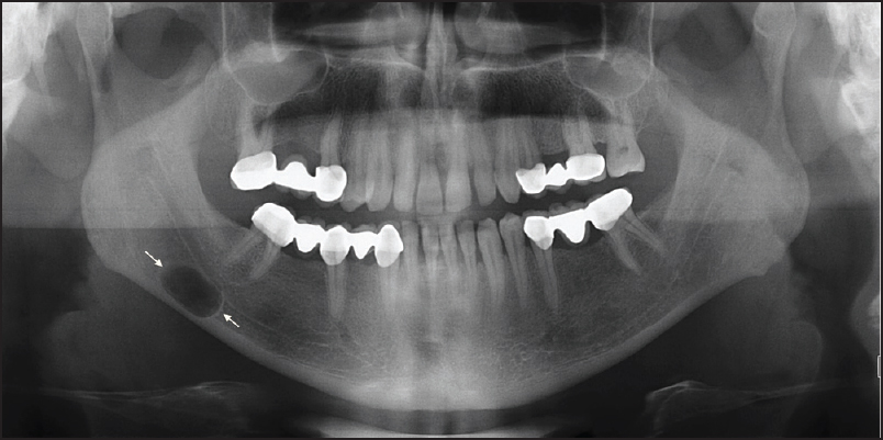
A radiolucent lesion was found incidentally on a dental panoramic radiograph in a 30-year-old man. The cyst was located in the mandible below the inferior alveolar canal. It was roughly oval in outline.

A Stafne cavity is a developmental depression or concavity that occurs in the border of the mandible, which is the lower jaw. It is important to note that Stafne cavities are not true cysts but can sometimes be mistaken for cysts on imaging studies like dental X-rays.

Stafne cavities are typically found in the posterior region of the mandible, near the angle of the jaw. They are more commonly seen in adult males and are considered to be a normal anatomical variation rather than a pathological condition.

These cavities are usually asymptomatic, which means they don’t cause any symptoms or problems for the patient. They are often discovered accidentally during routine dental X-rays or radiographic examinations.

The appearance of a Stafne cavity on an X-ray can resemble that of a cyst, but there are some differences. Unlike cysts, Stafne cavities do not cause expansion or erosion of the surrounding bone. They usually have a well-defined and smooth appearance. It’s also important to note that Stafne cavities are typically found on one side of the mandible and are usually symmetrical.

In most cases, treatment is not necessary for Stafne cavities because they are harmless and don’t cause any issues. However, if there is uncertainty about the diagnosis or if the lesion shows unusual features, further evaluation may be recommended. This can include additional imaging studies like a CT scan or even a biopsy to rule out any other potential pathological conditions.

It’s essential for dental professionals to be aware of Stafne cavities and their characteristic appearance on X-rays. This knowledge helps prevent confusion with other conditions and ensures appropriate management for the patient.

Carbohydrates and their Metabolism – NEET Pearls from Dentest and Pulse

  1. General formula of carbohydrate = (CH20)n
  2. Following are monosaccharides
    1. Glucose
    2. Fructose
    3. Galactose
  3. NON REDUCING SUGARS = trehalose
  4. REDUCING SUGAR = Maltose, lactose and cellobiose
  5. Pentose sugar present in heart cells = lyxose
  6. Monoscaarchirde glucose forms a part of the disaccharide sucrose 
  7. GALACTOSE
    1. not an oligosaccharide
    2. Monosaccharide with a maximum rate of absorption in intestine 
  8. SUCROSE 
    1. sugar characterised by its non-reducing property. It is also called cane sugar and table sugar 
    2. It cannot be metabolised 
    3. Invertase catalyzes the hydrolysis of = Sucrose into fructose and glucose**
  9. FRUCTOSE
    1. In hereditary fructose intolerance, one can see intolerance to = Fructose and Sucrose 
    2. A patient with hereditary fructose intolerance is deficient in = Aldolase enzyme
    3. Highest concentrations of fructose are found in = Seminal fluid
  10. LACTOSE 
    1. Milk sugar
    2. Hydrolysis of lactose yields glucose and galactose 
    3. Beta 1.4 glycosidic bond is present in = Lactose
  11. GLUCOSE
    1. Glucose, Maltose and Lactose = Exhibit inversion 
    2. Malatose
      1. is dissacride of = Glucose and glucose
      2. Glycosidic linkage = alpha 1 – 4 
    3. Epimeric Pair = D Glucose and D Galactose 
    4. Glucose monomers in glycogen are held by = Alpha 1-4 bonds, alpha 1-6 bonds
    5. Only sugar absorbed against concentration gradient = Glucose
    6. True blood sugar level measures the levels of = Glucose + Fructose
    7. Abnormal constituents of urine = Ketone + Glucose
    8. Renal threshold of glucose = 180 mg/dl 
    9. In prolonged starvation, the main energy source of brain = Ketone bodies
    10. GLUCOSE can be synthesised from = Glutamate, Asparate and Alanine
    11. The uptake of glucose by the liver increases followed by carbohydrate meal because = increase in phosphorylation of glucose by glucokinase
    12. Apart from liver, glucokinase is present in = Pancreatic islet cells
    13. Glucokinase = It is a Inducible enzyme
    14. In glucose solution at equilibrium, mutarotation results in a fixed optical rotation of = 52.5*
    15. Form of glucose predominatly seen is as = Beta D Glucopyranose
    16. Glycemic index is highest for = Glucose
    17. Amount of asymmetric carbon atoms in glucose = FOUR
    18. Number of stereoisomers of glucose is = 16
    19. Substrate used by RBC in fasting state = Glucose
    20. Important precursors of glucose in animals
      1. Pyruvate
      2. Lactate
      3. Glycerol 
    21. Major factor that determine weather glucose is oxidized by aerobic or anaerobic glycolysis = NADH and ATP/ADP Ratio
    22. Potassium Oxalate and Sodium Fluoride = added to the blood sample to estimate glucose
    23. Muscle cannot release glucose from glycogen because of the deficiency of = Glucose 6 phosophatase 
  12. GLYCOGEN
    1. Starch and Glycogen are polymers of = Alpha Glucose
    2. Amylose and amylopectin are constitutes of = Starch
    3. CARBOHYDRATE RESERVE OF THE BODY = glycogen 
    4. Glycogen breakdown = Glucose and Lactic acid
    5. Tissue with highest glycogen content = Liver
    6. An essential for the conversion of glucose to glycogen in the liver = UTP
    7. Muscle glycogen is mainly utilized for supplying energy to = Liver
  13. Ditery fiber is rich in = Cellulose
  14. The rate of absorption of sugars is highest for = Hexoses 
  15. Proteins and carbohydrates of glycoproteins are held by = Glycosidic bonds 
  16. Sialic acids are acetylated derivatives of = Neuraminic acid 
  17. GLYCOLYSIS
    1. The oxidation of glucose or glycogen to pyruvate and lactate by the EMF pathway is called = Glycolysis
    2. EMF pathway reaction takes place outside the mitochondria 
    3. Phosphofructokinase is the key rate-limiting enzyme of = Glycolysis 
    4. During the conversation of glycerol to pyruvic acid, the first glycolytic intermediate to form = Dihydroxy acetone phosphate 
    5. Allosteric inhibition with ATP effects and rate-limiting enzyme is = Phosphofructokinase**
    6. The enzyme involved in the first committed step of glycolysis = Hexokinase 
    7. Enzyme phosphofructokinase 1 is strongly activated by = Fructose 2,6 bisphosphate 
    8. Enzyme Phosphofructokinase is allosterically inhibited by = Citrate
    9. Whenever the cells ATP supply is depleted =  Phosphofructokinase enzyme activity is increased
    10. ATP is produced by the following enzyme = Pyruvate Kinase 
    11. Glycolysis enzyme inhibited by fluoride is = Enolase
    12. ENOLASE = Catalyzes the reversible degradation of 2 – phosphoglycerate to phosphoenolpyruvate 
    13. For glucose estimation in blood, the mode of transportation from primary health care to the laboratory is done by the addition of = Sodium Fluoride 
    14. Insulin acts on which enzyme = Glucokinase 
    15. Inhibition of glycolysis in the presence of oxygen is called as = Pasteur Effect 
    16.  The reverse of Pasteur effect = Crabtree effect 
    17. The main pathways of metabolism in the brain are = Glycolysis and Citric Acid Cycle 
    18. The end product of glycolysis under anaerobic conditions is = Lactic acid 
    19. Number of ATP molecules generated in the conversion of glycogen to lactate = 2 
    20. Following exercise, the level of lactic acid in blood during ventilation = Decreases 
    21. Ion which is important in the glycolysis = Magnesium 
    22. Pyruvate dehydrogenase and Alpha-ketoglutarate dehydrogenase = Multienzymee complex 
    23. Pyruvate Dehydrogenase complex contains = NAD, FAD, Co-A
    24. The enzyme which provides a link between glycolysis and citric acid cycle = Pyruvate Dehydrogenase
    25. Increase in pyruvate and lactate is seen in = Thiamine deficiency 
    26. Unique by product of lycolysis in RBC = 2,3 biphosphoglycerate
    27. Glycerol enters glycolysis via = Dihydroxyacetone phosphate
    28. Glycolytic pathway is located in = CYTOSOL
    29. Glycolysis is always anaerobic in = Erthrocytes
    30. Enzyme responsible for the conversion of glucose 1 phosphate to glucose 6 phosphate = Phosphoglucomutase
    31. Irreversible step of glycolysis involves
      1. Phosphofructokinase
      2. Pyruvate kinase
      3. Hexokinase 
    32. True statements regarding anaerobic glycolysis
      1. End product is lactic acid
      2. Net production of ATP is 2 
      3. Occurs in places like eyes, lens and RBC’s 
    33. In the glycolysis cycle, substrate-level phosphorylation takes place in = pyruvate kinase 
    34. Generation of ATP upt pyruvate in aerobic glycolysis = 7
    35. Cancer cells derive energy mainly from = Glycolysis 
  18. CITRIC ACID CYCLE 
    1. Enzymes concerned with the citric acid cycle are found in = Mitochondria 
    2. Kreb cycle occurs in = Aerobic conditions 
    3. Kreb cycle doesn’t occur in = RBC due to absence of mitochondria 
    4. In the TCA cycle, what is formed first = Citrate 
    5. In the TCA cycle, citrate is immediately converted into = Cisaconitate, after losing molecule of H20
    6. Acid formed in = Oxaloacetic acid 
    7. The correct sequential order of enzymatic reaction of kreb cycle when molecule acetyl-CoA enters the cycle = Citrate, ketoglutarate and oxaloacetate 
    8. In the TCA cycle, substrate-level phosphorylation takes place in = Succinyl CoA to succinate
    9. The enzyme involved in substrate-level phosphorylation = Succinyl CoA Synthetase 
    10. In the TCA cycle, substrate-level phosphorylation occurs at = Thiokinase
    11. High energy phosphate compound that is formed via substrate-level phosphorylation = GTP
    12. Succinyl CoA to Succinate = 1 ATP
    13.  1 molecule of glucose forms = 2 molecules of pyruvate 
    14. 1 molecule of acetyl CoA enzyme gives rise to = 12 ATP 
    15. Alpha Ketoglutarate = Metabolite which is used in the detoxification of ammonia in the brain 
    16. Acetly CoA can be converted into
      1. Fatty acids
      2. Cholesterol
      3. Ketone bodies
    17. Total Number of dehydrogenase = 4
    18. Final common pathway for the oxidation of carbohydrate, lipids and protein in human body is = TCA cycle 
  19. INSULIN
    1. Glycogen synthesis is increased by = Insulin 
    2. Insulin increases the following pathway
      1. Glycogen synthesis 
      2. Fatty acid synthesis 
      3. Protein synthesis 
    3. Insulin causes lipogenesis by
      1. Increasing acetyl CoA carboxylase activity 
      2. Increases the transport of glucose into the cells
      3. Decreases the intracellular cAMP level 
    4. Glucose transporter which is stimulated by insulin is located in = Skeletal muscle and adipose tissue 
    5. Activity of which of the following enzyme is not affected by insulin = Hexokinase
    6. Insulin = not polymer of glucose
    7. Homopolysaccharide made up of fructose is = Insulin
  20. GLYCOGENEIS 
    1. It requires
      1. Uridine diphosphate
      2. Glycogen synthetase
      3. Branching enzyme
  21. GLYCOGENOLYISIS 
    1. Hypoglycaemia is corrected by increasing the rate of live glycogenolysis = GLUCAGON
    2. The first product of glycogenolysis = Glucose 1 phosphate 
    3. Rate limiting step of glycogenolysis = Glycogen Phosphorylase 
    4. A deficiency of Phosphorylase = would impair the body’s ability to maintain blood glucose concentration during the first 24 hours 
    5. The conversation of glucose 6 P to G 1 P = example of isomerization 
    6. Adrenaline acts on the enzyme = phosphorylase
    7. Glycogenolysis in muscle does not raise blood sugar due to lack of = H-6-phosphatase
    8. Glucose can be synthesized from
      1. Tryptophan and Phenylalanine
      2. Glycerol
      3. Lactic acid and propionic acid 
  22. GLUCONEOGENSIS
    1. Mainly occurs in = Mainly in Liver and partly in Kidney, not in muscles**
    2. Liver and Muscle = involved in Cahill Cycle
    3. Substance for gluconeogenesis = Glycerol
    4. The key enzyme of gluconeogenesis = Pyruvate carboxylase 
    5. The compound that can give rise to glucose by gluconeogenesis = Lactate
    6. Amino acids that enter TCA cycle for gluconeogenesis = Phenylalanine, Tyrosine and Tryptophan 
    7. Glycerol is converted to glucose in = Liver 
    8. Major contribution towards hepatic gluconeogenesis = Lactate
    9. Hepatic gluconeogenesis is stimulated by = Glucagon and Epinephrine
      1. X = Aspartate
      2. Y = Oxaloacetate 
    10. Major contribution towards gluconeogenesis is by = Alanine and Glutamine
    11. Malate shuttle –  is important in = Gluconeogensis and Glycolysis 
    12. Substrates of gluconeogensis =
      1. Glucogenic amino acids
      2. Lactate
      3. Glycerol
  23. HMP Pathway 
    1. TRUE STATEMENTS
      1. HMP shunt is an alternative pathway for oxidation of glucose that occurs in the cytosol 
      2. It is characterized by the absence of production of ATP 
      3. It is active in the adipose tissue, liver and gonads 
      4. Oxidative phase generate NADPH 
      5. Non oxidative phase generates ribose precursors 
    2. Sites where HMP shunts can occur include
      1. Liver
      2. WBC
      3. Lactating mammary gland 
      4. Testes 
    3. Step in HMP pathway requiring TPP = Transketolase 
    4. HMP shunt is of great importance in cellular metabolism because it produces = NADPH 
    5. Dehydrogenases of HMP Shunt are specific for = NADP
    6. NADPH is the product of = HMP pathway 
    7. Enzymes which used NADP as coenzyme = Glucose 6 phosphate dehydrogenase = regulartory enzyme in HMP shunt
    8. First pentose formed in HMP shunt = Ribulose 5 phosphate
    9. Metabolites in HMP shunt are
      1. Sedoheptulose 7 phosphate
      2. Glyceraldehyde 3 phosphate
      3. Xylulose 5 phosphate 
  24. GLYCOGEN STORAGE DISORDERS
    1. Glucose 6 phosphate deficiency
      1. is seen in = Von Gierke’s disease = autosomal recessive 
      2. Defective cori cycle and increased mobilization of glycogen from liver is seen
      3. Hyper-ureicemia is feature of = type 1 glycogen disorder (Von Gierke’s disease)
    2. Type 2 glycogen disorder is due to deficiency of =
      1. 4 and 1,6 – glucosidase
      2. lysosomal alpha 1 
    3. McArdles disease is due to deficiency of = Myophosphorylase (Type 5)
    4. Beta Galactosidase is deficient in = Krabbe’s disease
    5. Limit detrin accumulate in cytosol = Type 3 – Cori’s disease**
    6. Pompe’s disease is due to defieceny = Acid Malatase
  25. Galactosaemia commonly is due to deficiency of = galactose 1 phosphate uridyl transferase
  26. Cytochromes are = iron containing porphyrins
  27. Main enzyme responsible for the activation of xenobiotics = Cytochrome P-450
  28. Most lipogenic = fructose
  29. Xylitol is = Natural five carbon sugar
  30. Glucose transporters present in the Beta cells of the islets of langerhans is = GLUT 2
  31. GLUT 4
    1. Is present in adipose tissue
    2. Facilitates diffusion of glucose
    3. Transferred from cytosol to the cell membrane by insulin
    4. Glucose transporter in myocyte stimulated by insulin is = GLUT4
  32. Prolonged carbohydrate deficiency leads to = Ketoacidosis 
  33. Glutathione
    1. Tripeptide
    2. Conjugates xenobiotics
    3. Co factor of various enzymes
  34. Sphingosine is present in = Ceramide
  35. Haworth structures refer to = Pyran and Furan forms of sugars 
  36. Glycosaminoglycan present in cornea as = Keratan Sulphate and Dermatan Sulphate
  37. Enantiomers – non superimposbale images of one another
    1. D mannose and L mannose 
    2. D Glucose and L glucose
  38. Specific rotation of beta D glucopyranose = +19*
  39. Hetropolysccaride among the following = Heparin
  40. Lactate formed in muscles can be utilized through = Cori cycle 
  41. Alpha Lactone is = Vitamin C
  42. Most prominent carbohydrate component of hemicelllulose = Arabinoxylan 
  43. Rothera’s test is for = Ketones
  44. Bacterial Glutamine synthethase = Enzyme catalyzes a reaction in which reduced nitrogen is introduced into cellular metabolism 
  45. When velocity of enzyme activity is plotted against substrate concentration = HYPERBOLIC CURVE 
  46. Glycoproteins = are important for white blood cell recognition
  47. Cytochrome C = recieves flavoproteins
  48. Anabolism and Catbolism are chemically linked in the form of = ATP
  49. Factors determining the activity of an enzyme
    1. Association with regulatory protein
    2. Sequestration
    3. Allosteric regulation 
  50. 6 – phosphogluconate dehydrogenase = catalyses the first step in the pentose phosphate pathway 
  51. Flux control coefficient = measure of the effect of an enzyme’s concentration on flux through a multi enzyme pathway 
  52. Frutose 2,6 bisphosphate = compounds are responsible for the coordinated regulation of glucose and glycogen metabolism 
  53. Elasticity Coefficent = measure of how responsive the enzyme is to changes in the concentration of metabolite 
  54. After Overnight fasting, levels of glucose transporters are reduced in = Adipocytes 
  55. Isomerase = enzyme that catalyzes the conversion of an aldose sugar to a ketose sugar 
  56. Insulin stimulated glucose uptake takes place in via GLUT 4 which is insulin dependent
    1. Heart 
    2. Skelteal Muscle
    3. Adipose tissue
  57. Transport of glucose in Liver = GLUT 2 = Insulin independent 
  58. Decrease BMR is seen in = Starvation 
  59. Caloric Value of Alcohol = 7Kcal/g

Is it better to treat maxillary incisors without dowel or ferrule?

The question of whether it is better to treat maxillary incisors with or without dowel or ferrule is a complex one that depends on various factors. Several studies have investigated the effects of ferrule and dowel on the fracture resistance of endodontically treated maxillary incisors.

One study found that endodontically treated maxillary incisors with a uniform 2 mm ferrule were more fracture-resistant than those with a uniform 1 mm ferrule [3].

Another study investigated the effects of using different diameters of parallel cast posts with or without ferrule on the overall fracture resistance of Cast Post and Core (CPC) and found that the use of a ferrule increased the fracture resistance of the CPC [1].

However, other studies have found that the presence of a ferrule did not significantly affect the fracture resistance of endodontically treated maxillary incisors [2][5].

The use of dowels and cores can also affect the fracture resistance of endodontically treated maxillary incisors. One study compared the fracture resistance of endodontically treated central maxillary incisors prepared with different types of dowel and core materials and found that the use of glass fiber dowels resulted in the highest fracture resistance [6].

Another study investigated the effect of a cervical cavity extending 1 mm apical to the cemento-enamel junction (CEJ) on fracture resistance and failure mode of maxillary central incisors that have been treated endodontically, present with complete and incomplete ferrules, and are restored with and without a fiber post [4].

In conclusion, the decision to treat maxillary incisors with or without dowel or ferrule depends on various factors, including the extent of tooth structure loss, the presence of a cervical cavity, and the type of dowel and core material used. It is important to consider each case individually and to use evidence-based techniques to ensure the best possible outcome.

REFERENCES

[1] (2019). Effects of Ferrule and Diameter of Parallel Cast Post and Core on Fracture Resistance. BDS, 4(22), 538-545. https://doi.org/10.14295/bds.2019.v22i4.1720 [2] (2017).

The influence of crown ferrule on fracture resistance of endodontically treated maxillary central incisors. Balkan J Dent Med, 3(21), 44-49. https://doi.org/10.1515/bjdm-2017-0006 [3] (2014).

Fracture resistance of endodontically treated maxillary central incisors with varying ferrule heights and configurations: In vitro study. J Conserv Dent, 2(17), 115. https://doi.org/10.4103/0972-0707.128038 [4] (2021).

Effect of Cervical Lesions on Fracture Resistance and Failure Mode of Maxillary Central Incisors Restored with Fiber Posts and Complete Crowns. Operative Dentistry, 6(46), 669-679. https://doi.org/10.2341/20-164-l [5] (2008).

The finite element analysis of the effect of ferrule height on stress distribution at post-and-core-restored all-ceramic anterior crowns. Clin Oral Invest, 2(13), 223-227. https://doi.org/10.1007/s00784-008-0217-5 [6] (2019).

Evaluation of Fracture Resistance and Sites of Failure of Different Dowel Core Restorations: An In-Vitro Study. TODENTJ, 1(13), 454-461. https://doi.org/10.2174/1874210601913010454

Diagnosis Demystified – Case 9/170

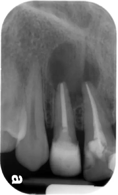
A well-circumscribed radiolucent lesion with a corticated outline was found on a radiograph related to the root of an upper lateral incisor. The tooth was not restored but was slightly discoloured.

A radicular cyst, also known as a periapical, apical, or dental cyst, is a type of cyst that develops around the root of a tooth. It is typically caused by chronic inflammation in the area, which is usually due to an infection or irritation of the dental pulp.

The development of a radicular cyst is influenced by osmotic pressure. Osmotic pressure refers to the movement of fluid from an area of lower solute concentration (inside the cyst) to an area of higher solute concentration (outside the cyst). This movement of fluid leads to the growth of the cyst, causing it to expand over time. As a result, radicular cysts are often rounded in shape and have a well-defined border or margin.

One of the common signs of a radicular cyst is tooth discoloration. The affected tooth may appear darker or grayer than the surrounding teeth. This discoloration is an indication that the tooth has lost its vitality or blood supply, which can occur when the cyst puts pressure on the nerves and blood vessels in the tooth’s pulp.

The development of a radicular cyst is usually triggered by periapical inflammation. Periapical inflammation refers to inflammation that occurs in the tissues around the apex (tip) of a tooth’s root. This inflammation can result from untreated dental caries (cavities), tooth fractures, or other factors that allow bacteria to enter the pulp and cause an infection.

It’s important to note that while radicular cysts can occur around any tooth, they are most commonly found in relation to the upper lateral incisor teeth. The exact reasons for this predilection are not entirely understood, but it may be due to the unique anatomy and susceptibility of these teeth to inflammation and infection.

Treatment of a radicular cyst typically involves removing the cyst surgically and addressing the underlying cause, such as root canal treatment or tooth extraction if necessary. It’s important for dental professionals to diagnose and manage radicular cysts promptly to prevent complications and maintain oral health.

I hope this explanation helps clarify the concept of a radicular cyst for you. If you have any further questions, feel free to ask!

Effect of C factor in bonding to root canal with or without fiber post

The C-factor, which is the ratio between bonded and non-bonded surfaces, has been found to have an effect on the bonding of fiber posts to root canal dentin. Studies have shown that a high C-factor, such as in the post hole, can lead to lower bond strength of the adhesive system to root canal dentin [3].

However, the use of intracanal medicaments such as CH, CH+CHX, and TAP have been found to enhance the pull-out bond strength of fiber posts to root canal wall as compared to the control group [1].

Other factors that have been found to influence the interfacial bond between fiber posts and root canal dentin include resin volume, adhesive system, root canal region, and the use of Er,Cr:YSGG laser irradiation [2][4][5].

Additionally, treatments such as Er:YAG laser irradiation with/without EDTA and the use of MTA have been found to enhance the bond strength of fiber posts to root canal dentin walls [7][6].

The effect of root canal irrigants such as CH and SH on the push-out bond strength of fiber posts cemented with an etch-and-rinse resin cement has also been studied, with CH showing a higher bond strength compared to SH [8].

Here are the references


[1] (2019). Effect of Intracanal Medicaments on Pull-Out Bond Strength of Fiber Post Cemented with a Self-Adhesive System. Eur J Dent, 03(13), 420-425. https://doi.org/10.1055/s-0039-1694313 [2] (2011).

Effects of C-factor and resin volume on the bonding to root canal with and without fibre post insertion. Journal of Dentistry, 6(39), 422-429. https://doi.org/10.1016/j.jdent.2011.03.007 [3] (2009).

Effect of relining on fiber post retention to root canal. J. Appl. Oral Sci., 6(17), 600-604. https://doi.org/10.1590/s1678-77572009000600012 [4] (2015).

Effect of Resin Cement Pre-heating on the Push-out Bond Strength of Fiber Post to Root Canal Dentin. J Dent Res Dent Clin Dent Prospects, 4(9), 233-238. https://doi.org/10.15171/joddd.2015.042 [5] (2012).

Effect of Er,Cr:YSGG pretreatment on bond strength of fiber posts to root canal dentin using a self-adhesive resin cement. Lasers Med Sci, 1(28), 65-69. https://doi.org/10.1007/s10103-012-1063-x [6] (2016).

Does MTA affect fiber post retention in repaired cervical root canal perforations?. Braz. oral res., 1(30). https://doi.org/10.1590/1807-3107bor-2016.vol30.0083 [7] (2013).

Efficacy of different treatments of root canal walls on the pull-out bond strength of the fiber posts. Lasers Med Sci, 2(30), 863-868. https://doi.org/10.1007/s10103-013-1457-4 [8] (2021).

Effect of Calcium Hypochlorite and Sodium Hypochlorite as Root Canal Irrigants on Push-Out Bond Strength of Fiber Post Cemented with Etch-and-Rinse Resin Cement: An In Vitro Study. fid. https://doi.org/10.18502/fid.v18i18.6325

Dental Caries Pathology – NEET Pearls from Dentest

  1. The widely accepted theory of dental caries is the acidogenic theory 
  2. Miller put forth the acidogenic theory of dental caries in the year = 1890
  3. The chemo-parasitic theory of dental caries is proposed by = MILLER
  4. The widely accepted theory of dental caries = Proteolytic chelation theory
  5. Environmental factors that contribute to dental caries include = nutrition, oral hygiene, fluoride and toothbrush abrasion 
  6. Sucrose = has been determined to be the most cariogenic carbohydrate 
  7. Streptococcus mutans synthesizes levans from = fructose
  8. Bacteria that adhere to tooth and cause caries is due to = the production of extracellular polysaccharides or dextrans 
  9. The extracellular polysaccharide synthesized by cariogenic streptococci in the presence of excess sucrose is best described as = dextran like glucan**
  10. Starch is considered to be less cariogenic than monosaccharides and disaccharides because = it does not diffuse through plaque
  11. Sorbitol acts in the prevention of dental caries by = microorganisms that lack the enzyme to metabolise sorbitol 
  12. Xylitol is a low-calorie sweetener that inhibits the growth of Streptococcus mutans
  13.  On average in the saliva of an adult man, the lactobacilli count is 70000 per ml 
  14. Pathology of dental caries is more closely related to = SUPRA-gingival plaque
  15. Pathology of periodontal disease is more closely related to = SUB- gingival plaque and Marginal plaque
  16.  Re-mineralisation of the tooth surface is associated with the following component of saliva = Histadine and Statherin
  17. STEPHAN’s CURVE
    1. the pH of the plaque = is related to the caries incidence and sugar intake of Stephan’s curve
    2. Stephan’s curve denotes the relationship between = plaque pH and glucose rinse 
    3. pH at 5.2-5.5 = initiation of caries begins
    4. TRUE STATEMENTS
      1. It depends on the amount of calcium and phosphorous in the saliva
      2. Bicarbonates are the chief buffer of saliva 
      3. At the highest pH, there is a net loss of minerals from the teeth
      4. pH = 5.5 
    5. The high viscosity of saliva is related to increasing caries in children is partially true 
    6. In a patient with reduced salivary flow, the caries incidence is = more than the patient with normal salivary flow 
    7. In carries free individuals, the saliva has = a high buffering capacity for acids
    8. The total caries experience in permanent teeth is greater in females than in males of the same age group. The reverse is applied to primary teeth
    9. Initiation of dental caries depends upon = the localization of acid over the tooth surface 
    10. For a bacterium to be seriously considered in the aetiology of dental caries it must = exist regularly in the dental plaque 
  18. MICROORGANISMS
    1. Streptococcus mutans
      1. It is considered to be a principal etiological agent of caries because it produces = organic acids and forms a gelatinous matrix 
      2. S. Sanguis, S.mitor, S.salivarius, S.milleri and Pepto streptococcus** = involved in the initiation of dental caries
      3. The enzyme glycosyl transferase secreted by S.mutans synthesizes glucans from = SUCROSE
    2. Lactobacillus acidophilus
      1. are numerous in caries lesions because they are = secondary invaders and help in the progression of caries, do not initiate smooth surface caries
      2. It is thought to be one of the etiological agents of dental caries because it is = both acidogenic and acidocuric
      3. The salivary peroxidase system is known to be effective against = lactobacillus acidophilus 
    3.  Organisms involved in deep smooth surface carries = Streptococcus mutans and Lactobacillus acidophilus 
    4. Actinomyces Viscosis and Lactobacillus = Initiates root surface caries***
  19. Smooth surface caries are characterized by the spread of caries in enamel and dentin as CONES, the alignment of the cone is = Apex to base 
  20. SELENIUM = Dental caries is higher in persons residing in seleniferous areas 
  21. Traces of molybdenum and vanadium in the diet may reduce the incidence of caries
  22. The anti-cariogenic mineral is = vanadium 
  23. In the early stages of the carious lesion. 
    1. Pioneer bacteria are seen in the area of = Dentinal Tubules/Dentin
    2. Early invading bacteria in carious lesion = Pioneer bacteria
    3. There is loss of = Interprismatic substance** of enamel with the increased prominence of rods 
    4. Accentuation of incremental lines of retizus is seen
    5. Clinically the earliest manifestation in an area of a calcification = resembles a smooth chalky white area 
  24. DENTINAL CARIES
    1. Microscopic zones of dentinal caries starting from DEJ = Bacterial invasion, de-calcification zone and zone of sclerosis 
    2. Miller’s liquefaction foci are present in = Dentinal caries in advanced cases
    3. The first zone to be found in dentinal caries = Miller’s liquefaction foci
    4. The clinical significance of sclerotic dentin is that = it is resistant to carries 
    5. Number of dentinal zones = 5
    6. Turbid dentin in carious tooth 
      1. Zone of bacterial invasion
      2. Zone which cannot be remineralised
      3. Zone in which collagen is irreversibly denatured
    7. Bacteria-free zone of dentinal caries = Zone of dentinal sclerosis 
  25. The lateral spread of caries is facilitated mostly by = dentino-enamel Junction 
  26. Mandibular 1st molar = Tooth in permanent dentition which is more susceptible to dental caries
  27. Mandibular 2nd Molar = Tooth in primary dentition which is more susceptible to dental caries
  28.  The maxillary arch is more frequently involved by caries than the mandible arch
  29. Probable reasons for a high incidence of dental caries in the teenage population relate more directly to = the frequency of sucrose intake
  30. The shape of the cervical form of dental caries = CRESCENT
  31. The most common surface involved in root carries in the maxillary arch is= the Proximal 
  32. Odontoclasia is = Linear enamel caries
  33. More number of microorganisms is seen = EARLY MORNING due to the long, overnight incubation period.
  34. Most pronounced effect on the oral microflora of reduction in the rate of salivary flow = Shift towards in rate of salivary flow
  35. Ammonia causes = a decrease in plaque formation
  36. Cavity formation in a tooth, due to dental caries is due to = Lateral spread of caries along DEJ and weakening of the outer covering enamel.
  37. Animals maintained in germ-free environments did not develop caries even when fed on a high carbohydrate diet given by = Orland and Fitzgerald
  38. Attachment of actinomyces species to the tooth surface is facilitated by fimbriae 
  39. The type of caries which are not clinically diagnosed but detected only on radiographs = OCCULT caries
  40. Linear enamel caries lesions in deciduous teeth predominate in = Maxillary anterior teeth