


Here is the google drive link – https://drive.google.com/file/d/1Mc0OOvAct65KCi2WRPT_Jt77Scdk-1oc/view?usp=sharing
Join our two-day hands-on workshop on porcelain veneers at Ivoclar Academy, Gurgaon, Delhi, India.
👉Dates: 27th and 28th October.
👉Limited to 15 participants only.
👉WhatsApp 8700383548 to register for the next batch.

To register, here is the website details – https://tmc.gov.in/m_conference_new/Conference/Onlinereg?confid=24290
Navigating the Antibiotic Puzzle in Maxillofacial Space Infections
Ah, the world of maxillofacial space infections, where microbes dance in the shadows, and antibiotics become our knights in shining armor. But, my dear readers, the antibiotic quest is no simple tale, for it’s a matter of choosing the right weapon against the unseen invaders.
🧫 Microbial Dance: The Cast of Characters
Antibiotics play a crucial role in the management of maxillofacial space infections. These infections can be caused by a wide variety of aerobic and anaerobic microorganisms (Mehedi et al., 2019). The choice of antibiotics should be based on the causative microorganisms and their susceptibility to different antimicrobial agents (Mehedi et al., 2019).
📋 Antibiotic Casting Call: The Right Players
In general, the main empiric antibiotics used for the treatment of oral and maxillofacial infections are amoxicillin-clavulanic acid, metronidazole, and erythromycin (Lee et al., 2022). However, it is important to note that the resistance to amoxicillin in dental infections can range from 9% to 54% (Lee et al., 2022).
🏥 Strategic Timing: Perioperative Antibiotics
In the management of maxillofacial space infections, the use of perioperative antibiotics is recommended to prevent postoperative infections (Lauder et al., 2010). The current standard of care is to administer antibiotics within 2 hours before surgery, as this has been shown to reduce the rates of surgical site infections (Lauder et al., 2010). However, the use of additional antibiotics outside the perioperative timeframe does not reduce the rate of postoperative infections (Lauder et al., 2010). It is worth noting that the use of additional antibiotics may be warranted in cases of severe facial trauma with multiple open fracture wounds (Lauder et al., 2010).
🕶️ The Antibiotic Hero: Clindamycin Takes Center Stage
In terms of antibiotic efficacy, a study conducted by found that clindamycin was the most effective single antibiotic, with a sensitivity rate of 90% in cases of orofacial space infections (Mehedi et al., 2019). Other effective single antibiotics included erythromycin (50%) and azithromycin (40%) (Mehedi et al., 2019). However, it is important to note that most orofacial space infections are caused by mixed microorganisms, making it difficult to treat them with a single empirical antibiotic (Mehedi et al., 2019).
🦠 Antibiotic-Resistant Drama: A Growing Plot Twist
The emergence of antibiotic-resistant bacteria is a growing concern in the management of maxillofacial infections. It has been reported that the overuse, abuse, and misuse of antibiotics contribute to the development of antibiotic-resistant bacteria (Yuvaraj, 2015). However, clinical observations have shown that the presence of penicillin-resistant strains in mixed microflora of odontogenic maxillofacial infections does not adversely affect the outcome of treatment when penicillin is prescribed as an adjunct to surgical drainage (Yuvaraj, 2015).
💊 Beyond Antibiotics: Multifaceted Strategies
In addition to antibiotic therapy, other treatment modalities may be used in the management of maxillofacial space infections. These include surgical drainage of the abscess, removal of the source of infection (such as extraction or endodontic therapy of the offending tooth), and the use of herbal anti-edematous agents to reduce post-operative swelling (Dongol et al., 2022; Dar-Odeh et al., 2018).
🔍 In Conclusion: The Script for Success
As the final act approaches, remember that the script for success depends on understanding the microbial ensemble and their antibiotic preferences. Perioperative antibiotics are the opening act, but the choice should be tailored to the situation. Keep an eye on the looming specter of antibiotic-resistant bacteria and let responsible stewardship guide the way.
RESEARCH ARTICLES WITH DOWNLOADABLE LINKS
REFERENCES
Dar-Odeh, N., Abu-Hammad, S., & Abu-Hammad, O. (2018). Herbal anti-edematous agents for certain cases of facial cellulitis of odontogenic origin. clinical recommendation.. The International Arabic Journal of Antimicrobial Agents, 8(3). https://doi.org/10.3823/825 Dongol, A., Bhattarai, N., Yadav, A., Acharya, P., Mahato, V., & Jaisani, M. (2022). Microbial flora and their antibiotic susceptibility in oral and maxillofacial infections at bpkihs: a prospective observational study. Journal of Bp Koirala Institute of Health Sciences, 5(1), 9-14. https://doi.org/10.3126/jbpkihs.v5i1.43381 Lauder, A., Jalisi, S., Spiegel, J., Stram, J., & Devaiah, A. (2010). Antibiotic prophylaxis in the management of complex midface and frontal sinus trauma. The Laryngoscope, 120(10), 1940-1945. https://doi.org/10.1002/lary.21081 Lee, H., Moon, S., Oh, J., Choi, H., Park, S., Kim, T., … & You, J. (2022). Eskape pathogens in oral and maxillofacial infections. Journal of Oral Medicine and Pain, 47(1), 52-61. https://doi.org/10.14476/jomp.2022.47.1.52 Mehedi, A., Chowdhury, G., Rab, A., & Haider, I. (2019). Evaluation of efficiency of conventional empirical antimicrobial regimen for the management of maxillofacial fascial space infection. Journal of Armed Forces Medical College Bangladesh, 11(2), 47-54. https://doi.org/10.3329/jafmc.v11i2.39823 Yuvaraj, V. (2015). Maxillofacial infections of odontogenic origin: epidemiological, microbiological and therapeutic factors in an indian population. Indian Journal of Otolaryngology and Head & Neck Surgery, 68(4), 396-399. https://doi.org/10.1007/s12070-015-0823-x
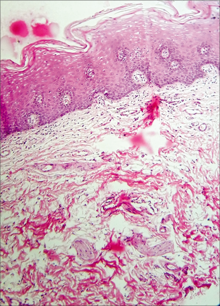
Sections show buccal mucosa in which there is mild epithelial atrophy with parakeratosis. The pattern of epithelial maturation is regular and the overall architecture is preserved. The rete processes are flattened and bands of hyaline collagen best seen in Van Geison stained sections are present in the lamina propria. A mild chronic inflammatory infiltrate is present in the subepithelial tissue.
Our adventure starts with a visit to the microscopic realm of the buccal mucosa – the inner lining of the cheek. Imagine a bustling cityscape with layers of epithelial cells, each playing its role in maintaining the oral harmony. But wait, something’s not quite right here!
🔬 Clue 1: The Atrophy Enigma
The buccal mucosa seems to be undergoing a transformation – a mild epithelial atrophy. It’s as if the cells are shrinking, losing some of their vitality. Parakeratosis is in play too, where these cells are holding onto their nuclei longer than they should. It’s like they’re not quite ready to grow up and shed their immature ways.
📜 Clue 2: The Architectural Anomaly
Despite the changes, the overall architectural blueprint of the buccal mucosa remains intact. The maturation of the epithelial cells follows a regular pattern, almost like well-practised dancers performing a choreographed routine. The rete processes – the finger-like projections that interlock the layers – appear flatter than usual. It’s as if they’re tired and can’t stand as tall as they used to.
🔍 Clue 3: The Mysterious Collagen Chronicles
Ah, now for a fascinating twist! Van Geison stained sections reveal bands of hyaline collagen lurking in the depths of the lamina propria – the supporting layer beneath the epithelium. These collagen bands are like secretive cobwebs, weaving a mysterious tale of their own. Their presence hints at something more profound beneath the surface.
🔥 Clue 4: The Inflammatory Intrigue
As our investigation deepens, we stumble upon an unexpected guest – a mild chronic inflammatory infiltrate. It’s almost like a small group of protesters voicing their concerns beneath the epithelial cityscape. What could they be protesting? What’s causing this subtle turmoil?
🚀 The Grand Reveal: Unveiling Submucous Fibrosis
Now, my fellow detectives, armed with our clues and insights, it’s time for the big reveal! The answer to this intriguing riddle is none other than Submucous Fibrosis.
🕵️♂️ Unraveling the Mystery
Submucous Fibrosis is a condition often linked to the chewing of paan (betel), a common practice in certain cultures. In this condition, dense collagenous bands sneakily weave their way into the lamina propria – that’s the collagen we spotted earlier! These bands tighten their grip, causing limitations in mouth opening and even trouble with swallowing.
But wait, there’s more! The potential consequences get even more serious. With these collagenous infiltrators running amok, there’s a risk of dysplasia – that’s abnormal cell growth – and even the development of oral cancer.
A 65-year-old man went through some serious stuff. 🙌 He had a mandibular rim resection and neck dissection to tackle squamous-cell carcinoma in the floor of his mouth and ventral tongue. 🦠 The pathologist’s notes spill the beans – the tumor was mainly hanging out in the mouth floor, measuring 28mm wide and 11mm deep. And guess what? Out of 48 lymph nodes from the neck dissection, 5 were playing host to some sneaky squamous-cell carcinoma guests, 2 even decided to venture out of their capsules! 🏃♂️💥 So, what’s the verdict?
So, in fancy doctor lingo aka Union for International Cancer Control, they call it pT2 when the tumor’s between 2 and 4cm big, and pN2b when you got a bunch of nodes involved but none are bigger than 6cm. 🤷♂️
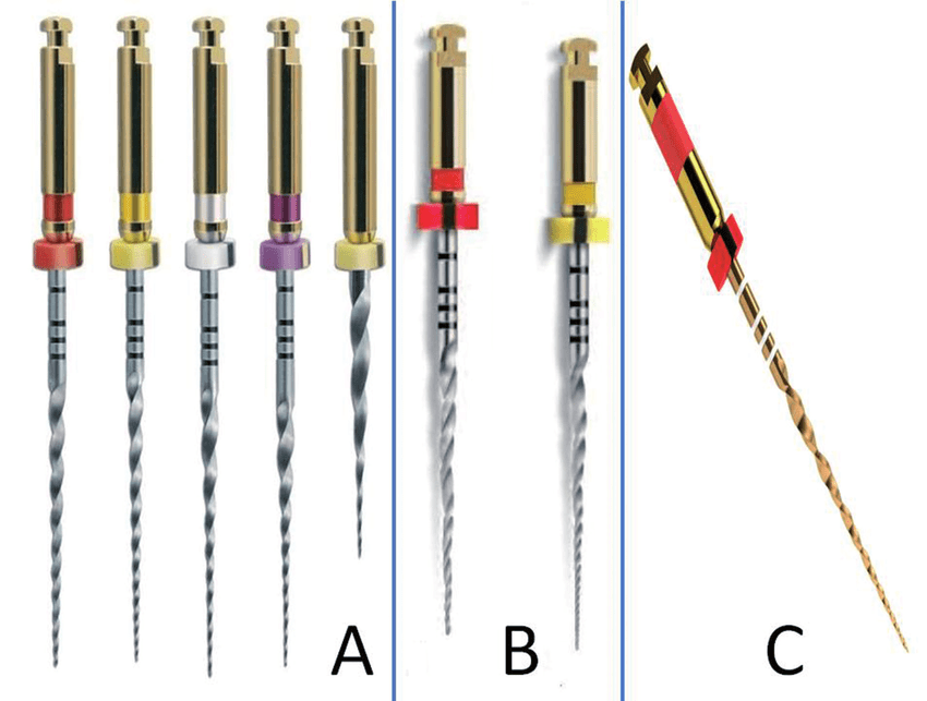
Fracture strength of roots instrumented with ProTaper Universal, ProTaper Next, and ProTaper Gold systems has been the subject of several studies. These studies have evaluated the influence of different factors on the quality of root canal preparation and the incidence of instrument fractures.
Duque et al. (2017) took a deep dive into the influence of NiTi wire in these systems on root canal preparation quality. Spoiler alert: it matters! 🧐
One study by Duque et al. (2017) aimed to evaluate the influence of the NiTi wire in ProTaper Universal (PTU) and ProTaper Gold (PTG) instrument systems on the quality of root canal preparation. The study used high-definition microcomputed tomography to scan twelve mandibular molars with separate mesial canals. The PTU and PTG instruments were used to shape twelve mesial canals each. The canals were scanned after preparation with F2 and F3 instruments of the PTU and PTG systems. The study found that the NiTi wire in both systems had an influence on the quality of root canal preparation.
Machado et al. (2018) did some detective work, analyzing when and where instrument fractures occurred. Surprise, surprise – operator experience played a role! 👨⚕️🔍
Another study by Machado et al. (2018) retrospectively analyzed the incidence of ProTaper Universal System instrument fractures. The study analyzed charts, clinical record cards, and radiographs of endodontic treatments performed using the ProTaper Universal System. The study found a low incidence of instrument fractures and identified the arch, group of teeth, and root thirds in which these fractures occurred.
In a similar study by (Machado et al., 2018), the incidence of ProTaper Universal System instrument fractures was evaluated in relation to the arch, group of teeth, and root thirds. The study aimed to evaluate the null hypothesis that complexity results in greater rotary flexure of the instrument, thereby concentrating the forces of stress that may cause premature failure of the NiTi alloy.
Analyzing the incidence of fracture of ProTaper Universal System instruments, Machado et al. (2018)found that the experience of the operators was one of the main reasons associated with higher instrument fracture rates. The study compared the fracture rates with previous studies and identified the operators’ experience as a contributing factor.
Siddique et al. (2020) went into battle against bacteria, testing these systems for their antibacterial prowess. XPendo Shaper emerged as the hero! 🦠💪
Siddique et al. (2020) conducted a clinical trial to compare the antibacterial effectiveness of three rotary file systems, including ProTaper Next, ProTaper Gold, and XPendo Shaper. The study used real-time polymerase chain reaction to evaluate the antibacterial effectiveness in root canals of teeth with asymptomatic apical periodontitis. The study found that XPendo Shaper showed better intracanal bacterial reduction than ProTaper Gold.
In another study by (Siddique et al., 2020), the effectiveness of ProTaper Next in bacterial reduction was affirmed by various in vitro studies. The study compared ProTaper Next with other file systems and found that ProTaper Next had higher bacterial reduction than Twisted file, ProTaper Universal, and manual techniques.
Milani et al. (2022) tested the resilience of teeth prepared with these systems and found that they didn’t all hold up the same. Different strokes for different folks, or in this case, teeth! 😬💥
The fracture resistance of roots prepared with ProTaper Universal, ProTaper Next, and ProTaper Gold rotary files was compared in a study by (Milani et al., 2022). The study evaluated the fracture resistance of fifty-six single-canal premolar teeth prepared with the three file systems. The study found differences in the fracture resistance of the teeth prepared with the different file systems.
Devi et al. (2021) looked for cracks induced by these systems during root canal prep. ProTaper got caught red-handed causing cracks in 25% of cases! 😱
Devi et al. (2021) conducted an in vitro study to assess dentinal defects induced by ProTaper Universal, ProTaper Gold, and Hyflex electric discharge machining (EDM) rotary file systems during root canal preparation. The study observed cracks in 25% of the roots instrumented with ProTaper at the apical root surface. The study compared the incidence of dentinal microcracks resulting from the use of different file systems.
Miguéns-Vila et al. (2017) inspected micro-cracks caused by ProTaper NEXT and ProTaper Universal and found that the control group had squeaky-clean roots! 😁🔍
The incidence of dentinal micro-cracks resulting from the use of ProTaper NEXT and ProTaper Universal systems was evaluated in a study by (Miguéns-Vila et al., 2017). The study used LED transillumination to analyze dentinal micro-crack formation at different points in the root canal. The study found no root defects in the control group.
Yusufoğlu et al. (2019) compared fracture resistance after using ProTaper and One Shape systems, showing that round cross-sectioned root canals are less likely to break a sweat! 💪🦷
The fracture resistance of roots enlarged with ProTaper and One Shape rotary systems was compared in an in vitro study by (Yusufoğlu et al., 2019). The study evaluated the fracture resistance of roots filled with different sealers. The study found that a round cross-sectioned root canal resulted in more homogeneous stress distribution and increased fracture resistance.
Agrawal et al. (2022) put these systems to the fatigue test, with Hyflex EDM emerging as the Iron Man of files! 🦾💥
Agrawal et al. (2022) conducted a study to compare the fracture resistance of Hyflex EDM, Neolix Neo NiTi, ProTaper Next, and ProTaper Gold files. The study evaluated the cyclic fatigue resistance of the different file systems and found that Hyflex EDM OneFile instruments showed greater cyclic fatigue resistance than ProTaper Gold Primary instruments, as well as ProTaper Next and Neolix NiTi.
In conclusion, several studies have evaluated the fracture strength of roots instrumented with ProTaper Universal, ProTaper Next, and ProTaper Gold systems. These studies have examined factors such as the influence of the NiTi wire, incidence of instrument fractures, antibacterial effectiveness, dentinal defects, and fracture resistance. The results of these studies provide valuable insights into the performance and characteristics of these rotary file systems.
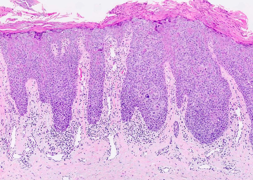
Sections show oral mucosa. In the oral epithelium there is basal-cell crowding and hyperplasia. Atypical mitotic figures are present throughout the thickness of the oral epithelium. The squamous cells show nuclear and cellular pleomorphism, and keratin whorls are present. The rete ridges are drop shaped and individual cell keratinisation is present in some areas.
The diagnosis here is 🔍 Carcinoma in situ! 🦠
In simple terms, it’s like a full-blown drama show happening in the oral epithelium! 🎭 The cells are misbehaving – basal-cell crowding, hyperplasia, and atypical mitotic figures are causing chaos! 🤯🔬
The squamous cells are like divas with nuclear and cellular pleomorphism, and there are even keratin whorls for added glam! 💁♀️✨
The rete ridges are shaped like drops, adding some artistic flair, and individual cell keratinisation is stealing the spotlight! 💅🌟
Now, here’s the twist – severe epithelial dysplasia is often considered a prelude to this drama, and together they’re sometimes known as Squamous Intraepithelial Neoplasia Grade 3 (SIN 3)! 📜🌆
Hey, dental explorers! 🌟 Let’s journey into the captivating realm of implant cast accuracy and the intricate interplay between implant angulations and impression techniques! 🦷🔬
The literature is buzzing with studies unveiling the secrets of digital vs. conventional implant impressions and the impact of those pesky implant angles on accuracy.
📚 Basaki et al. (2017) dived into digital vs. conventional impressions. For digital magic, angulation wasn’t a game-changer. But in the conventional realm, the material, connection type, and a dash of angulation might have stirred the accuracy potion.
Basaki et al. (2017) conducted a study comparing the accuracy of digital and conventional implant impression approaches. They found that for a digital impression approach, where material strain is not a concern, implant angulation did not have a significant influence on impression accuracy. This finding is consistent with previous studies that have also reported no effect of implant angulation on digital impression accuracy. However, the lack of influence on the error in the conventional approach could be potentially explained by the moderate angulation, choice of the impression material, and implant connection type.
🔍 Martínez-Rus et al. (2013) took on multiple implant systems and found metal-splinted direct technique ruling the accuracy charts. The journey went from most accurate to less-so with acrylic resin-splinted, indirect, and unsplinted direct methods.
Martínez-Rus et al. (2013) evaluated the effect of four implant-level impression techniques on the accuracy of definitive casts for a multiple internal connection implant system with different implant angulations and subgingival depths. They found that the metal-splinted direct technique produced the most accurate casts, followed by the acrylic resin-splinted direct, indirect, and unsplinted direct techniques. The study also reported that the accuracy of impressions for internal connection implants decreased as the divergence angle between implants increased.
🔬 Elshenawy et al. (2018) faced angulated implants head-on. Distortion danced with angulation, and unsplinted vs. acrylic resin-splinted techniques fought it out. Up to 15°, direct methods scored in accuracy.
Elshenawy et al. (2018) conducted a study comparing the dimensional accuracy of casts obtained from three impression techniques for three definitive lower casts with implants at different angulations. They found that implant angulation affected the impression accuracy, with increased angulation resulting in increased distortion. The study also reported that the direct unsplinted technique and direct acrylic resin-splinted technique exhibited more accuracy compared to the indirect technique when angulation of implants increased up to 15°.
🌐 Arora et al. (2019) explored parallel vs. angulated implants in the splinted vs. nonsplinted battle. The crown? Splinted technique won in angulated implants, staying true to parallel’s perfection.
Arora et al. (2019) evaluated the accuracy of implant casts generated with splinted and nonsplinted impression techniques with multiple parallel and nonparallel implants. They found that the splinted technique in angulated implants exhibited greater accuracy compared to the nonsplinted technique in parallel implants. This finding was consistent with previous studies that reported less accurate impressions with angulated implants than parallel implants.
📊 Parameshwari et al. (2018) simulated unilateral partially edentulous scenarios. The pick-up technique charmed for multiple angulated implants.
Parameshwari et al. (2018) investigated the effects of implant angulation, type of impression material, and tray selection on impression accuracy in simulated master casts of unilateral partially edentulous situations. They found that there was no statistically significant difference in the accuracy of pick-up non-splinted and transfer techniques when there were three or fewer implants, but the pick-up technique produced superior accuracy for multiple implants with implant angulation more than 20 degrees.
In a nutshell, the harmony of implant cast accuracy is a symphony composed of angulation, impression technique, material, and the magic of digital vs. conventional. Remember, digital might be less angulation-sensitive. And whether you’re picking up or transferring, the choice matters in the land of precision! 🧙♀️🏰
Arora, A., Upadhyaya, V., Parashar, K., Malik, D. (2019). Evaluation Of the Effect Of Implant Angulations And Impression Techniques On Implant Cast Accuracy – An In Vitro Study. The Journal of Indian Prosthodontic Society, 2(19), 149. https://doi.org/10.4103/jips.jips_337_18 Basaki, K., Alkumru, H., Souza, G., Finer, Y. (2017). Accuracy Of Digital Vs Conventional Implant Impression Approach: a Three-dimensional Comparative In Vitro Analysis. The International Journal of Oral & Maxillofacial Implants, 4(32), 792-799. https://doi.org/10.11607/jomi.5431 Elshenawy, E., Alam-Eldein, A., Elfatah, F. (2018). Cast Accuracy Obtained From Different Impression Techniques At Different Implant Angulations (In Vitro Study). International Journal of Implant Dentistry, 1(4). https://doi.org/10.1186/s40729-018-0118-6 Martínez-Rus, F., García, C., Santamaría, A., Özcan, M., Pradíes, G. (2013). Accuracy Of Definitive Casts Using 4 Implant-level Impression Techniques In a Scenario Of Multi-implant System With Different Implant Angulations And Subgingival Alignment Levels. Implant Dentistry, 3(22), 268-276. https://doi.org/10.1097/id.0b013e3182920dc5 Parameshwari, G., Chittaranjan, B., Sudhir, N., Ck, A., Taruna, M., M, R. (2018). Evaluation Of Accuracy Of Various Impression Techniques and Impression Materials In Recording Multiple Implants Placed Unilaterally In A Partially Edentulous Mandible- An In Vitro Study. Journal of Clinical and Experimental Dentistry, 0-0. https://doi.org/10.4317/jced.54726
