The angulation of the Twin Block appliance’s inclined planes underwent three distinct stages of development, each driven by clinical observations and biomechanical reasoning.
Stage 1 — 90° (Initial Design)
The earliest Twin Block appliances, developed by W.J. Clark, featured bite blocks angulated at 90° to the occlusal plane. Patients were required to consciously posture the mandible forward to occlude the blocks. However, many patients struggled to maintain this forward position and habitually returned to their original distal occlusal position, causing the flat-surfaced blocks to stack on top of each other. This resulted in a significant posterior open bite, a complication seen in approximately 30% of early Twin Block cases.
Stage 2 — 45° (Functional Correction)
To resolve the compliance problem, the angulation was modified to 45° to the occlusal plane. This immediately guided the mandible forward more passively, eliminating the stacking issue. A 45° angle provides an equal downward and forward force component to the lower dentition, promoting both vertical and sagittal growth stimuli. Clark continued using this angulation clinically for approximately 8 years before the next modification.
Stage 3 — 70° (Current Standard)
After the prolonged use of the 45° design, the angulation was increased to 70° to the occlusal plane — the current standard configuration. This steeper angle introduces a more horizontal force component, theoretically encouraging greater forward (sagittal) mandibular growth rather than a combined downward-forward stimulus. The 70° angle is now incorporated into the standard Twin Block design with maxillary and mandibular acrylic base plates.
Angulation at a Glance
Angulation
Rationale
Limitation
Angulation
Rationale
Limitation
90°
Original design; edge-to-edge block contact
~30% posterior open bite; poor compliance
45°
Equal forward + downward force vector
Used for 8 years; less horizontal growth stimulus
70°
More horizontal force; greater forward mandibular growth
May reduce mandibular postural guidance
Clinically, if a patient struggles to maintain the forward mandibular posture with a 70° design, it is advisable to revert to a 45° angulation to facilitate easier maintenance of the protruded position.
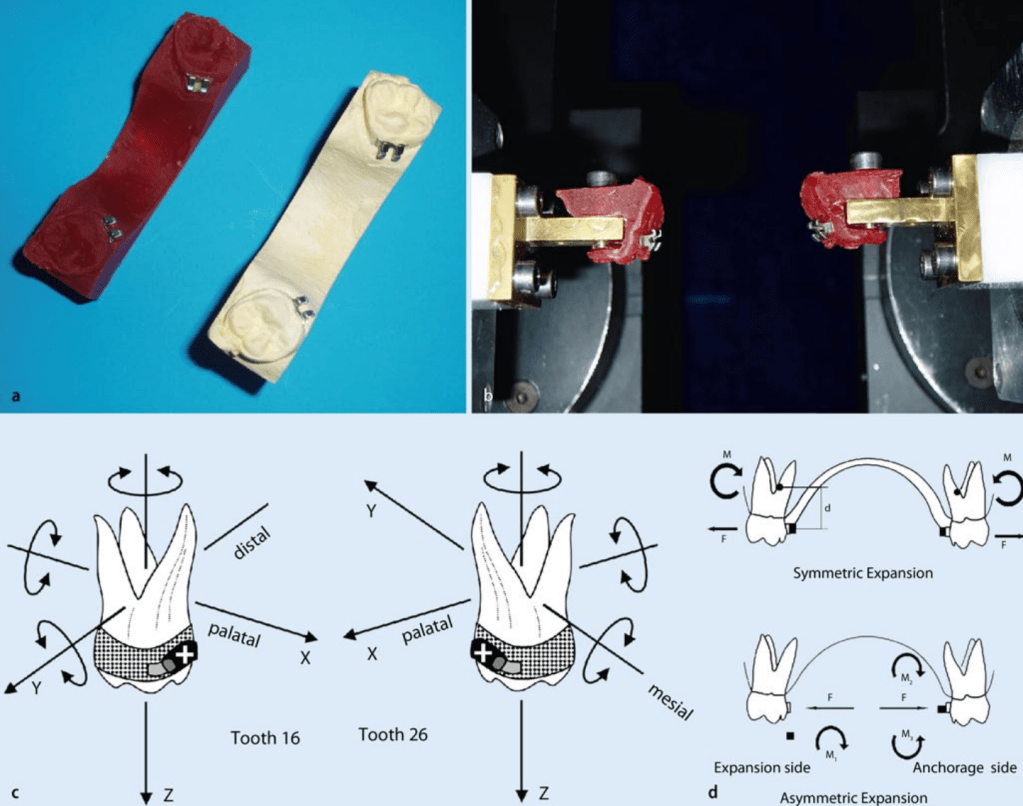
Posterior crossbite is one of the most common transverse discrepancies encountered in orthodontic practice. A transpalatal arch (TPA) is a deceptively simple appliance — but when activated using Burstone biomechanics, it becomes a powerful tool capable of producing controlled symmetric or asymmetric molar expansion.
Understanding force systems, moment-to-force ratios, and side effects is essential if one wants to use this appliance predictably.
This article walks through the biomechanics, clinical application, and outcomes of a Burstone-type TMA transpalatal arch.
1. Why Molar Transverse Position Matters
Correct positioning of maxillary first molars is critical for:
Functional occlusion
Arch coordination
Midline stability
TMJ health
Untreated transverse maxillary deficiency may cause:
Posterior crossbite
Functional mandibular shift
Midline deviation
TMJ strain
Posterior crossbite prevalence:
Unilateral: ~9%
Bilateral: ~4%
Quick Viva Pause
Q: Why is unilateral crossbite more problematic than bilateral crossbite?
A: Because it frequently causes functional mandibular shift, leading to asymmetry and midline deviation.
2. What is a Burstone-Type Transpalatal Arch?
A transpalatal arch (TPA) connects the maxillary first molars across the palate.
It can be used in two modes:
Mode
Purpose
Passive
Anchorage reinforcement / stabilization
Active
Tooth movement
The Burstone system differs from traditional TPA systems.
Key Differences
Feature
Burstone TPA
Goshgarian TPA
Attachment
Lingual bracket
Lingual sheath
Wire material
TMA
Stainless steel
Force magnitude
Lower
Higher
Control
High precision
Less controlled
TMA wires produce ~60% lower force compared to stainless steel, improving control and reducing unwanted side effects.
Viva Pause
Q: Why is TMA preferred over stainless steel in Burstone TPA?
Answer
Lower load-deflection rate
Greater formability
More controlled force delivery
Reduced risk of excessive tipping
3. Recommended Activation
Typical parameters reported:
Parameter
Value
Activation
3–10 mm
Expansive force
1.5–4 N
Wire dimension
0.032 × 0.032 TMA
A 10 mm activation produces approximately 4 N expansion force.
However, force depends on:
Wire length
Loop configuration
Height of arch
Patient anatomy
Viva Pause
Q: What happens if the TPA height increases?
Answer
The moment-to-force ratio changes, altering the type of tooth movement.
4. Types of Expansion Using TPA
1. Symmetric Expansion
Both molars move buccally.
Used for:
Bilateral posterior crossbite
Narrow maxilla
2. Asymmetric Expansion
One side expands more than the other.
Used for:
Unilateral crossbite
This is achieved by creating moment differential between molars.
Biomechanical Principle
Side
Force System
Crossbite side
Force → tipping movement
Anchorage side
Force + counter-torque
This allows unilateral expansion without significant movement of the anchorage molar.
Viva Pause
Q: Why is tipping used on the crossbite side?
Answer
Because tipping requires less force than bodily movement, making unilateral correction easier.
5. Biomechanics of Burstone TPA
The appliance generates:
Force component
Effect
Expansive force
Buccal movement
Moment
Crown tipping
Vertical force
Minor extrusion/intrusion
The center of resistance of molars lies approximately:
7 mm apical to the bracket level in the furcation region.
Viva Pause
Q: Why does TPA cause buccal crown tipping?
Answer
Because the force is applied away from the center of resistance, creating a moment that tips the crown buccally.
6. Clinical Outcomes (Study Findings)
Symmetric Expansion
Parameter
Result
Mean expansion
~4.5 mm
Buccal tipping
~10°
Treatment time
12 weeks
Vertical side effects
Minimal
Expansion occurred primarily due to buccally directed forces acting at the crown level.
Viva Pause
Q: What is the main disadvantage of symmetric TPA expansion?
Answer
Buccal crown tipping of molars, which may require later torque correction.
7. Asymmetric Expansion Outcomes
For unilateral crossbite:
Parameter
Crossbite Side
Anchorage Side
Tooth movement
~2.5 mm
~0.8 mm
Torque
Higher
Lower
Vertical movement
Minimal
Minimal
Thus effective unilateral expansion was achieved in all patients.
Viva Pause
Q: Why does the anchorage side show less movement?
Answer
Because counter-torque increases moment-to-force ratio, resisting tipping.
8. Side Effects
Vertical Effects
Movement
Magnitude
Intrusion
~0.6 mm
Extrusion
~0.8 mm
These are considered clinically insignificant.
Sagittal Effects
Minor:
Mesial rotation of molars
Minimal sagittal displacement
Viva Pause
Q: What is the most common rotational side effect?
Answer
Mesial rotation of molars
9. Why Simulation Systems Were Used
The study used Orthodontic Measurement and Simulation System (OMSS).
Purpose:
Measure force systems
Predict tooth movement
Compare simulation vs clinical outcomes
Findings:
Simulated movements were highly consistent with clinical results.
Viva Pause
Q: Why can’t simulation fully replicate real orthodontic tooth movement?
Answer
Because it cannot account for:
Mastication
Occlusal contacts
Soft tissue forces
Material fatigue
Biological variability
10. Clinical Pearls for Orthodontists
1. TPA is not just an anchorage appliance
It can produce controlled molar movement.
2. Shape matters
Force depends on:
Height
Length
Configuration
3. Perfect force systems are difficult
Even identical activation may produce different forces due to anatomical variation.
4. Tipping is expected
Crossbite correction usually occurs by molar tipping rather than bodily movement.
5. Torque correction may be needed later
After expansion, clinicians may need to:
Add counter-torque
Use archwire adjustments
Rapid Revision Table
Feature
Symmetric Expansion
Asymmetric Expansion
Indication
Bilateral crossbite
Unilateral crossbite
Force system
Equal bilateral forces
Differential moment
Mean expansion
~4.5 mm
~2.5 mm on affected side
Crown tipping
Present
Controlled
Side effects
Minimal
Minimal
Ultimate Viva Questions (PG Level)
Basic
1. What is the function of a transpalatal arch?
Anchorage control
Molar rotation control
Transverse expansion
Intermediate
2. Why is TMA preferred in Burstone TPA?
Lower load-deflection rate
Better formability
More controlled forces
Advanced
3. How does asymmetric TPA correct unilateral crossbite?
By generating different moment-to-force ratios on each molar.
Clinical
4. What is the most common side effect of TPA expansion?
Buccal crown tipping.
Biomechanics
5. Why does tipping occur with TPA?
Force acts away from center of resistance, generating a moment.
Final Takeaway
The Burstone-type TPA is a biomechanically sophisticated appliance capable of producing:
Controlled symmetric molar expansion
Targeted asymmetric correction of unilateral crossbite
Minimal side effects
When understood biomechanically, it transforms from a simple wire into a precise orthodontic force delivery system.
If you’ve ever wondered why one functional appliance seems to “work better” than another — or why your supervisor prefers the Herbst over the Activator — the answer might lie in a single, elegant metric: the coefficient of efficiency.
What Is the Coefficient of Efficiency?
Imagine two appliances, both claiming to stimulate mandibular growth. One achieves 6 mm of supplementary elongation in 12 months. Another achieves the same 6 mm, but takes 24 months. Are they equally effective? Technically yes — but practically, no.
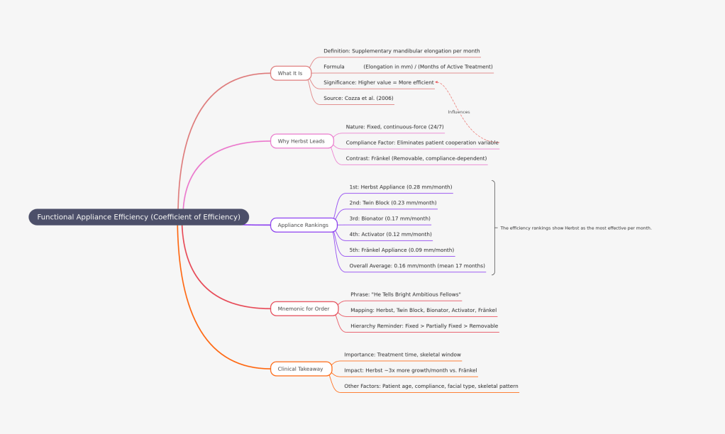
This is exactly the problem that Cozza, Baccetti, Franchi, De Toffol, and McNamara Jr. sought to address in their landmark 2006 systematic review published in the American Journal of Orthodontics and Dentofacial Orthopedics. They proposed a simple but powerful formula:
Coefficient of Efficiency=Months of active treatmentSupplementary mandibular elongation (mm)
In plain terms — how many millimetres of extra jaw growth does the appliance produce per month of wear? The higher the number, the more efficient the appliance.
The Rankings: Who Wins?
Cozza et al. analyzed 22 studies (4 RCTs + 18 CCTs) spanning literature from 1966 to 2005. Here’s how the five major functional appliances stacked up:
Rank
Appliance
Coefficient of Efficiency
🥇 1st
Herbst Appliance
0.28 mm/month
🥈 2nd
Twin Block
0.23 mm/month
🥉 3rd
Bionator
0.17 mm/month
4th
Activator
0.12 mm/month
5th
Fränkel Appliance
0.09 mm/month
The overall average across all appliances was 0.16 mm/month, with a mean active treatment duration of approximately 17 months.
Why Does the Herbst Appliance Lead?
The Herbst appliance is a fixed, continuous-force device — it works 24/7, regardless of patient cooperation. This relentless, round-the-clock mandibular advancement is the primary reason it tops the efficiency chart at 0.28 mm/month.
In contrast, the Fränkel appliance sits at the bottom (0.09 mm/month) — not because it’s biologically inferior, but because it is a tissue-borne, removable appliance heavily dependent on patient compliance. Worn only part of the day, its per-month output naturally dilutes.
The lesson? Compliance is a hidden variable in efficiency. Fixed appliances eliminate this variable; removable ones are at its mercy.
Think: “He Tells Bright Ambitious Fellows” — going from the most efficient to the least.
Or simply associate the appliance type with compliance demand:
Fixed (Herbst) = Highest efficiency
Partially fixed (Twin Block) = Second
Removable (Bionator, Activator, Fränkel) = Lower, in descending order
The Clinical Takeaway
For busy orthodontic practices where treatment time matters — especially in growing patients with a closing window of skeletal opportunity — choosing a more efficient appliance can make a meaningful difference. A patient treated for 18 months with a Herbst gains the equivalent of roughly 3× more supplementary mandibular growth per month compared to a Fränkel wearer.
That said, efficiency isn’t everything. Patient age, compliance, facial type, and skeletal pattern all factor into appliance selection. But the next time someone asks “which functional appliance works best?” — you now have the data to give a precise, evidence-based answer.
Reference: Cozza P, Baccetti T, Franchi L, De Toffol L, McNamara JA Jr. Mandibular changes produced by functional appliances in Class II malocclusion: a systematic review. Am J Orthod Dentofacial Orthop. 2006 May;129(5):599.e1-12. PMID: 16679196.
In orthodontics, one of the greatest clinical advantages you can develop is predictability. The ability to anticipate how a patient will respond to treatment—especially functional appliance therapy—can transform your treatment plan, appliance choice, and patient counseling. Yet many students focus on memorizing appliance designs while overlooking the cephalometric predictors that actually determine whether treatment will succeed.
One of the most valuable—but often underemphasized—predictive tools lies in understanding mandibular morphology and growth potential, particularly concepts such as the Stutzman angle and the Co–Go–Me angle.
The Landmark Study That Shifted Prognostic Thinking
A pivotal investigation by Lorenzo Franchi and Tiziano Baccetti evaluated pretreatment cephalometric predictors of mandibular growth response in Class II patients treated during peak pubertal growth.
They analyzed 51 patients who underwent functional therapy with Twin Block or Herbst appliances at CS3 (peak growth stage). Importantly, their outcome measure was actual mandibular growth increase, not merely occlusal correction—making the findings especially clinically meaningful.
The Co–Go–Me Angle: A Powerful Prognostic Indicator
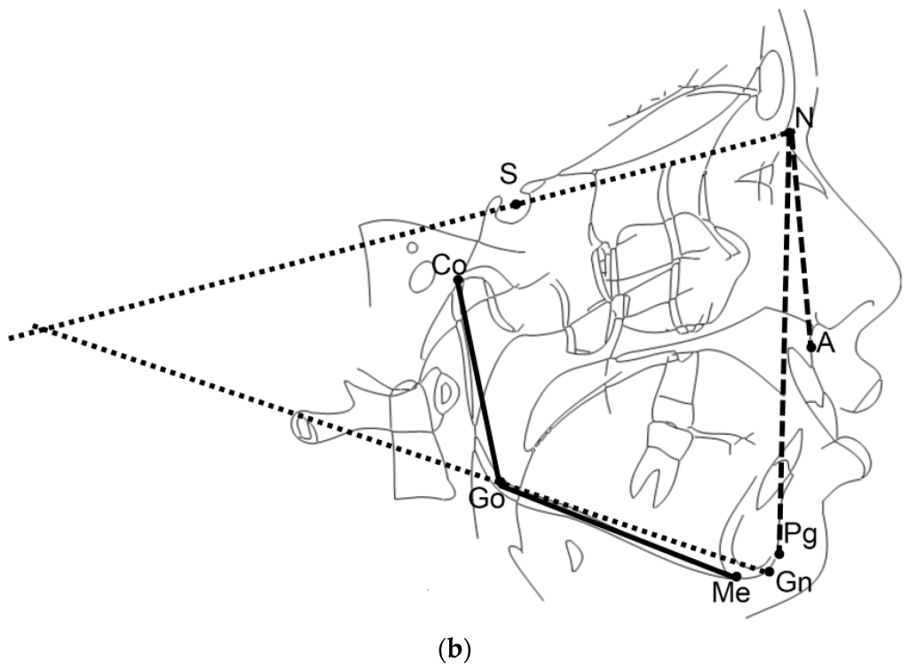
The mandibular angle Co–Go–Me (condylion–gonion–menton) has emerged as a highly practical predictor of treatment response.
< 125–125.5° → Favorable prognosis
> 125.5° → Poor prognosis
Interpretation Table
Value
Prognosis
Clinical Meaning
< 125.5°
Favorable
Strong mandibular growth potential
> 125.5°
Unfavorable
Limited skeletal response expected
Patients with smaller Co–Go–Me angles typically demonstrate greater mandibular growth during functional appliance therapy.
Additional Cephalometric Features That Predict Success
A strong skeletal response is more likely when the patient also presents with:
High Jarabak ratio (greater posterior vs anterior facial height)
Together, these features indicate a horizontal growth pattern, which is biologically more responsive to mandibular advancement therapy.
Viva one-liner: Co–Go–Me < 125° with low MP angle, low basal plane angle, and high Jarabak ratio indicates good prognosis for functional appliance therapy in Class II patients.
Memory Hook
Low angle = Grower → Treat confidently with functional appliance
The Stutzman Angle: Direction Matters as Much as Amount
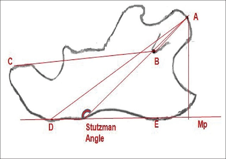
While Co–Go–Me predicts how much growth may occur, the Stutzman angle provides insight into how the mandible grows.
Definition: The Stutzman angle is formed between:
the condylar process axis (line from the most posterosuperior condylar point to the midpoint of the mandibular foramen), and
the mandibular plane
Clinical Significance
This angle reflects directional growth and biologic response, not just magnitude. It is especially useful for monitoring treatment progress over time.
Change
Meaning
Clinical Interpretation
Increase (Opening)
Condylar axis elongates/rotates
Active growth or forward positioning
No change
Minimal structural change
Limited skeletal response
Decrease (Closing)
Remodeling
Stabilization after advancement
Clinical rule: Opening = growth or advancement Closing = remodeling or stabilization
Why These Predictors Matter
Understanding these angles allows clinicians to move beyond trial-and-error treatment. Instead of hoping a functional appliance will work, you can predict response before treatment begins, improving:
Case selection
Treatment timing
Appliance choice
Patient counseling
Clinical confidence
In modern orthodontics, success isn’t just about mechanics—it’s about biologic forecasting. And mastering predictors like the Co–Go–Me and Stutzman angles gives you that edge.
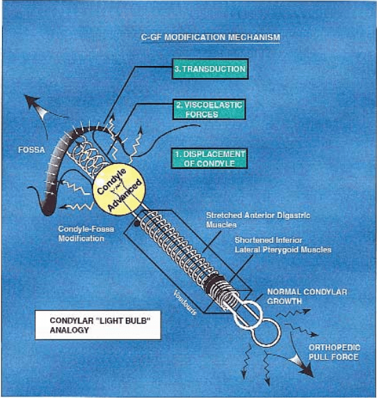
If you’ve ever wondered how functional appliances actually stimulate mandibular growth, this is the idea that changes everything. Not muscles. Not magic. Not forced growth.
Instead — growth is relative.
Let’s break it down so clearly that you’ll remember it even during a 3 AM exam panic.
The Big Idea in One Line
Mandibular advancement doesn’t create new growth — it redirects existing growth potential through biomechanical signaling.
Why This Hypothesis Was Needed
For years, people believed that forward posturing appliances worked mainly because muscles became hyperactive and stimulated bone growth.
But that didn’t fully explain:
why growth changes occur even when muscles adapt
why both condyle and glenoid fossa remodel together
why relapse can occur when advancement stops
So researchers proposed the Growth Relativity Hypothesis — most notably explained by Voudouris.
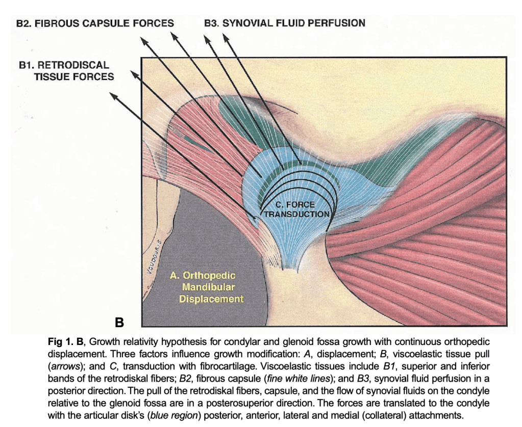
The Three Forces That Actually Drive Growth
Think of mandibular advancement like stretching a spring-loaded system. Three biological forces start working simultaneously:
1️⃣ Displacement — The Trigger
When a functional appliance holds the mandible forward:
the condyle is physically displaced from its original fossa position
the joint must adapt to this new relationship
👉 Displacement = switch turns ON
2️⃣ Viscoelastic Tissue Pull — The Driver
Non-muscular tissues stretch:
retrodiscal tissues
capsule
ligaments
synovial structures
These tissues behave like elastic bands trying to pull the condyle back.
👉 This pull generates continuous biological signals.
3️⃣ Transduction Through Fibrocartilage — The Builder
The stretched forces don’t stay localized.
They spread through:
condylar fibrocartilage
glenoid fossa lining
This mechanical signaling stimulates:
bone apposition
remodeling
adaptive growth
👉 Transduction = signal converted into growth
The Golden Principle
Growth is not increased. It is redirected.
The condyle and fossa simply:
grow relative to their new displaced relationship
They are adapting — not overgrowing.
The Light-Bulb Memory Trick 💡
Imagine condylar growth as a light bulb with a dimmer switch:
Appliance activation → brightness increases
Tissue stretch → keeps light on
Appliance removal → light dims
You don’t create electricity. You just turn the dial.
Why Relapse Happens (And Students Forget This!)
After appliance removal:
stretched tissues recoil
muscles regain original balance
joint tries returning to old position
If retention isn’t managed → relapse tendency
The One Sentence You Should Write in Exams
Condylar and glenoid fossa growth during mandibular advancement is governed by displacement, viscoelastic tissue forces, and fibrocartilage force transduction, producing adaptive remodeling rather than true growth stimulation.
Memorize that line and you can answer:
theory questions
viva questions
mechanism questions
comparison questions
Ultra-Simple Analogy (Final Memory Lock 🔒)
Functional appliance = moving a plant toward sunlight You didn’t make the plant grow. You just changed where it grows.
Definition: Viscoelasticity describes the combination of viscous (fluid-like) and elastic (solid-like) properties exhibited by biological tissues. It primarily applies to elastic tissues such as muscles, but the concept extends to all non-calcified tissues.
Key Concepts:
It concerns both viscosity and flow of synovial fluids and elasticity of soft tissues including:
Retrodiskal tissues
Fibrous capsule
TMJ ligaments and tendons
Lateral pterygoid muscle (LPM) perimysium
Other non-muscular, non-mineralized soft tissues
Essentially, it explains how these tissues deform under stress and recover when the stress is removed, with a time-dependent response.
Historical Notes:
The concept faced opposition from Herren (1953), Harvold (1974), and Woodside (1973) to the original Anderson–Haupl theory, which had a different interpretation of joint tissue adaptation.
Stages of the Viscoelastic Reaction
The viscoelastic reaction proceeds through five sequential stages:
Emptying of blood vessels – initial vascular response to stress.
Pressing out interstitial fluid – displacement of tissue fluids to redistribute pressure.
Stretching of fibres – collagen and elastic fibers undergo elongation.
Elastic deformation of bone – bone matrix responds elastically under load.
Bioplastic adaptation – long-term remodeling and adaptation of supporting tissues.
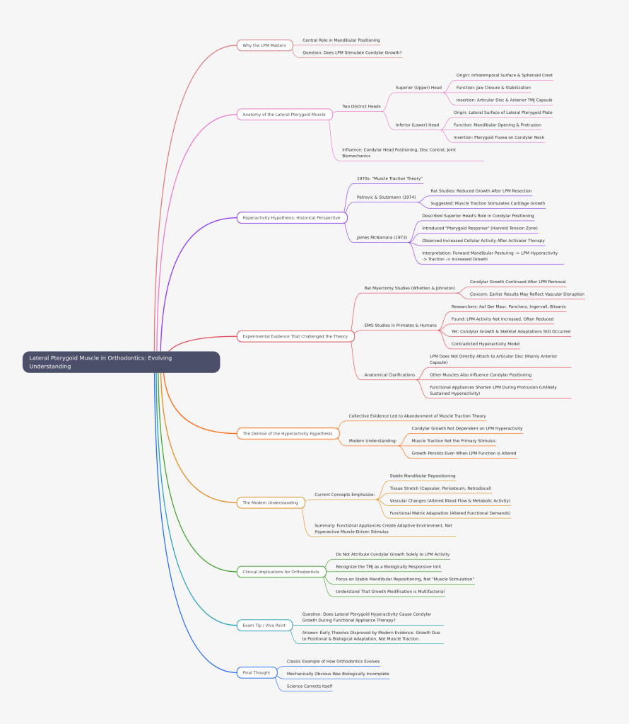
In orthodontics, few topics have sparked as much debate as the role of the lateral pterygoid muscle (LPM) in functional appliance therapy. Once considered the prime driver of condylar growth through “hyperactivity,” the LPM has since undergone a scientific re-evaluation.
Let’s explore how our understanding evolved.
Why the Lateral Pterygoid Matters
The LPM plays a central role in mandibular positioning, particularly during protrusive and lateral movements. Because functional appliances posture the mandible forward, early researchers naturally questioned:
Does the lateral pterygoid muscle stimulate condylar growth through traction?
To understand the controversy, we must first revisit its anatomy.
Anatomy of the Lateral Pterygoid Muscle
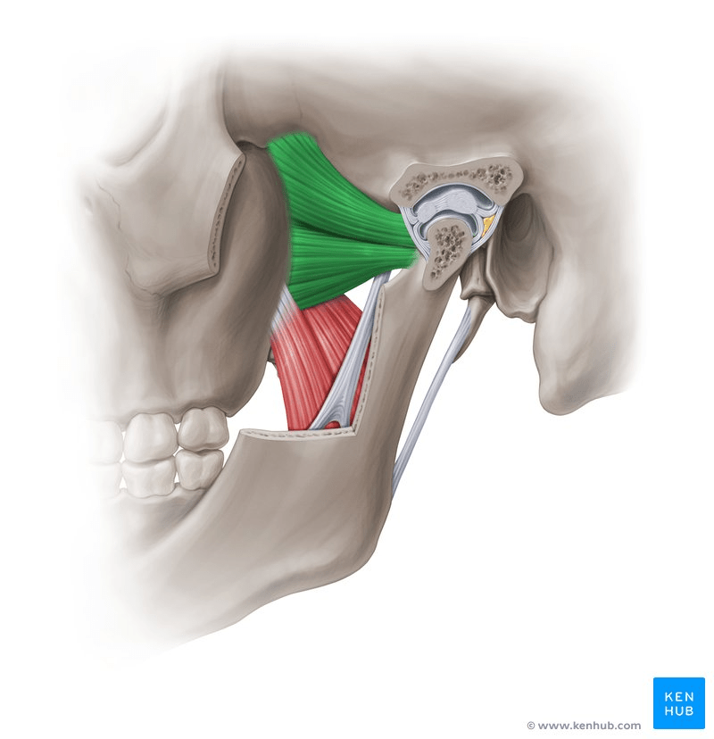
The LPM has two distinct heads:
🔹 Superior (Upper) Head
Origin: Infratemporal surface and crest of the greater wing of the sphenoid
Function: Active during jaw closure and stabilization
Insertion: Primarily into the articular disc and anterior capsule of the TMJ
🔹 Inferior (Lower) Head
Origin: Lateral surface of the lateral pterygoid plate
Function: Active during mandibular opening and protrusion
Insertion: Pterygoid fovea on the condylar neck
Both heads converge posteriorly and influence condylar head positioning, disc control, and joint biomechanics.
The Hyperactivity Hypothesis: A Historical Perspective
In the 1970s, the “muscle traction theory” dominated thinking.
🔬 Petrovic & Stutzmann (1974)
Rat studies showed reduced condylar growth after LPM resection.
Suggested that muscle traction stimulates condylar cartilage growth.
📚 James McNamara (1973)
Described the role of the superior head in condylar positioning.
Introduced the concept of the “Pterygoid Response” (also called the Harvold Tension Zone).
Observed increased cellular activity above and behind the condyle following activator therapy.
The interpretation? Forward mandibular positioning → LPM hyperactivity → Traction on condyle → Increased growth.
It seemed biologically elegant and mechanically convincing.
Experimental Evidence That Challenged the Theory
Science, however, demands replication and scrutiny.
🧪 Rat Myectomy Studies (Whetten & Johnston)
Condylar growth continued even after LPM removal.
Raised concerns that earlier results may have reflected vascular disruption rather than true traction effects.
📈 EMG Studies in Primates and Humans
Researchers such as:
Auf Der Maur
Pancherz
Ingervall
Bitsanis
found that during functional appliance therapy:
LPM activity was not increased
In many cases, LPM activity was actually reduced
Yet condylar growth and skeletal adaptations still occurred
This contradicted the hyperactivity model.
Anatomical Clarifications
Further anatomical studies revealed:
The LPM does not directly attach to the articular disc as previously thought.
Its attachment is mainly to the anterior capsule, not firmly to the disc.
Other muscles (temporalis, masseter) also influence condylar positioning.
Functional appliances actually shorten the LPM during protrusion, making sustained hyperactivity biomechanically unlikely.
This was a critical turning point.
The Demise of the Hyperactivity Hypothesis
The collective evidence led to abandonment of the muscle traction theory.
Today we understand:
✔ Condylar growth is not dependent on LPM hyperactivity ✔ Muscle traction is not the primary stimulus ✔ Growth persists even when LPM function is altered
So what explains the skeletal changes?
The Modern Understanding
Current concepts emphasize:
🔹 Stable Mandibular Repositioning
Forward posturing alters spatial relationships within the TMJ.
🔹 Tissue Stretch
Capsular tissues, periosteum, and retrodiscal tissues experience adaptive stretch.
🔹 Vascular Changes
Altered blood flow and metabolic activity contribute to remodeling.
🔹 Functional Matrix Adaptation
Growth is influenced by altered functional demands, not isolated muscle traction.
In short:
Functional appliances create an adaptive environment — not a hyperactive muscle-driven stimulus.
Clinical Implications for Orthodontists
For postgraduate students and clinicians:
Do not attribute condylar growth solely to LPM activity.
Recognize the TMJ as a biologically responsive unit.
Focus on stable mandibular repositioning rather than “muscle stimulation.”
Understand that growth modification is multifactorial — muscular, skeletal, vascular, and biomechanical.
Exam Tip / Viva Point
If asked: “Does lateral pterygoid hyperactivity cause condylar growth during functional appliance therapy?”
Answer:
Early theories supported this view, but modern experimental and EMG evidence disproves it. Condylar adaptation occurs despite reduced LPM activity, suggesting growth is due to positional and biological adaptation rather than muscle traction.
Final Thought
The story of the lateral pterygoid muscle is a classic example of how orthodontics evolves.
What once seemed mechanically obvious was biologically incomplete.
And that’s the beauty of science — it corrects itself.
For decades, functional appliances like the Twin Block and Herbst have been mainstays in the treatment of Class II malocclusions due to mandibular retrognathism. As orthodontic students, we are often taught what these appliances do—but not always how or why their effects change over time.
This is where the concept of Growth Relativity becomes essential.
The Traditional Question: Do Functional Appliances Really Grow the Mandible?
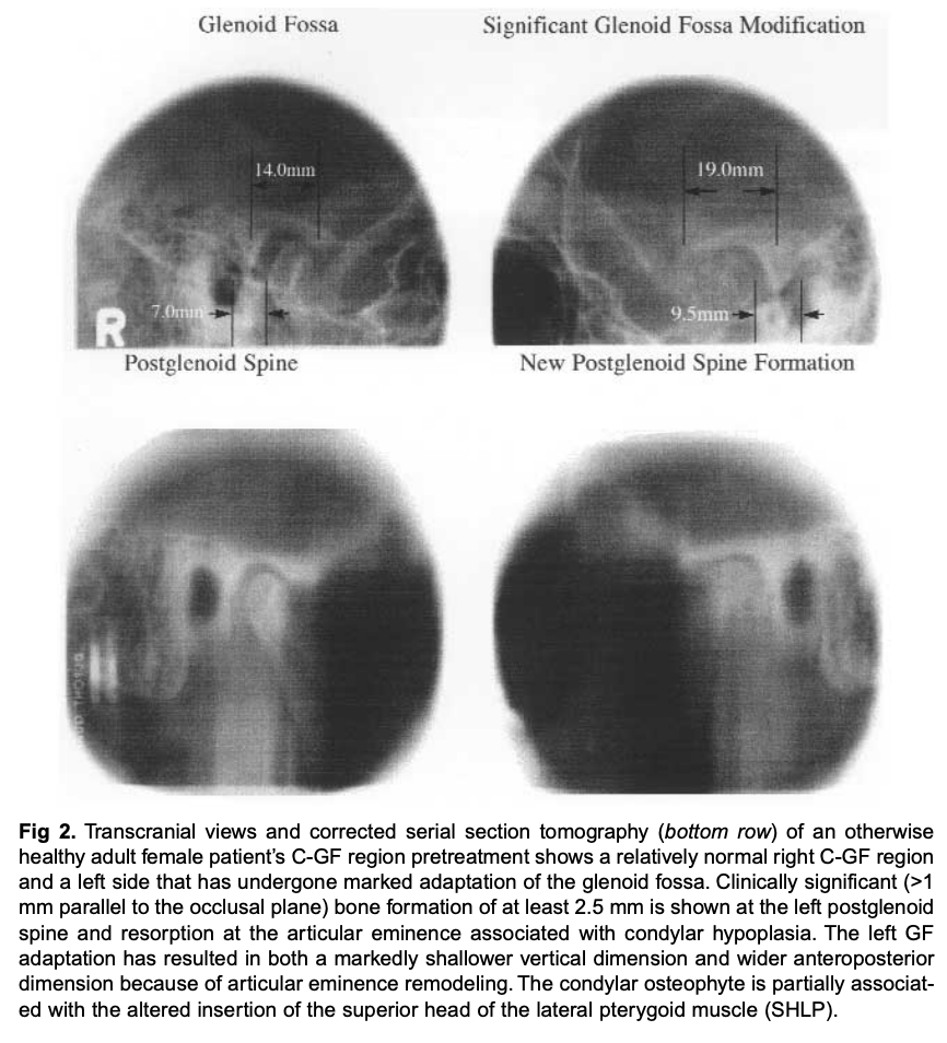
A common question in orthodontics is whether functional appliances can truly stimulate mandibular growth beyond genetic potential. Short-term studies often show promising results—forward positioning of the mandible, improved facial profile, and apparent condylar changes. However, long-term studies consistently demonstrate that many of these effects reduce or relapse after appliance removal.
This discrepancy highlights an important principle: 👉 Not all growth observed during treatment is permanent growth.
Growth Relativity: A More Realistic Biological Explanation
The Growth Relativity hypothesis proposes that condylar and glenoid fossa changes during functional appliance therapy are relative, adaptive, and time-dependent, rather than permanent growth stimulation.
According to this concept, three major factors influence condyle–fossa modification during mandibular advancement:
Mandibular Displacement Forward positioning of the mandible alters the spatial relationship between the condyle and the glenoid fossa.
Viscoelastic Tissue Stretch Non-muscular tissues—such as the retrodiskal tissues, fibrous capsule, ligaments, and synovial fluid—are stretched during advancement. These tissues exert biologically significant forces on the condyle and fossa.
Force Transduction via Fibrocartilage The unique fibrocartilaginous cap of the condyle acts as a conduit, allowing forces to be transmitted and “radiate” to areas where new bone formation may occur—even at a distance from the original soft tissue attachment.
Why the Condyle Is Not an Epiphysis
Unlike long bone epiphyses, the mandibular condyle:
Is covered by fibrocartilage, not hyaline cartilage
Lacks a strong intrinsic growth-driving mechanism
Responds more to functional and environmental influences
As a result, condylar changes during functional therapy are adaptive responses, not genetically programmed growth spurts.
The Light Bulb Analogy
A helpful way to visualize Growth Relativity is the light bulb on a dimmer switch:
🔆 During active treatment: Mandibular advancement “turns up the light.” Condylar and glenoid fossa remodeling becomes more active.
🔅 During retention: Once the appliance is removed, muscle activity returns, the condyle reseats, and the “light dims.”
💡 Long-term: Growth activity returns close to baseline levels.
This explains why short-term gains may not be fully maintained unless carefully managed.
Clinical Implications for Twin Block and Herbst Appliances
Understanding Growth Relativity changes how we use these appliances in practice.
Twin Block
Intermittent force
Requires good patient compliance
Allows vertical control
Stepwise mandibular advancement is preferred to avoid tissue overload
Herbst Appliance
Continuous force
Compliance-free
Higher risk of condylar compression if poorly designed
⚠️ Condylar compression should be avoided, as it may reduce adaptive remodeling and increase the risk of TMJ problems.
Why Relapse Happens
Relapse occurs due to:
Release of stretched viscoelastic tissues
Reseating of the condyle into the fossa
Reactivation of masticatory muscle forces
This reinforces the idea that functional appliances reposition structures—they do not permanently override biology.
Key Takeaway for Orthodontic Students
Functional appliances are powerful tools—but only when used with biological realism.
✔ They produce relative, adaptive skeletal changes ✔ They rely heavily on soft tissue biomechanics ✔ Long-term stability depends more on growth timing, appliance design, and retention, not just advancement
Understanding Growth Relativity helps us move beyond appliance mechanics and toward biologically intelligent orthodontics.
 Dentowesome