Written by – Sanjana Agrawal
SOURCE – RAMYA RAGHU TEXTBOOK
Written by – Sanjana Agrawal
SOURCE – RAMYA RAGHU TEXTBOOK
Nausea ,vomitting are common go to side effects of a drug, when external examiner asks during viva of a medical student. But, these symptoms can reveal many underlying conditions and diseases apart from the side effects of many drugs.
Nausea is imminent desire to vomit and often precedes or accompanies vomitting. vomiting is the forceful expulsion of the gastric contents through mouth.
PATHOPHYSIOLOGY:
1. Activation of the CTZ ,sends the impulses to the vomiting centre in brainstem,
2. Gastric fundus and gastroesophageal sphincter relaxes inturn cause raise in intra-abdominal pressure increase,due to increase in abdominal and diaphragm muscle contraction causes the gastric contents to enter the oesophagus ,
3. Increased intra-thoracic pressure results in further movement of food material to mouth
4.Reflex elevation of the soft palate and closure of glottis protect the nasopharynx and trachea and completes the vomiting
Causes of vomiting:
  1. DRUGS:
              -Cancer chemotherapy
              -Antibiotics
              -Cardiac antiarrhythmics
              -Digoxin
              -Oral hypoglycemics
              -Oral contraceptives
2.Metabolic and endocrine causes:
               -Pregnancy
               -Uremia
               -Ketoacidosis
               -Thyroid and parathyroid disease.
               -Adrenal insufficiency
3.Toxins:
              -Liver failure
              -Ethanol
4.Labyrinthine disease
             - Motion sickness
             - Labyrinthitis
             - Malignancy
5. Bowel obstructing disorder
6. Cardiopulmonary disorders and cardiomyopathies
7.Enteric infections
      -viral
      -Bacterial
8.Inflammatory dieases.
      - cholecystitis.
      -Pancreatitis
      -Appendicitis
      -Hepatitis.
9.Intracerebral disorders
      -Intracerebral disorders
      -Malignancy
      -Abscess
      -Hydrocephalus
10. Post operative vomiting
11.Impaired motor functions.
Careful clinical examination, will reveal the underlying
 cause for nausea and vomiting, like ,vomiting that occurs
 predominantly in the morning is often seen in pregnancy,
 uremia, and alcoholic gastritis;projectile vomiting suggests
 increased intracranial pressure,vomiting during or shortly
 after a meal may be due to psychogenic causes or peptic
 ulcer disease.
complications, can be Rupture of the esophagus,hematemesis
from a mucosal tear,dehydration, malnutrition, dental caries
and erosions, metabolic alkalosis, hypokalemia,
and aspiration pneumonitis.
Treatment is mainly based on the treatment of the underlying cause,
  -Antihistamines such as meclizine and dimenhydrinate are effective
for nausea due to inner ear dysfunction
  -Anticholinergics such as scopolamine are effective for nausea
   associated with motion sickness
  -Haloperidol and phenothiazine derivatives such as
    prochlorperazine are often effective in controlling mild nausea
    and vomiting, but sedation, hypotension, and parkinsonian
    symptoms are common side effects.
  -Metoclopramide may be superior to the phenothiazines in treating
   severe nausea and vomiting.IV metoclopramide may be effective
   as prophylaxis against nausea when given before chemotherapy.
  -Ondansetron and granisetron, serotonin receptor blockers,
   and glucocorticoids are used for treating nausea and vomiting
    associated with cancer chemotherapy.
  -Aprepitant, a neurokinin receptor blocker, is effective at
    controlling nausea from highly emetic drugs like cisplatin
Written by – Sanjana Agrawal
SOURCE – GHOMS TEXTBOOK
Written by – Sanjana Agrawal
SOURCE – GHOMS TEXTBOOK
1
2
References: Shafers Textbook Of Oral Pathology 7Ed, Image source: Google
Contd…Page 2👇🏻
Vegetations are more likely to form where valvular damage already exists. In many cases, the initial inflammation is caused by catheter-induced abrasion or prosthetic devices.
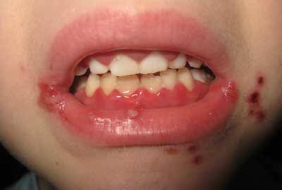
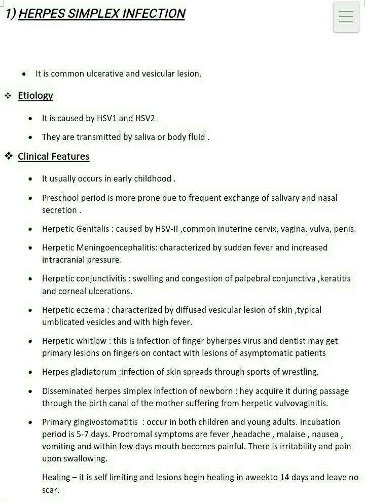
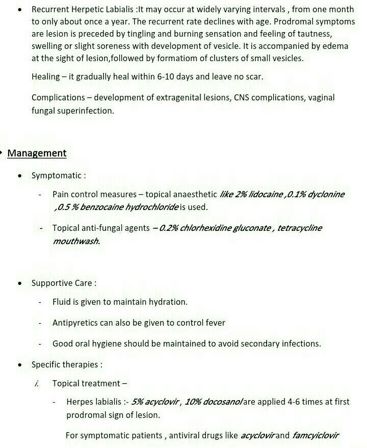
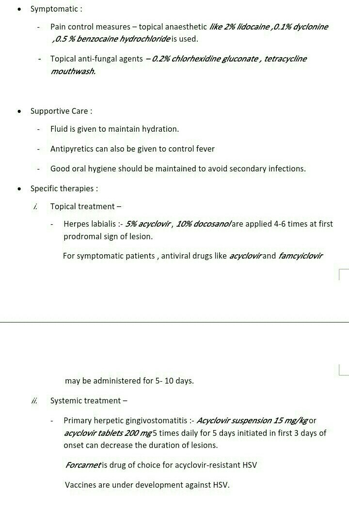
Oral disease transmitted by droplet spread/contact with the lesion.
• Incidence – Children & young adults. <6 months of age: Rare (Due to presence of circulating Ab. in the infant derived from the mother)
• Clinical Manifestations:
➡️ Within a few days mouth becomes painful with gingiva being inflamed, edematous & erythmatous.
➡️ Involvement of lips, tongue, buccal mucosa, palate, pharynx & tonsils.
Yellowish fluid-filled vesicles develop
⬇️
Rupture
⬇️
Shallow, ragged, extremely painful ulcers (grey membrane, erythmatous halo)
➡️ Ulcers heal within 7-14 days with No scar formation
➡️ Isolation of HSV-1 from these lesions after onset: (2 – 6 days)
➡️ Trigeminal for HSV-1 (Virus remain latent until reactivated in the ganglia)
➡️ Viral DNA to Host DNA – lifelong infection
➡️ Incubation Period: 2-20 days
➡️ While most children will be asymptomatic, diagnosis of children with symptoms is made based on clinical presentation of erythematous gingiva, mucosal hemorrhages, and clusters of small erupted vesicles throughout the mouth.
➡️ The condition is highly contagious and complications range from indolent cold sores to dehydration and even life-threatening encephalitis.
➡️ Symptomatic relief primarily involves pain management and oral fluids to prevent dehydration until the viral infection subsides.
➡️ Acyclovir is a well established antiviral drug used effectively for the treatment of herpes simplex infections, chickenpox (shortened fever time), and shingles. It is also used frequently for children with immunodeficiency.
➡️ The current recommended dose of oral acyclovir is 40 to 80 mg/kg a day, divided in 3 or 4 doses, for 7 days. Caregivers should be aware of potential adverse effects of acyclovir such as headache, malaise, and vomiting.
Dr. Mehnaz Memon🖊
References: Shafers Textbook Of Oral Pathology 7Ed, Internet
Written by – Sanjana Agrawal
SOURCE – RAMYA RAGHU TEXTBOOK
Written by – Sanjana Agrawal
SOURCE – RAMYA RAGHU TEXTBOOK

