Circulatory Shock

Key Points:

  • Shock is the state in which reduced cardiac output, vasodilation, or low blood volume leads to tissue hypoperfusion and cell death.
  • Four types of shock:
    – Distributive, which is the most common form
    – Hypovolemic
    – Cardiogenic
    – Obstructive (which is the least common).

Be aware that these types of shock can co-exist in a patient.

  • Three stages of shock:
    – Nonprogressive (aka, compensated)
    – Progressive
    – Irreversible
  • Treatment can generally be summarized with the acronym VIP:
    – Ventilate (administer oxygen)
    – Infuse (administer fluids)
    – Pump (administer vasoactive agents that facilitate circulatory pumping).

Be aware that the treatments can vary by type of shock and the underlying causes; for example, in some cases, fluid infusion can cause more harm than good.

  • Reperfusion injury: Reperfusion of the tissues can cause additional injury due to increased production of superoxide and hydroxyl radicals.
  • Multiple organ dysfunction syndrome: Aka, multiple organ failure describes the progressive dysfunction of two or more organs.

Stages of Shock

  • Nonprogressive stage is characterized by physiological adaptations that compensate for the effects of shock and allow for recovery.
    – Compensation relies on negative feedback mechanismsthat respond to low tissue perfusion by increasing cardiac output and/or increasing arterial pressure to ensure adequate blood supply.
  • Progressive stage is characterized by positive feedback mechanisms in which shock facilitates the progression of shock.
    – Reduced blood supply to the myocardium weakens the heart’s ability to contract.
    – Sustained lack of blood to the brain leads to depression of the vasomotor center, which leads to loss of sympathetic activation.
    – As a result of slow flow and buildup of metabolic wastes, blood in the small vessels begins to agglutinate and obstruct flow.
    – Hypoxic capillaries become leaky, and release fluid into the tissues; this exacerbates low blood volume and cardiac output.
    – Ischemic tissues and/or bacteria release toxins that cause further cell damage.
    – As a result of hypoxemia, the tissues begin to break down, especially in the liver, lungs, and myocardium.
    – Because the tissues are malfunctioning, acidosis develops. Lactic acid is especially elevated as tissues shift to anaerobic metabolism, which is why we measure lactic acid levels in suspected cases of shock.
  • Irreversible stage is fatal – a patient cannot be saved, even with medical intervention, because the high-energy compounds necessary for cellular functioning are depleted.
    – Thus, early recognition and treatment of shock is crucial for saving a patient’s life.

Signs and symptoms

Consider how each one is predictable given the causes and effects of shock.

  • Shock leads to low arterial pressure
    – However, be aware that hypotension may be minimal,initially, due the body’s physiological responses to reduced blood pressure, such as increased sympathetic activity.
  • Altered mental states, such as confusion, can develop when brain doesn’t receive sufficient oxygen.
  • Tachycardia can occur due to increased sympathetic activity.
  • Lactic acid levels rise because the cells shift to anaerobic metabolism.
  • Skin changes are common, and depend on the cause of shock, but can produce clamminess, flushing, cold, or other changes.
  • Oliguria is common due to reduced renal perfusion.

Types & Causes of Shock
Two important things to keep in mind are:

  • Different types of shock can overlap.
  • Discovery of the underlying cause is key to treatment.

Volume-Related Causes
Distributive Shock: shift in distribution of blood volume.

  • Septic shock is the most common form of all shock types; it is the result of bacterial infection.
  • Anaphylactic shock is the result of a severe allergic reaction; bee stings, foods such as peanuts, latex, and some medications are among the most common causes of anaphylactic shock.
  • Neurogenic shock can occur; this is most often the result of trauma to the CNS.
  • Hemodynamic changes:
    – Each of these types of distributive shock induce significant vasodilation.
    To remember this, recall that vasodilation is triggered by various toxins (i.e., bacteria), inflammation (i.e., allergy), and suppression of the sympathetic nervous system (i.e., CNS).
    – Distributive shock is often associated with normal, or even high, cardiac output due to high venous return (which is, itself, due to the vasodilation). As we’ll see, high cardiac output is unique to distributive shock.
    – Preload is reduced due to capillary leakage and transfer of fluid from the intravascular to the extravascular tissues.
  • Skin changes:
    – Distributive forms of shock often produce flushing of the skin
    – More specifically, the skin of patients in anaphylactic shock are often warm and itchy (think of an allergic reaction); patients with septic shock experience fever and chills; and, patients in neurogenic shock typically have dry skin.
  • Treatment: Administration of fluids and vasopressors to increase cardiac output and arterial pressures; epinephrine is given for anaphylactic shock.

Hypovolemic shock: loss of blood volume

  • Associated with internal or external hemorrhaging, severe burns, very low fluid intake, and vomiting or diarrhea (recall that dehydration, and, therefore, hypovolemic shock are particularly common in sick children in low-income countries).
  • Hemodynamic changes:
    – Preload and cardiac output are reduced; to compensate for low blood volume, systemic vascular resistance increases (via vasoconstriction and fluid retention).
  • Skin Changes: Cold, clammy skin
  • Treatment: administration of fluids and controlling the bleeding or fluid loss.

Output-Related Causes
Cardiogenic shock: structural/mechanical problems in the heart.

  • Results form cardiac defects that produce abnormal contractility, rhythm, or structures; thus, common causes are myocardial infarction, arrhythmias, valve dysfunctions, and drugs.
  • Hemodynamic changes: Dysfunctional pumping reduces cardiac output, and, as in hypovolemic shock, systemic vascular resistance increases in response to low arterial pressure. Preload, however, may increase.
  • Skin: Cold and clammy.
  • Treatment: Underlying causes must be treated, and vasopressors or inotropes may be provided to improve blood pressure and flow.

Obstructive shock: extracardiac causes, including cardiac tamponade, pulmonary embolism, and tension pneumonothorax.

  • Cardiac output is reduced as a result of extracardiac obstructions that inhibit ventricular filling or emptying. Systemic vascular resistance increases, but preload is variable.
  • Skin: Cold and clammy.
  • Treatment: Remove the obstruction, administer medications to improve blood pressure and flow.
Hypertension: Treatments

Overview

  • Antihypertensives aim to reduce cardiac output and/or total peripheral resistance.
    – For a review of how cardiac output and total peripheral resistance, please see our tutorial on hypertension pathophysiology.
  • Lowering blood pressure in hypertensive patients reduces their risk of cardiovascular disease and cerebrovascular events.
    – Recent guidelines recommend a target blood pressure of less than 130/80 mmHg.
  • The following lifestyle modifications are typically suggested:
    – Changes in diet, increased physical activity, stress reduction, smoking and alcohol cessation or reduction, and weight loss.
    – DASH:
    Dietary changes to reduce hypertension are encapsulated by the Dietary Approaches to Stop Hypertension (DASH) plan, which recommends reductions in sodium and emphasizes whole grains, fruits, vegetables, low-fat dairy, fish, poultry, and legumes, nuts, and seeds.
  • In many individuals, however, lifestyle modifications are inadequate or even inappropriate for reducing blood pressure; these patients will need antihypertensive medications.
  • Initial treatment may rely on a single medication, depending on the stage of hypertension.
  • However, many patients ultimately require two or more drugs with complementary actions to reach their target blood pressure.
  • Individuals vary in their responses to antihypertensive medications, and that specific recommendations are made for some populations.
    – For example, African Americans, the elderly, and patients with certain medical conditions may respond differently to an antihypertensive drug than the rest of the populations.
  • Resistant hypertension is when an individual’s blood pressure remains elevated above the target goal, despite concurrently using three or more antihypertensive medications, including a diuretic.

Thiazide and thiazide-like diuretics

  • These drugs act on the distal convoluted tubule of the nephron to prohibit sodium and water reabsorption; sodium and water are excreted in the urine, so blood volume and blood pressure are reduced.
    – Often a first line choice, particularly in salt-sensitive individuals
    – They are associated with hypokalemia.
  • Chronic use causes vasodilation, which also contributes to reduction in blood pressure; the exact mechanism by which these diuretics cause vasodilation is uncertain.

Renin-Angiotensin System

Two drugs that block the actions of angiotensin II, which is a powerful vasoconstrictor that also triggers the release of other blood pressure mediators, including aldosterone.

  • Briefly illustrate the renin-angiotensin system:
    – The liver releases angiotensinogen.
    – The kidneys release renin, which transforms angiotensinogen to angiotensin I.
    – Then, as angiotensin I circulates in the blood, especially in the pulmonary blood, it encounters angiotensin-converting enzyme (ACE), which is released from vascular endothelial cells.
    – Angiotensin converting enzyme, as its name suggests, converts angiotensin I to angiotensin II.
    – Angiotensin II binds with arterial receptors and induces vasoconstriction.

Angiotensin-converting enzyme inhibitors

  • ACE inhibitors prohibit the formation of angiotensin II by blocking the actions of angiotensin-converting enzyme.
  • First-line drugs.
  • Can cause hyperkalemia.
  • Angiotensin II also breaks down bradykinin, which is an important vasodilator; thus, angiotensin-converting enzyme inhibitors effectively increase bradykinin levels, which ultimately enhances vasodilation.
    – Increased bradykinin is associated with cough and angioedema.

Angiotensin-receptor blockers

  • Block the arterial receptors for angiotensin II.
  • Like ACE inhibitors, they prevent angiotensin II from increasing blood pressure.
  • Also like ACE inhibitors, they are associated with hyperkalemia.
  • However, since they don’t prohibit the formation of angiotensin II, they don’t effect bradykinin, so patients don’t experience cough and angioedema.

Three “blockers” that act directly on the heart and/or vasculature.

Calcium channel blockers prevent calcium binding:

  • In the heart, receptors are located at the sinoatrial and atrioventricular nodes, as well as in the cardiac tissue; thus, calcium channel blockers reduce conduction velocity, contractility, and heart rate.
  • In the vasculature, prevention of calcium blocking reduces vasoconstriction.
  • Calcium channels are considered a first line treatment, particularly for African Americans, in whom other antihypertensive drugs are often less effective.
  • Calcium channel blockers are associated with swelling in the lower extremities, rash, flushing, and dizziness.

Beta blockers prevent norepinephrine and epinephrine binding

  • In the heart, like calcium channel blockers
  • Third generation beta blockers also produce vasodilation.
  • Beta blockers block renin secretion from the kidney, which blocks the formation of angiotensin II and elevates bradykinin levels.
  • Commonly reported side effects include fatigue, cold hands/feet, depression, sleep disturbances, and erectile dysfunction.
  • Furthermore, some beta blockers can trigger bronchospasm in patients with asthma and chronic obstructive pulmonary disease.

Alpha blockers prevent norepinephrine from binding

  • In the vasculature, this reduces vasoconstriction.
  • Orthostatic hypotension is common, particularly in the elderly.

Hypertension: Pathophysiology

  • Mean arterial pressure is determined by cardiac output and total peripheral resistance (aka, systemic vascular pressure).
    – Thus, hypertension, which is elevated blood pressure, is the result of increased cardiac output and/or increased total peripheral resistance.
    – Cardiac output is the product of heart rate and stroke volume.
    – Stroke volume is determined by preload and contractility.
    – Blood volume contributes to preload, by way of increased venous return.
    – The degree of sodium and water retention in the kidneyscontributes to blood volume.
    – Degree of vasoconstriction, particularly of the small arteries and arterioles, is a significant determinant of total peripheral resistance.

Key mediators of blood pressure implicated in primary and/or secondary hypertension
– Notice that many of these mediators effect both cardiac output and total peripheral resistance, but be aware that some effects may be more significant in hypertension development than others.

  • Posterior pituitary secretes antidiuretic hormone (aka, vasopressin),
    – Vasoconstrictor that also increases sodium and water retention in the kidneys.
    – Increased sodium and water retention results in increased blood volume, and, therefore, increased cardiac output.
  • Aldosterone is secreted by the adrenal cortex and has similar effects.
  • Angiotensin II, which is a product of the renin-angiotensin-aldosterone system, has direct and indirect effects on blood pressure:
    – Like antidiuretic hormone and aldosterone, it triggers vasoconstriction and increases sodium and water retention.
    – Angiotensin II also stimulates the release of norepinephrine, antidiuretic hormone, and aldosterone, further enhancing vasoconstriction and sodium/water retention.
    – Multiple antihypertensive drugs work against the effects angiotensin II.
  • Norepinephrine is a vasoconstrictor that also increases heart rate and contractility.
  • Vascular remodeling: hypertension produces damage and inflammation that leads to vascular remodeling, which alters local mediators.
    – Endothelin, which is a key vasoconstrictor, is elevated in remodeled vessels.
    – Secretion of local vasodilators, such as nitric oxide, is reduced.
  • Vasodiators: nitric oxide, prostaglandins, histamine, and bradykinin.
    – Bradykinin is broken down by angiotensin II; thus, angiotensin II not only induces vasoconstriction, it removes a vasodilator.
    The relationship between angiotensin II and bradykinin contributes to the effectiveness of drugs that inhibit angiotensin-converting-enzyme – when circulating levels of angiotensin II are reduced, bradykinin levels can rise.
  • Genetic and epigenetic factors, diet, physical activity levels, and other environmental or biological factors can affect blood pressure by acting on the various components of this diagram.
    – For example, we can now understand how individuals who are salt-sensitive or have aldosterone-secreting tumors develop hypertension via elevated blood volume and preload.
  • Hypertensive crisis occurs when blood pressure is dangerously high, typically exceeding 180/120mmHg.
    – Hypertensive urgency: no end-organ damage
    – Hypertensive emergency: end-organ damage has occurred
  • Symptoms of hypertensive emergency include severe headache with confusion and impaired vision, chest pain and shortness of breath, nausea/vomiting, anxiety, and seizures.

Hypertension: Overview

  • Hypertension is characterized by sustained elevated blood pressure.
  • Hypertension is common worldwide, and, according to updated guidelines, approximately 46% of Americans 20 years and older have hypertension.
    – Many of these individuals are unaware of their status, which is why hypertension is sometimes referred to as a “silent killer.”
  • Hypertension predisposes patients to cardiovascular disease,which is one of the most common causes of death in both men and women worldwide.

Review of blood pressure and its determinants
Review Cardiac Cycle

  • Systolic pressure is the highest arterial pressure, reached after blood is ejected from the left ventricle.
    – 120 mmHg
  • Diastolic blood pressure is the lowest arterial pressure, reached during ventricular relaxation.
    • 80 mmHg
  • Mean arterial pressure is determined by Cardiac Output and Total Peripheral Resistance.
    • 93 mmHg
  • Cardiac output refers to the amount of blood ejected by the left ventricle in in one minute
  • Total Peripheral Resistance refers to the resistance of the systemic arteries to blood flow (total peripheral resistance is also referred to as systemic vascular resistance, SVR).
  • Hypertension occurs when the cardiac output and/or total peripheral resistance increases.

Classification of Hypertension: 2017 guidelines by the American College of Cardiologists and American Heart Association.

  • Healthy/normotensive: Systolic blood pressure less than 120 mmHg, and diastolic pressure less than 80 mmHg.
  • Elevated: Systolic blood pressures between 120 and 129 mmHg, and diastolic pressures less than 80 mmHg.
  • Stage 1 hypertension: Systolic pressures between 130 and 139 mmHg, OR diastolic pressures between 80 and 89 mmHg.
  • Stage 2 hypertension: Systolic pressures above 140 mmHg, OR diastolic pressures above 90 mmHg.
  • The higher value determines the stage of hypertension if the systolic and diastolic values fall in different categories.
    • For example, if a patient’s systolic pressure is 135 mmHg and but diastolic pressure is 75 mmHg, it would be classified as Stage 1 hypertension.
  • Blood pressure fluctuates throughout the day and in response to various situations, so multiple measurements need to be taken in and out of the health clinic, and during waking and sleeping.
    One frustrating aspect of measuring blood pressure is that the presence of a health care professional can affect the blood pressure! This may be due to sympathetic activation in response to anxiety or other factors.
  • “White coat hypertension” occurs when an untreated patient has high blood pressure in the presence of a medical professional, but is otherwise normotensive.
    • “White coat effect” is the same phenomenon, but in patients who are under treatment for high blood pressure.
  • “Masked hypertension” is when an untreated patient is normotensive in the presence of a medical professional but is hypertensive otherwise;
    • “Masked uncontrolled hypertension” is the same phenomenon, except in patients who are being treated for hypertension.

Primary Hypertension

  • Accounts for 90-95% of all adult cases.
  • In primary hypertension, there is no single identifiable cause. Instead, one or more of the following factors contribute to high blood pressure.
    • Some variables are modifiable, and many are interrelated.
  • Genetic and epigenetic factors can contribute to development of high blood pressure.
  • Obesity contributes to hypertension via various direct and indirect mechanisms; for example, some propose that dysfunction in the sympathetic nervous system and kidneys contribute to high blood pressure in obese patients.
  • Sedentary lifestyles which are common in societies where we spend several hours per day at our desks. — — — Increasing physical activity confers multiple protective effects on the cardiovascular system.
  • Diet is a significant predictor of hypertension
    • Particularly salt intake and salt sensitivity.
      Salt sensitivity refers to how efficiently individuals excrete salt and, therefore, avoid
      elevations in blood pressure. Exactly what determines salt sensitivity is uncertain, but African-Americans, the elderly, and post-menopausal women seem to be particularly susceptible to elevations in blood pressure due to lower rates of salt excretion.
  • On the other hand, inadequate intake of other nutrients,including calcium, potassium, vegetable proteins, and fiber may also contribute to hypertension.
  • Alcohol as well as electronic and tobacco cigarettes are associated with hypertension due to their effects on the sympathetic nervous system and other regulators of blood pressure.
  • Chronic stress, including psychosocial stress, also contributes to hypertension; this may be due to activation of the sympathetic nervous system and/or other physiological reactions to stress.
  • Population differences
    • Hypertension prevalence varies by population, and are likely due to differences in both genetic and environmental factors.
    • There are dramatic differences in the prevalence of hypertension in United States subpopulations, due to both genetic and environmental differences.
    • Hypertension prevalence is highest in non-Hispanic African Americans, American Indians, and Native Alaskans as compared to non-Hispanic Caucasian Americans and Hispanic Americans.
    • Hypertension prevalence also varies by age and sex:
      In general, hypertension prevalence increases with age.
      Premenopausal women typically have lower blood pressures than do age-matched men or post-menopausal women.
      However, post-menopausal women have blood pressures equal to or higher than their age-matched male peers.

To illustrate this, we draw a simple graph that shows that, as men and women age, blood pressure increases; after menopause, women’s blood pressure levels exceed that of men’s.

Secondary Hypertension

  • Accounts for 5-10%* of adult hypertension.
  • In secondary hypertension, an underlying condition causes the elevation in blood pressure.
  • Renovascular hypertension occurs when renal artery stenosis prevents blood from reaching the kidneys; in response to low oxygen levels, the kidneys release hormones that increase blood pressure.
    • Notice that the adaptive physiological response to low oxygen levels is, in this case, contributing to pathology.
    • Two key causes of renal artery stenosis and renovascular hypertension are atherosclerosis and fibromuscular dysplasia.
  • Obstructive sleep apnea
  • Primary aldosteronism is characterized by adrenal glands that release excess aldosterone, which leads to increased sodium and fluid retention, and, therefore, increased blood volume and pressure.
    • Two key causes of primary aldosteronism and hypertension are aldosterone-producing adenomas and bilateral idiopathic hyperaldosteronism.
  • Renal parenchymal diseases also lead to hypertension due to inadequate blood volume regulation by damaged renal tissue.
  • Drugs
    • Examples: caffeine, NSAIDS, hormonal contraceptives (especially those with synthetic estrogen), decongestants, cocaine, amphetamines, and some herbal agents.
  • Pregnancy:pregnant women can develop gestational hypertension, which can lead to pre-eclampsia (aka, toxemia).
    • Hypertension is a significant cause of morbidity and mortality in both mothers and their neonates, and women who are hypertensive prior to pregnancy require special attention.

Additional Causes of Secondary Hypertension

  • Pheochromocytoma
  • Coarctation of aorta (children)
  • Cushing syndrome
  • Hyperparathyroidism

Vasculitis – Large Vessel

Overview

  • Large vessel vasculitides involve the aorta and its large branches.
  • Vasculitides of all sizes are characterized by blood vessel inflammation, sometimes with necrosis, and can lead to ischemia and organ damage.
  • Vessels and organs affected vary by the specific vasculitic disorder.
  • General symptoms and signs of vasculitides are due to systemic inflammation, including fever, arthritis, arthralgia, fatigue, and weight loss. As we’ll see, specific end-organ damage varies by the vessels affected.
  • Typical treatment includes corticosteroids and immunosuppressants.

Giant Cell Arteritis aka, temporal arteritis or Horton disease.

  • It is a granulomatous disease.
  • It specifically affects the aorta and its large branches, especially the external carotid arteries.
    – Thus, affected vessels include those of the head and neck, such as the superficial temporal artery, which is commonly involved.
  • Common symptoms include:
    – Raised, tender temporal artery, which is sometimes visible on the patient’s forehead
    – Vision loss
    – Neck and/or jaw pain
    – Tongue necrosis
  • Indicate that giant cell arteritis is most common in women over 55 years of age, and especially those of Northern European descent.
  • It is associated with polymyalgia rheumatica in up to 50% of cases.
  • Serious complications include aortic aneurysm, stroke, and blindness; thus, treatment should be provided as soon as possible.

Histopathology:

We show a normal large artery, for comparison.

  • Giant cell arteritis
    – Key elements of the pathogenesis of giant cell arteritis, according to a very generalized model.
    Dendritic cells in the adventitia are activated, perhaps by viruses or other stimuli;
    Activated dendritic cells recruit and activate CD4+ T cells
    CD4+ T cells release pro-inflammatory cytokines and recruit and activate pro-inflammatory macrophages.
    Macrophages secrete growth factors and facilitate migration and proliferation of vascular smooth muscle cellswithin the intima, *which promotes its hyperplasia.
    Additionally, the macrophages form multinucleated giant cells, which secrete various harmful substances, such as ROS, NO, and matrix metalloproteinases (MMP), which destroys the tunica media and internal elastic lamina.
    – In summary, the histopathology of giant cell arteritis is characterized by:
    Chronic inflammation
    Granuloma formation
    Destruction of the tunica media and elastic laminae
    Tunica intima remodeling and hyperplasia
    Lumen occlusion – which, as we’ve seen, can lead to downstream ischemia and tissue damage.

Takayasu arteritis, aka, pulseless disease

  • A granulomatous disease that affects the aorta and its large branches.
  • Thickening and fibrosis of the vessel walls narrows the lumen.
    – In the image, we can see the large, bulging aorta and the aortic arch branches.
  • The resulting ischemia produces:
    – A weak or absent pulse in the extremities (hence the alternative name, “pulseless disease”)
    – Different blood pressures in the upper extremities (that is, right vs. left), and vascular bruits
    – Claudication in the limbs
    – Chest pain
    – And, possible hypertension, due to renal artery stenosis.
  • Possible complications include aneurysm, aortic regurgitation, and retinopathy.
  • Takayasu arteritis is most common in women younger than 40 years old, and seems to be more common in women of Asian descent.

Vasculitis – Medium & Variable Vessel Sizes

Overview:

  • Vasculitides are characterized by blood vessel inflammation with possible necrosis, ischemia, and organ damage.
  • The vessels and organs affected vary by the specific vasculitic disorder.
  • General symptoms and signs are due to systemic inflammation,which can cause fever, arthritis, arthralgia, fatigue, and weight loss.
  • Skin lesions are associated with small and medium vessel vasculitides.
  • Treatments include corticosteroids and immunosuppressants.

MEDIUM VESSEL VASCULITIDES

Polyarteritis nodosa is characterized by necrotizing inflammation that most commonly involves the medium muscular arteries, especially at their branch points.

  • The resulting ischemia can affect multiple organ systems:
    – Nervous system involvement often produces mononeuritis multiplex or asymmetric polyneuropathy; sensory and motor deficits of the median, ulnar, and fibular nerves are common.
    – Renal involvement can lead to hypertension, oliguria, and even renal failure.
    – Gastrointestinal involvement can produce pain and malabsorption; when larger vessels, such as the celiac trunk or its branches are affected, fatal aneurysms can develop.
    – Skin manifestations can take various forms, including livedo reticularis (which presents as a purplish lace-like pattern), ulcers, subcutaneous nodules, or even gangrene.
    – Heart failure can occur due to coronary artery obstruction.
  • Polyarteritis nodosa can be systemic or cutaneous.
  • Some secondary forms are related to hepatitis B and C infections, or to rheumatoid arthritis.
  • Most commonly affects men over 50 years of age.

Kawasaki disease, aka, mucocutaneous lymph node syndrome

  • Characterized by proliferative inflammation.
  • Tends to involve the medium muscular arteries.
  • Predilection for the coronary arteries, which are affected in approximately 20% of cases
    – In fact, it is a key cause of acquired heart disease in children.
    – Large coronary aneurysms can be fatal, as they can cause myocarditis, cardiac tamponade, or, as we’ve illustrated here, coronary artery thrombosis.
  • Typically presents as a self-limiting, acute febrile illness in children:
    – Fever
    – Conjunctivitis
    – Erythematous macular rash
    – Edema and desquamation, particularly of the limbs
    – Cervical lymphadenopathy
    – A red, inflamed throat
    – Strawberry tongue with dry, fissured lips.

VARIABLE VESSEL SIZE VASCULITIDES

Behçet disease

  • A chronic, relapsing vasculitis that affects vessels of all sizes.
  • Mucosal inflammation is a common manifestation of the disorder, and often causes oral and genital lesions; ocular inflammation can produce uveitis or hypopyon.
  • Cutaneous manifestations vary, and include pus-filled bumps, palpable purpura, erythema nodosum, and superficial thrombophlebitis.
  • Typical age of onset is around 20 years old.
  • Behçet disease is equally common in men and women, though it’s often worse in men.

Buerger disease

Don’t confuse Buerger disease with Berger’s disease, a nephropathy caused by IgA accumulation.

  • Characterized by inflammation and occlusive thrombosis of the medium and small arteries and veins
  • Most commonly affects the extremities; signs and symptoms typically begin distally and move proximally.
  • Ischemia from vessel occlusion often produces numbness, coldness, or tingling in the extremities, with claudication, then, pain at rest.
  • Affected limbs are often cold, sweaty, and cyanotic, and ulcers form that progress to gangrenous.
  • Smoking is a major cause of Buerger disease, and smoking cessation is key to remission.

Vasculitis – Small Vessel

Key Points

  • Vasculitides are characterized by blood vessel inflammation with possible necrosis, ischemia, and organ damage.
  • The vessels and organs affected vary by the specific vasculitic disorder – the ones we’ll address in this tutorial affect the small arteries, veins, and capillaries.
  • General symptoms are due to systemic inflammation, and include fever, arthritis, arthralgia, fatigue, and weight loss.
  • Cutaneous manifestations can occur in isolation.
  • Treatments include corticosteroids and immunosuppressants.

ANTI-NEUTROPHIL CYTOPLASMIC ANTIBODIES (ANCA) – ASSOCIATED VASCULITIDES

Granulomatosis with polyangiitis aka, Wegener’s

  • Characterized by necrotic granulomatous inflammation.
  • Granulomas comprise giant cells, plasma cells, lymphocytes, neutrophils, and eosinophils.
  • The respiratory tract is typically involved, which can lead to sinusitis, otitis media, rhinorrhea, and epistaxis; the mucosa can become granular and crusted.
    – Erosion and destruction of the nasal septum can case the nasal bridge to collapse.
    – Destruction of the tracheobronchial tree can lead to stenosis and airway obstruction.
    – When the lungs are involved, patients can develop a cough, difficulty breathing, hemoptysis, and hemorrhaging.
  • The kidneys are also commonly affected, leading to necrotizing crescent focal glomerulonephritis and thrombosis.
  • Cutaneous manifestations vary, and include palpable purpura, livedo reticularis, ulcers, and the formation of deep, tender nodules.
  • The eyes, nervous system, heart, and musculoskeletal system can also be involved.
  • Granulomatosis with polyangiitis most commonly occurs in Caucasians; the average age of onset is 40 years.

Eosinophilic granulomatosis with polyangiitis aka, Churg-Strauss

  • ANCA-associated disorder characterized by necrotizing granulomatous inflammation.
  • Manifests as both vascular and extravascular necrotizing granulomas with (as its name suggests) eosinophil blood and tissue infiltrations.
    Three stages of eosinophilc granulomatosis with polyangiitis:
  • The allergic stage is often marked by adult-onset asthma, which can develop years before full presentation of the disorder.
    – Patients may also develop sinusitis, hemoptysis, and pulmonary infiltrates as part of the allergic stage.
  • The eosinophilic stage is characterized by blood and tissue infiltration of eosinophils.
  • The vasculitic stage can affect multiple organ systems:
    – Nervous system involvement is associated with multiple mononeuropathy.
    – Cutaneous involvement manifests as nodules or papules on the extensor surfaces, especially around the elbows.
    – GI involvement can produce pain, diarrhea, and bleeding.
    – Cardiac involvement often leads to inflammation and/or cardiomyopathies.
  • Be aware that the kidneys are less commonly affected than in the other ANCA-associated vasculitides.
  • Disease onset is usually during the late 40s.

Microscopic polyangiitis

  • Characterized by necrotizing pauci-immune inflammation without granulomas.
  • Renal involvement is associated with glomerulonephritis and rapid progression to renal failure.
  • Purpuric rash is common.
  • The lungs are less commonly affected, but, when they are, alveolar hemorrhage and fibrosis can be serious.
  • Microscopic polyangiitis usually develops in patients 50-60 years old.

NON-ANCA-ASSOCIATED SMALL VESSEL VASCULITIDES

Immunoglobulin A-associated vasculitis aka, Henoch-Schonlein purpura

  • Occurs when IgA immune complexes are deposited in the small vessels.
  • Patients develop palpable purpura, especially on the lower extremities, arthralgias, abdominal pain, dark stools, and focal glomerulonephritis.
  • It’s common and self-limited in children, whereas, in adults, it often becomes chronic.

Cryoglobulinemia

  • Occurs when cryoglobulins in the blood clump at cold temperatures.
  • Patients experience notable fatigue, palpable purpura in the legs, arthralgias in the knees and hands, glomerulonephritis, and PNS disturbances.
  • Type I cryoglobulinemia is associated with B-cell lymphoproliferative disorder.
  • Types II and III (aka, mixed cryoglobulinemia) is assoc. with Hepatitis C virus.

Autoimmune Disorders

  • The autoimmune disorders systemic lupus erythematosus and rheumatoid arthritis are also associated with small-vessel vasculitis.

Atherosclerosis

  • Atherosclerosisis a common and clinically significant form of arteriosclerosis that involves the inflammatory response and lipid accumulation within the vessel walls.
  • Arteriosclerosis means “hardening of the arteries.”
  • Atherosclerosis is a specific form of arteriosclerosis; it affects the elastic and medium to large muscular arteries.
    — If blood flow through one of these arteries, such as the aorta or carotid arteries, is obstructed, downstream effects can include ischemia and organ failure.
  • Anatomy
  • Three layers of the vessel wall:
    — The tunica intima comprises a thin layer of endothelial cells, which play an active role in the inflammatory response.
    — The tunica media comprises smooth muscle cells, which are responsible for moderation of the vessel lumen via dilation and constriction;
    — The outermost layer is the adventitia.
  • Our histological sample has a large thrombus blocking the lumen.
  • Sample also has a large atherosclerotic plaque that has developed between the tunica media and endothelial layers.
    — Cholesterol crystals create a “cleft”-like appearance.
    — Foam cells are macrophages that have engulfed lipid droplets.
    — Fibrinous cap; as we’ll see, the integrity of this cap determines the stability of the plaque, and, therefore, its clinical consequences.
  • We also have a gross image with a large atherosclerotic plaque at the bifurcation of common carotid artery.

PATHOGENESIS OF PLAQUE FORMATION

  1. Plaque formation is triggered by endothelial injury and response; recall that endothelial activation involves the release of cytokines, chemokines, coagulation factors, and other pro-inflammatory molecules.
    — Injury triggers increased permeability and subsequent recruitment of leukocytes and platelets, which migrate into the intima. Injury can include a variety of forces including hemodynamic stress, inflammation, bacteria, viruses, etc.
  2. Macrophage activation and smooth muscle cell recruitment forms fatty streaks within the intima.
    — Intima is now crowded by macrophages and smooth muscle cells; these cells and other inflammatory molecules were recruited by cytokines released as part of endothelial activation.
    — The active smooth muscle cells proliferate and synthesize extracellular matrix proteins; importantly, collagen is deposited.
    — The smooth muscle cells and macrophages engulf lipid droplets, trapping them in the intima; consequently, indicate that the macrophages become the foam cells we identified in our histological samples.
  3. Fibrofatty plaque formation results in a mature atheroma; be aware that not all fatty streaks progress to this stage.
    — Plaque comprises a fibrous cap and necrotic core:
    The cap comprises smooth muscle cells, collagen fibers, and other proteins.
    The necrotic core comprises extracellular lipids, intracellular lipids housed in foam cells and smooth muscle cells, T lymphocytes, and cellular debris.
    — The potential for the plaque to become thrombotic or break free and embolize depends in large part on the composition and resulting stability of the fibrous cap, and the hemodynamic stresses that act on it.
    Smooth muscle cells in the cap promote collagen deposition, and, therefore, stability; but, inflammatory cells, which are also present, promote cap degradation. Thus, their relative proportions determine plaque vulnerability.

TRIGGERS, MAINTENANCE, AND CLINICAL CONSEQUENCES OF ATHEROSCLEROSIS

  • Key culprits of endothelial injury that ultimately lead to plaque formation include hemodynamic disturbances and hypercholesterolemia.
    — Hemodynamic disturbances that produce turbulent flow increase the risk of plaque formation.
    Thus, plaques commonly form where arteries bifurcate, such as where the common carotid artery splits to form internal and external branches – refer to our gross image in the blue box. Another common site of formation is along the posterior wall of the abdominal aorta, which is also subject to turbulent stresses.
    — Hypercholesterolemia is characterized by high levels of cholesterol in the blood; recall that cholesterol is synthesized by the liver and ingested in the diet, and is transported in the blood as lipoprotein complexes.
    Chronic hypercholesterolemia damages endothelial cells and impairs the vasodilator activity of nitric oxide.
    Additionally, LDLs, which are the lipoproteins complexes that deliver cholesterol to the peripheral tissues, accumulate within the intima of the arteries; indeed, as we saw in the histological sample and our diagrams, cholesterol is a major component of atherosclerotic plaques. Within the plaques, cholesterol has toxic and pro-inflammatory effects that damage the vessel and promote plaque growth.
    — Thus, risk factors for atherosclerosis include family history (via polygenic mechanisms) and other conditions, such as diabetes mellitus, that induce hypercholesterolemia.
  • Review hyperlipidemia, which contributes to atherosclerosis.
  • Statins are often prescribed to lower cholesterol levels, as they reduce its production by the liver.
    — Other risk factors for atherosclerosis include cigarette smoking and age.
  • Role of macrophages and chronic inflammation in atherosclerosis
    — Recall that chronic inflammation occurs when tissue injury and repair attempts overlap.
    — Accumulation of macrophages, cholesterol crystals, and other substances within the vessel wall triggers the inflammatory response.
    In turn, cytokines, particularly IL-1, are released and recruit additional immune cells, including macrophages and T lymphocytes, which promotes a positive feedback cycle of inflammation.
    — Thus, inflammatory conditions are now considered a risk factor for atherosclerosis; clinical assessment may include measurement of C-reactive proteins, which are markers of inflammation that have been associated with atherosclerosis.
    Research to assess the effectiveness of anti-inflammatory therapies in atherosclerosis is ongoing.
  • Significant clinical consequences of atherosclerosis
    — Coronary artery disease (CAD) and heart attack; stroke; aortic aneurysm; and, peripheral vascular disease (PVD).
    — Vessel occlusion and/or aneurysm are responsible for these complications: we show in a histological sample that plaque formation can dangerously narrow or even completely obstruct blood flow (aka, stenosis); write that obstruction of blood flow can lead to ischemia and organ failure.
    — Aneurysm occurs when the vessel wall bulges outward; we show in a histological sample that fatty materials and debris can migrate between the tunics of the vessel wall, leading to dissection (aka, separation).
    From the patient’s perspective, dissection is experienced as a painful tearing sensation; ultimately, mural remodeling and wall weakening can allow vessel rupture and bleeding.
    — We also show an image of a necrotic foot, the result of peripheral vascular disease.

CHEST XRAY INTERPRETATION

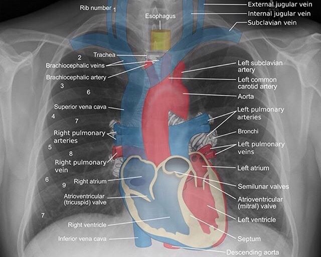
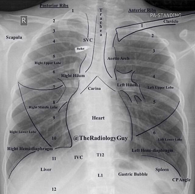
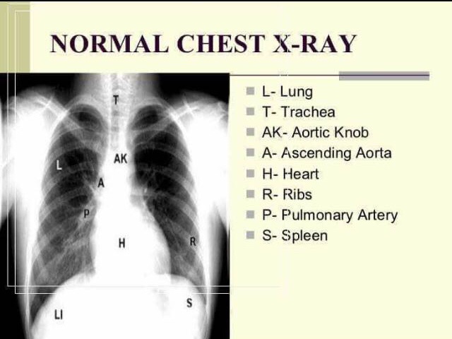
1) NORMAL CHEST XRAY

•THE CHEST X-RAY•

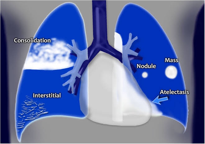
Airway: The trachea is located in the midline. Inspect the trachea and right and left main bronchi. If the trachea is pushed to the left or right look for a cause. Either mass effect pushing the trachea or volume loss (atelectasis) pulling on it.

Breathing: The lobes of the lungs. 3 on the right (upper, middle, lower) and two on the left (upper and lower). Look for consolidation, nodules, masses. Things replacing normal aerated lung (black) will show up as white.

Cardiac: look at the heart size (should be less than 50% of the diameter of the chest). Make sure the borders are clear and sharp. The right atrium makes up the right heart border and the left ventricle makes up the left heart border on the chest X-ray. If the border is obscured, think pneumonia or mass.

Diaphragm: should be well defined to the costophrenic angle. Becomes obscured by lower lobe pneumonia and pleural effusions. Always look for free air under the diaphragm.

Everything else:

  • Bones – look for fractures and destructive lesions
  • Hilar contours – look for enlargement caused by lymphadenopathy or masses
  • Vascular structures – enlarged pulmonary arteries and aortic aneurysms.

Page 1 of 14

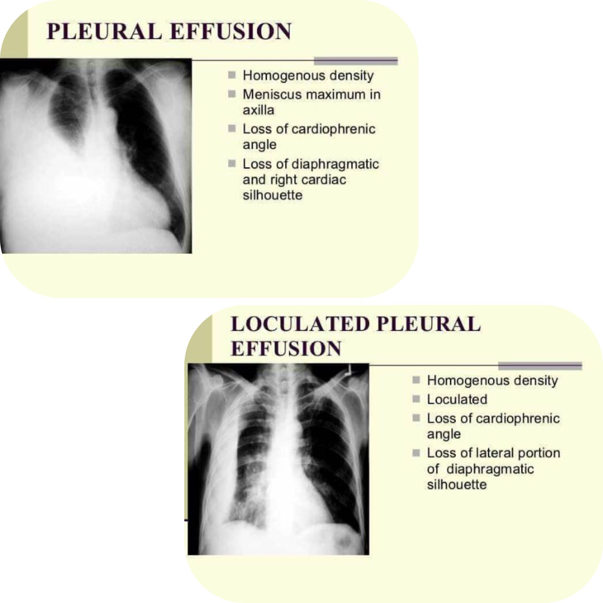
2) PLEURAL EFFUSION

⬇️ Vertical Dimension

1) Mediastinal shift to opposite side

2) Blunting of costophrenic angle

3) Homogenous opacity (Presence of fluid)

4) Ellis S-shaped curve (Concave border)

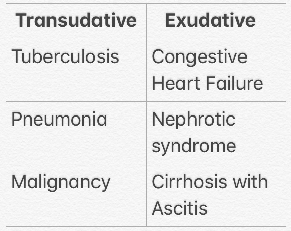
▪️Causes:

▪️Treatment:

➡️ Both treatment and prognosis are completely dependent on the underlying cause:

  • an infected effusion should be drained with an intercostal drain
  • a large effusion secondary to heart failure or in a patient with cirrhosis may respond to diuretics
  • an effusion secondary to malignancy may require pleurodesis following drainage

Page 2 of 14

Read More »