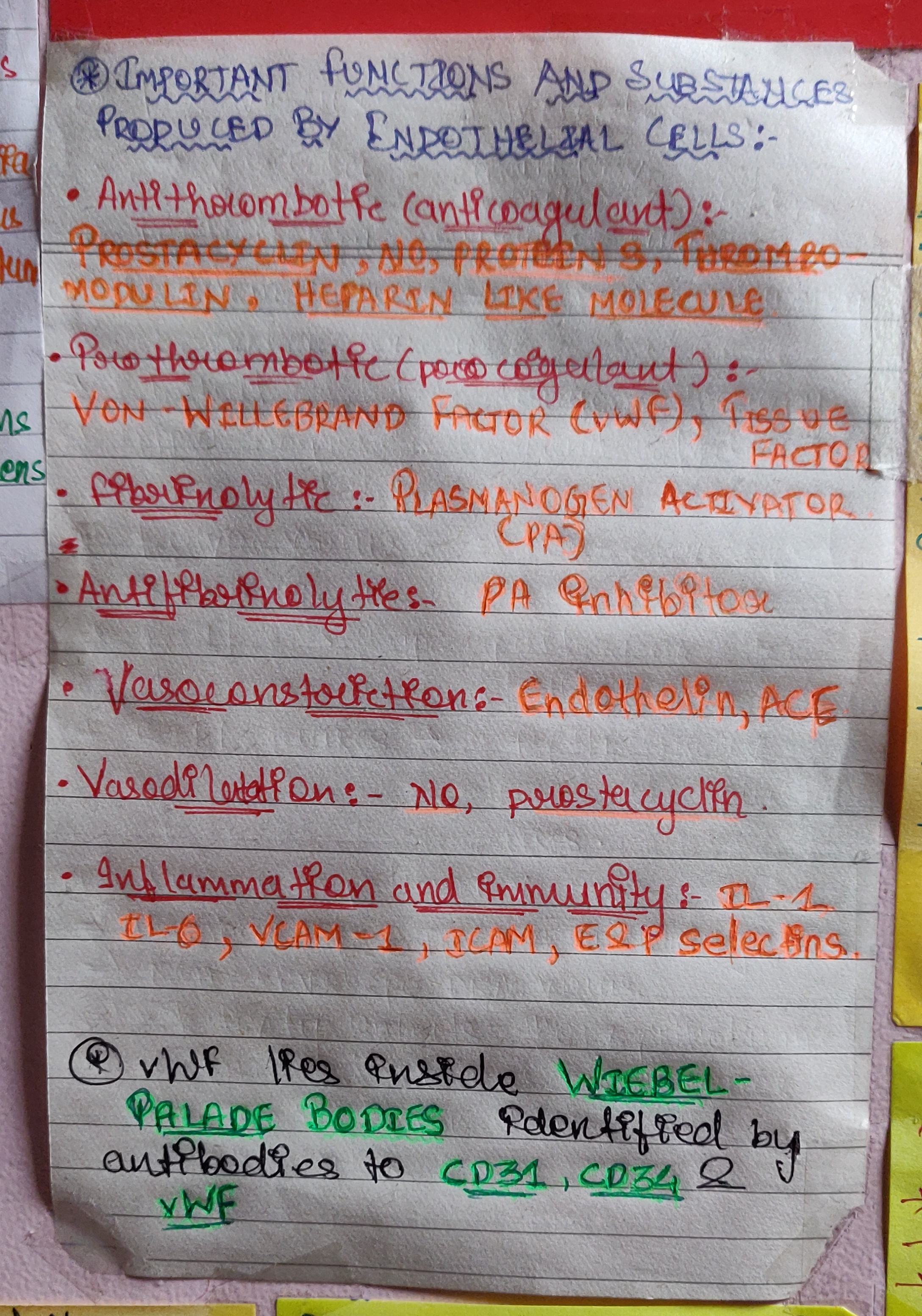
Source: Dental Pulse MCQS for Dental PG Entrance Examinations Volume-1. 11th edition
Source: Dental Pulse MCQS for Dental PG Entrance Examinations Volume-1. 11th edition
DENTIN HYPERSENSITIVITY :It is characterized by short , sharp pain arising from exposed dentin in response to stimuli typically thermal , chemical, osmotic, evaporative, tactile and which cannot be ascribed to any other form of dental defect or pathology.
ETIOLOGY :
ENAMEL LOSS :
CEMENTAL LOSS:
DEVELOPMENT OF DENTIN HYPERSENSITIVITY
There are two phases in the devlopment of dentin hypersensitivity .
Several theories have been put forward to explain the dentin hypersensitivity.
CLINICAL FEATURES:
DIAGNOSIS :
CASE HISTORY :
CLINICAL EXAMINATION :
PREVENTION :
MANAGEMENT :
DESENSITIZATION BY OCCLUDING DENTINAL TUBULES :
DESENSITIZATION BY BLOCKING PULPAL SENSORY NERVES
REFERENCE: STURDEVANTS 7TH EDITION



REGARDS,
Kriti Naja Jain 🙂
References:-
White & Pharow 7th e
Written by- Dr. Urusa I Inamdar
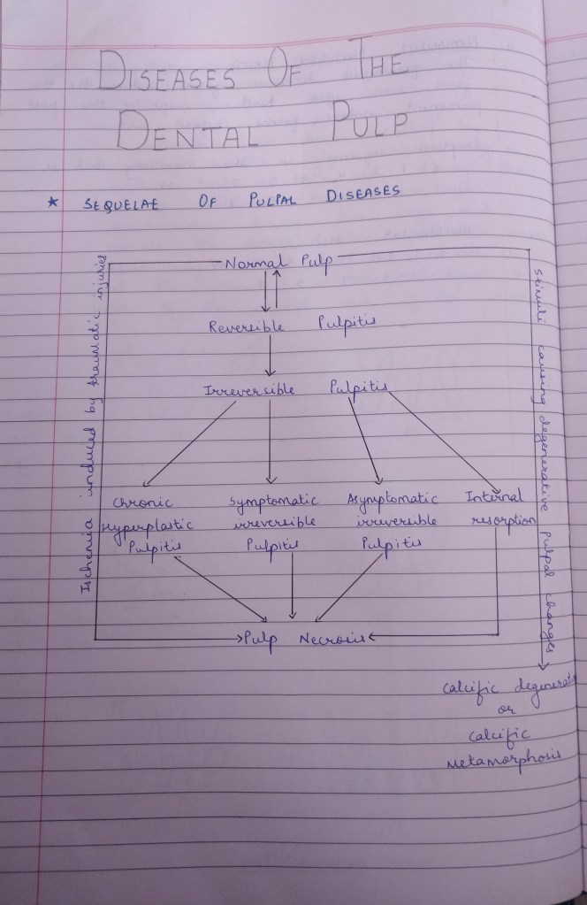

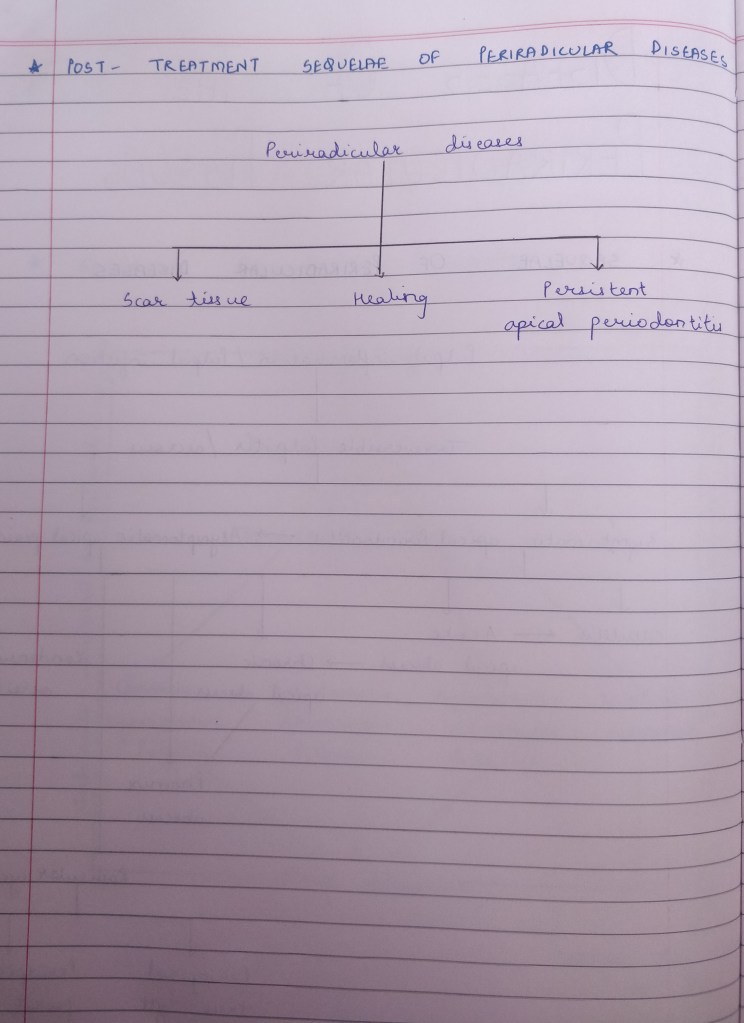
Reference:
Grossman’s Endodontic Practice (13th edition)

source – Nancy 13th edition

source – Nancy 13th edition
One of the basic goals of conservative dentistry is to preserve the health of the pulp as it is subjected to many insults such as caries, trauma, operative procedures, restorative materials.
Pulpal irritants can be classified as microbial, mechanical, chemical, thermal.
NEED FOR PULP PROTECTION
PULP PROTECTIVE AGENTS
CLASSIFICATION
CAVITY SEALERS: These are materials that provide a protective coating to the walls of the prepared cavity .They are applied to all the walls of the cavity and seal the interface between the restoration and the tooth . Eg: cavity varnish and resin bonding agents.
CAVITY VARNISH: A varnish is a natural gum or a synthetic resin dissolved in an organic solvent . Once it is applied to tooth surface the organic solvent evaporates leaving behind a protective film. Varnish is used beneath amalgam restoration to seal the amalgam tooth interface until corrosion product form to reduce the marginal gap. Thickness of varnish is 2-5 micrometer . They does not provide thermal protection and are contraindicated for GIC .
RESIN BONDING AGENTS: Currently the best method for the bonding composite resin to the tooth structure. Apart form adhesion it also provides cavity sealing. They are also employed for bonding amalgam restorations to reduce micro leakage .
CAVITY LINERS :They are used to provide a barrier against the passage of irritants from the cements or other restorative materials and to reduce the sensitivity of freshly cut dentin . They are usually suspension of calcium hydroxide in a volatile solvent . TYPES : TYPE III GIC , TYPE IV ZOE . Thickness is 0.5 mm
CAVITY BASES: A base is a layer of cement placed beneath the permanent restoration to encourage recovery of the injured pulp and to protect it against numerous types of insults. TYPES : high strength bases and low strength bases .
HIGH STRENGTH BASES provide thermal protection for pulp and mechanical support for the restoration . Eg : zinc phosphate , zinc polycarboxylate, RMGI, GIC
LOW STRENGTH BASES have minimum strength and rigidity . act as a barrier to irritation , chemicals and to provide therapeutic effect to pulp.Eg calcium hydroxide . ZnOE .
REFERENCE : STURDEVANT’S 7TH EDITION
ASHTINDER 🖊
SOURCE- RANGARAJAN TEXTBOOK
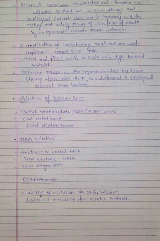
SOURCE- RANGARAJAN TEXTBOOK
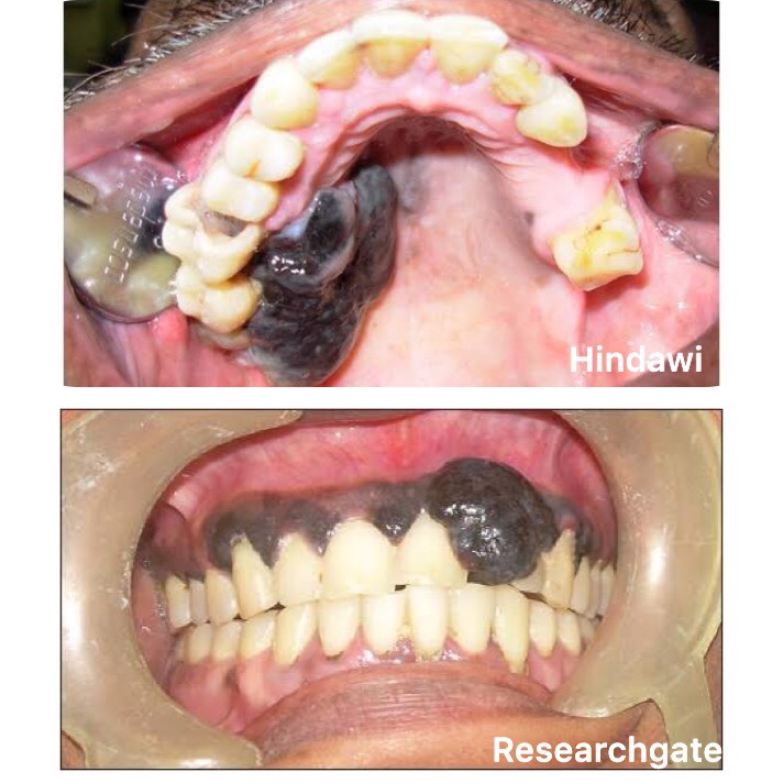
A neoplasm of epidermal melanocytes and third most common cancer of skin.
▪️Arises in preexisting mole. Appearance – Large, flat, spreading lesion; deeply pigmented Nodule
(i) Congenital Nevi
https://dentowesome.wordpress.com/2020/05/18/oral-nevi/
(ii) Dysplastic Nevi (Atypical Mole)
(iii) Lentigo Maligna: Also called as Hutchinson’s freckle, is a tan or black on the skin that looks like a freckle.
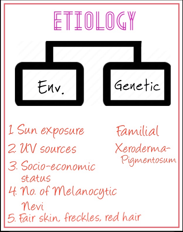
• Genes in the development of Melanoma:
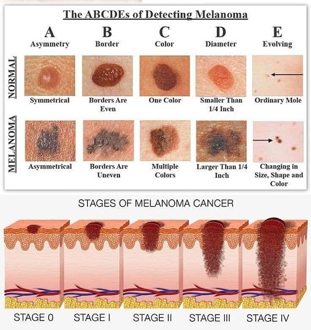
• Phases in the growth of Melanoma:
1. Superficial spreading Melanoma:
2. Lentigo Maligna Melanoma:
3. Nodular Melanoma:
4. Acral Lentiginous:
➡️ Metastases that cause symptoms but cannot be removed may be treated with radiation, immunotherapy, targeted therapy, or chemotherapy.
Dr. Mehnaz Memon🖊
References: Shafer’sTextbook Of Oral Pathology; Textbook Of Surgery by S.Das

