
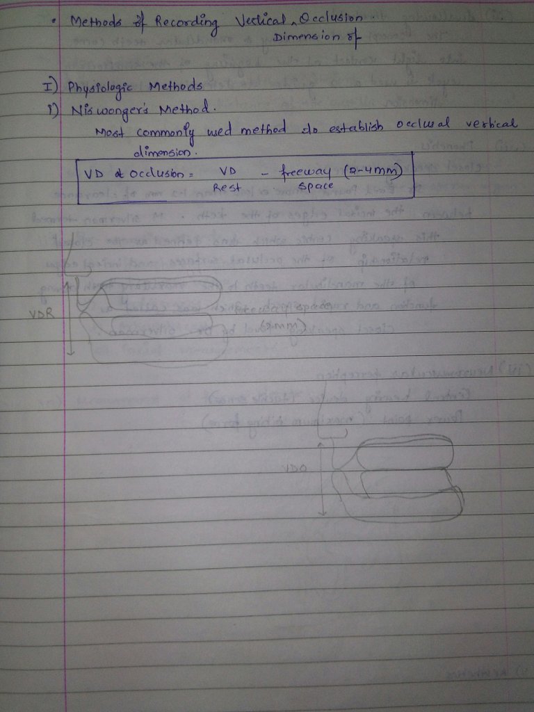
Source – Rangarajan textbook
Source – Rangarajan textbook
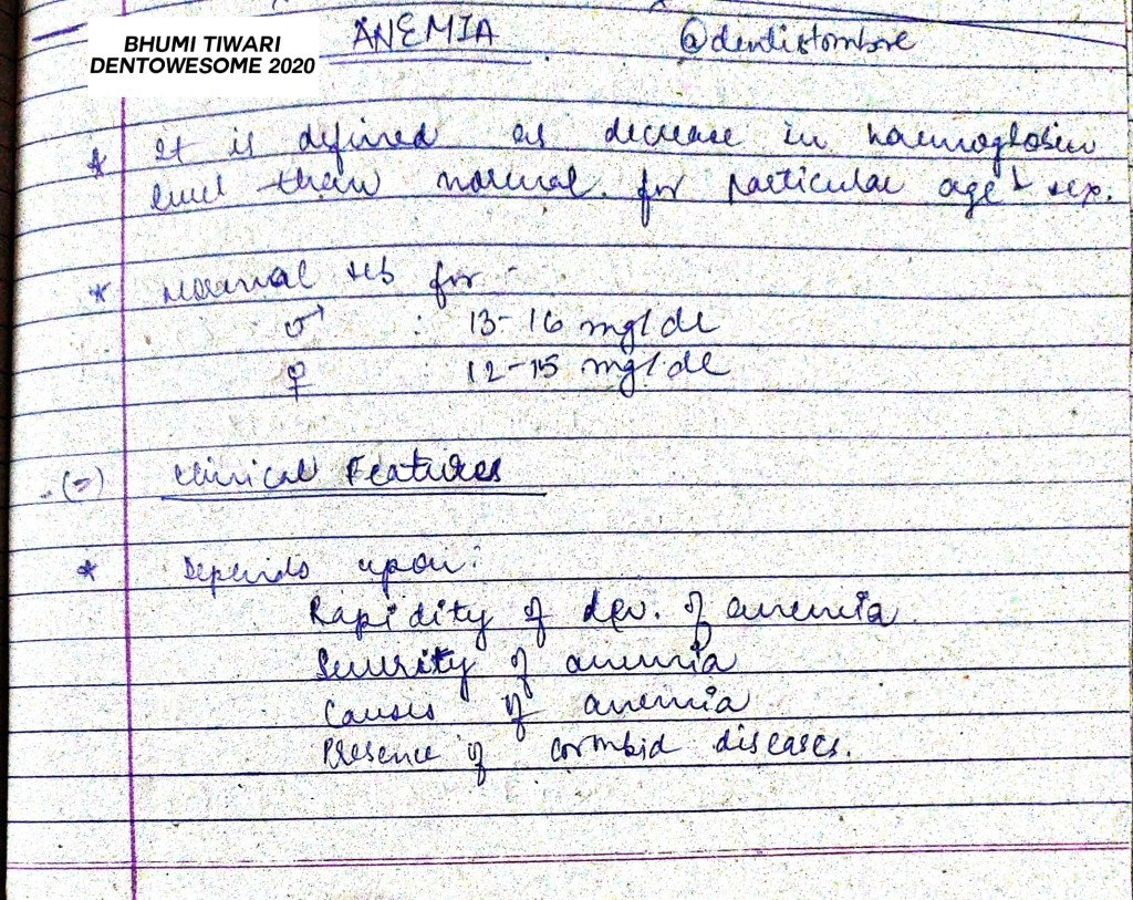
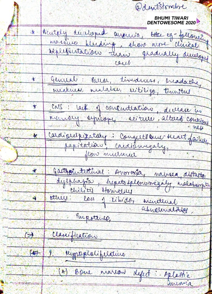


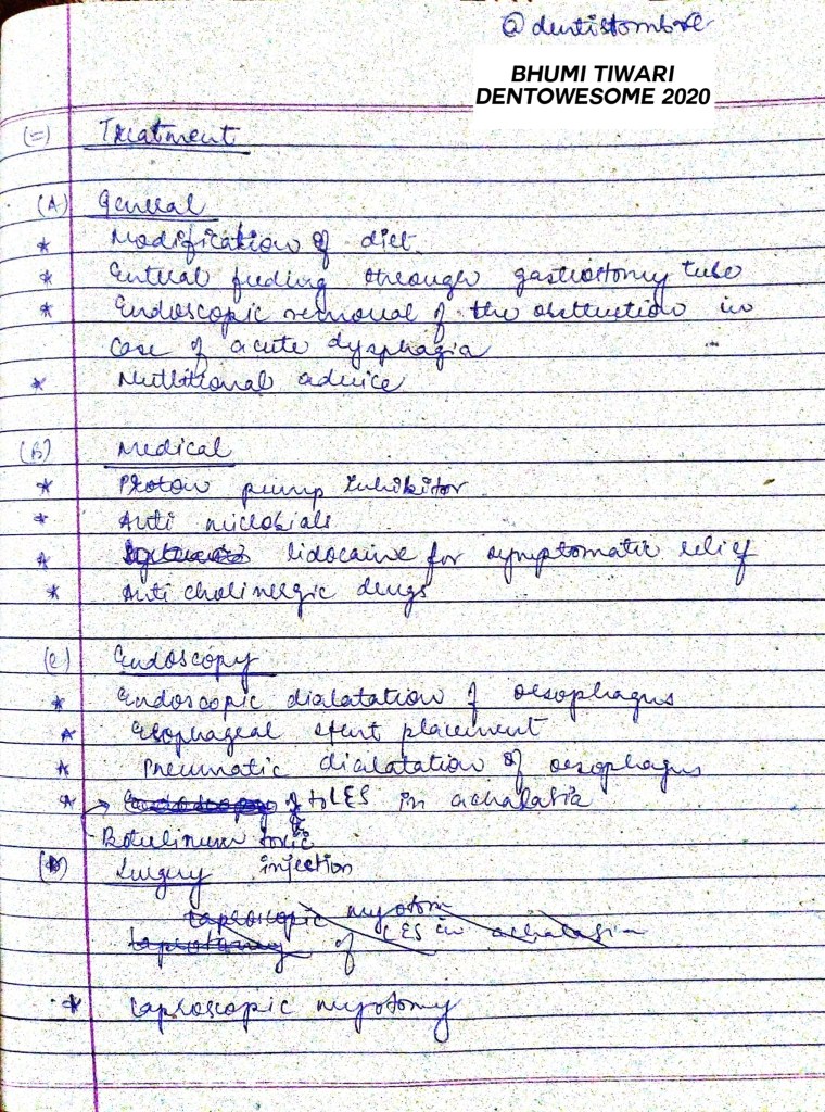
source: AK Tripathi- Essentials of Medicine for Dental Students, 2nd edition
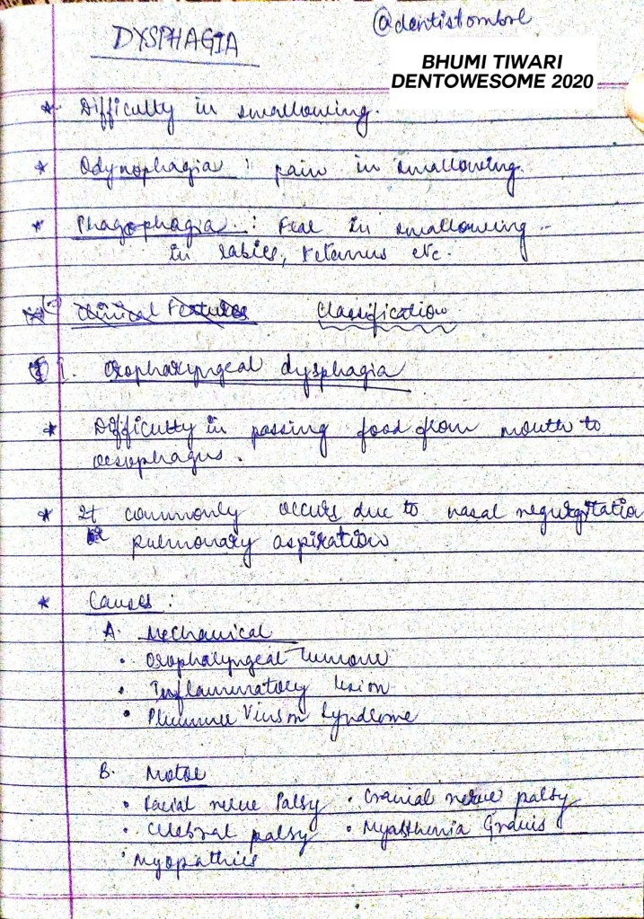

source: AK Tripathi- Essentials of Medicine for Dental Students, 2nd edition
Several modifications have been proposed for in class II cavity design for amalgam restorations . These modifications are changes from the classic preparation design . They are indicated for those situations which are not ideal. Modifications are based on following factors:
MODIFICATIONS :
2. SLOT PREPARATION : It is similar to class V amalgam cavity preparation except that it is done on the proximal surface of the tooth. It is indicated in proximal root surface caries with gingival recession. The design is similar to that of the slot preparation except it is approached from the facial aspect without disturbing the contact area.
3. CONSERVATIVE DESIGNS : It is recommended in all posterior teeth where caries incidence is very low and all occlusal pits and fissures are not involved. The conservative design preserves all the remaining sound tooth structures such as transverse ridge or oblique ridge and thereby protects the cuspal strength .
4. MODIFICATIONS TO PRESERVE ESTHETICS : To preserve the esthetics in the critical area during a mesio-occlusal cavity preparation , the facial wall of the mesial box should be prepared straight , i.e parallel to the long axis of the tooth rather than gingivally divergent . Another modification is to avoid breaking the facial contact whenever caries is limited only to the mesio-lingual embrassure.
5. MODIFICATIONS OF ROTATED TEETH : Depending on the degree of rotation , the proximal box is displaced facially or lingually.
6. ADJOINING RESTORATIONS : The intersecting margins of the two restorations -should be at right angles . Care should be taken so as to prepare the outline of the new cavity without weakening the amalgam margin of the existing restoration .
7. MODIFICATIONS FOR ABUTMENT TOOTH: The facial and lingual walls of the proximal box must be extended more so that the entire rest seat can be prepared in the amalgam without encroaching on the occlusal margins.
The pulpal floor is deepend an extra 0.5 mm apical to the region of the rest seat so as to provide an adequate thickness of amalgam.
8. CUSP CAPPING : It is indicated where there are extensive caries underneath the cusps and facio-lingual extensions of the occlusal preparation is more than 2/3rd the distance between the facial and lingual cusp tips. For cusp capping 1.5 to 2 mm of cusp reduction is necessary . The reduced cusps must meet the adjacent unreduced cusp at 90 degree cavosurface angle to provide both adequate edge strength of amalgam.
References: STURDEVANTS 7TH EDITION
LASER: Light Amplification by Stimulated Emission of Radiation .Laser dentistry is easier, more precise ,less traumatic for patients . Lasers provide a state-of-the-art approach for modern dentistry.
DISCOVERY : It was discovered by Shallow and Towns on 1958. First working laser was built by Maiman of Hughes research laboratory in 1960.
COMPONENTS OF LASER :
BASIC PRINCIPLES OF LASERS
LASER TISSUE INTERACTION :
TYPES OF LASERS :
1.BASED ON WAVELENGTH :
2. BASED ON TARGET TISSUE
LASER APPLICATION IN CONSERVATIVE DENTISTRY :
REFERENCES: STURDEVANTS 7TH EDITION
Written by – Dr. Urusa I Inamdar
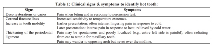
A tooth that is difficult to anesthetize is known as a ‘hot tooth ‘. This is most commonly encountered in a mandibular first molar tooth wherein after the anesthetic block , the patient may describe profound numbness of the ipsilateral lip and tongue but still may experience acute pain during the access opening procedure.
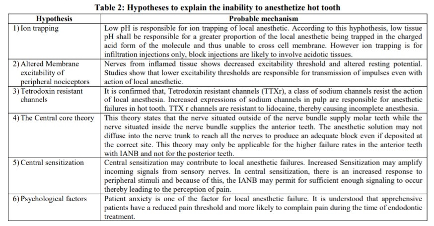
There is a special class of sodium channels on C – fibers, known as tetrodotoxin-resistant (TTXr) sodium channels. The expression shifts from TTX- sensitive to TTXr during neuroinflammatory reactions and the TTXr sodium channels play a role in sensitizing C-fibers and creating inflammatory hyperalgesia. One of the clinically significant characteristics of these sodium channels is that they are relatively resistant to lidocaine. These channels are five times more resistant to anesthetic than TTX- sensitive channels. Hot tooth may be explained by the fact that the TTXr sodium channels have not been adequately blocked by the anesthetic.
Bupivacaine has been found to be more potent than lidocaine in blocking TTXr channels and may be the anesthetic of choice when treating ‘hot tooth.’ Supplemental intraligamentary or intraosseous injections are most helpful to ensure profound local anaesthesia.
References:
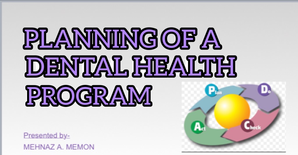
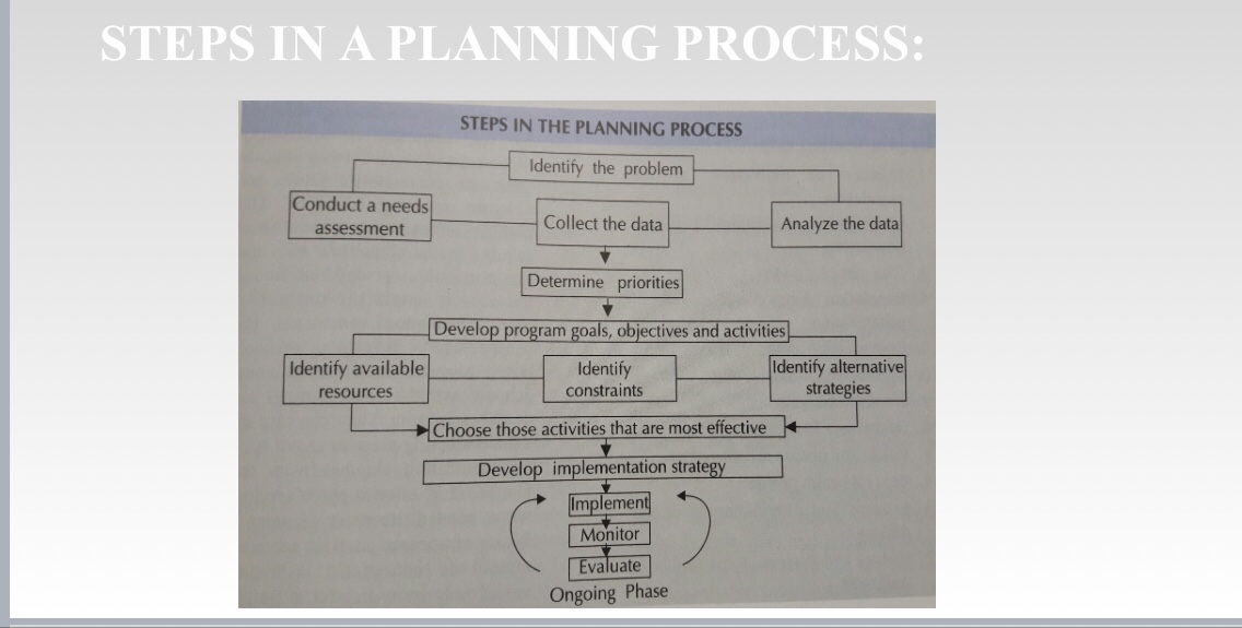
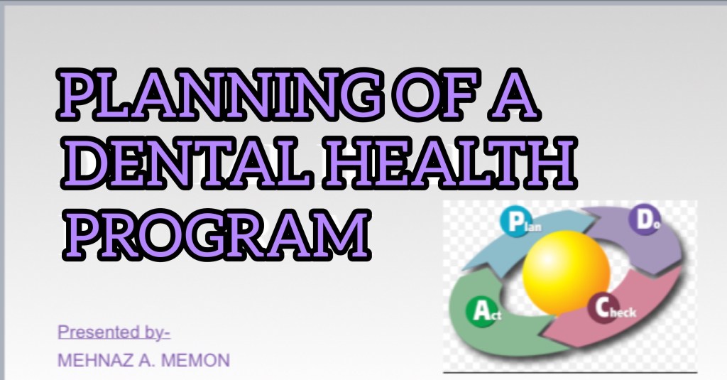
STEPS IN THE PLANNING PROCESS👇🏻
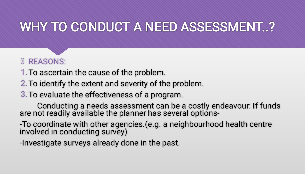

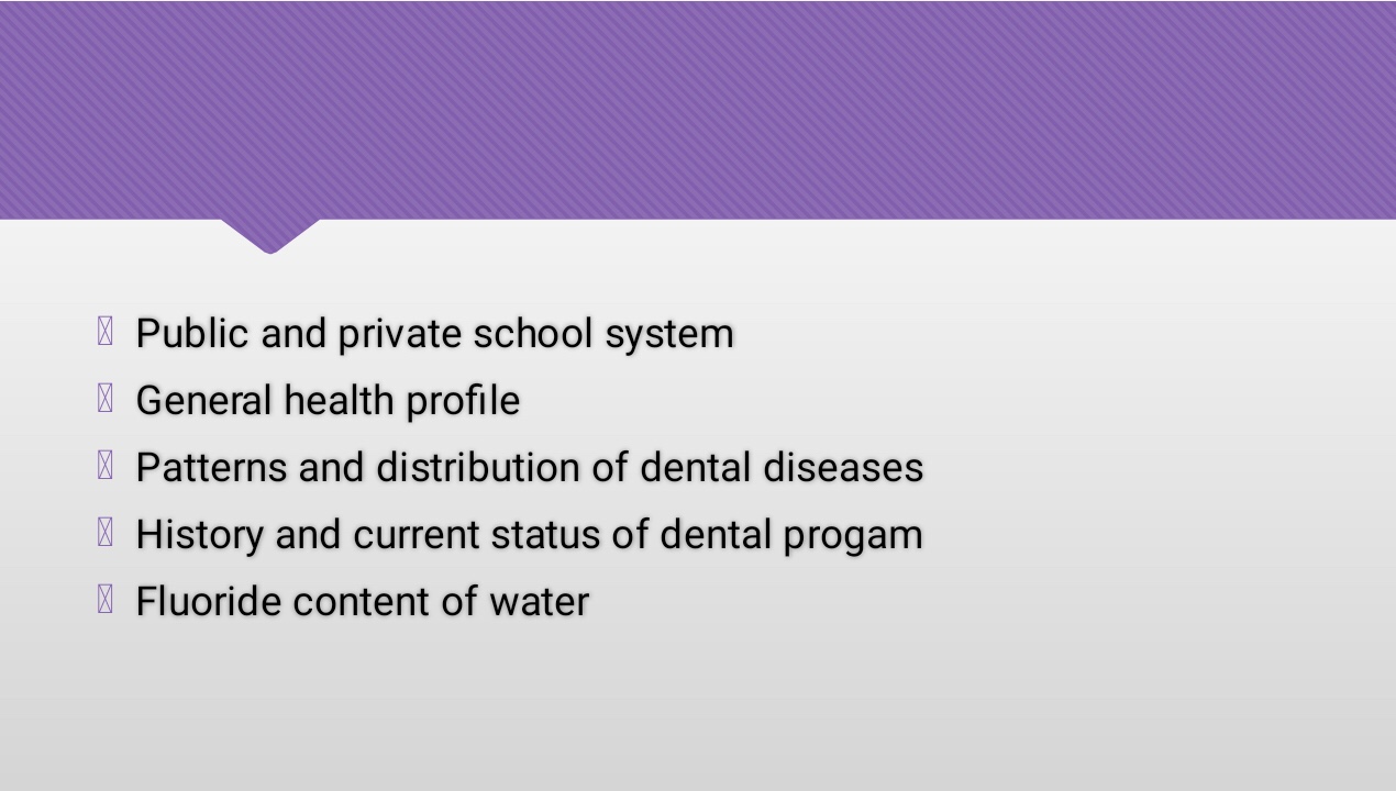
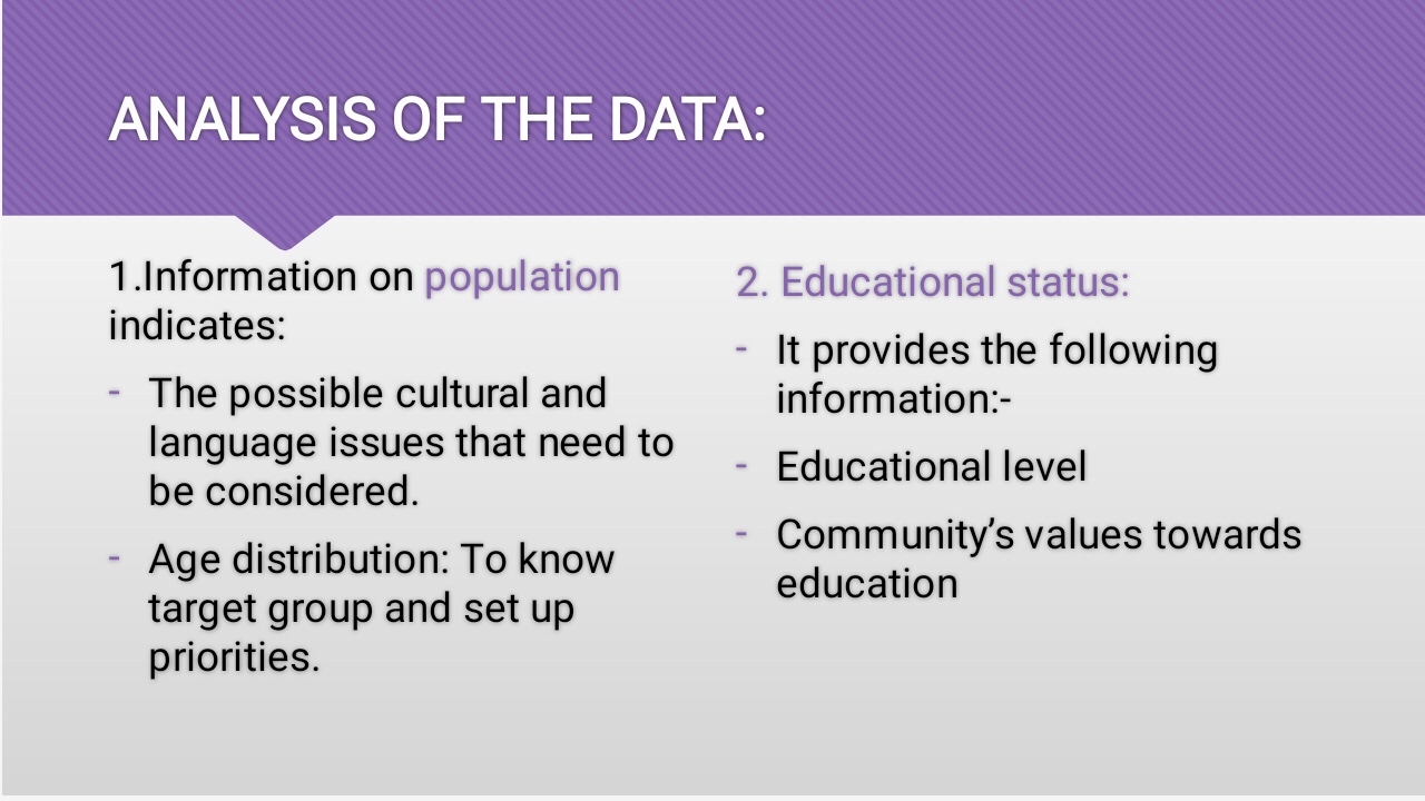





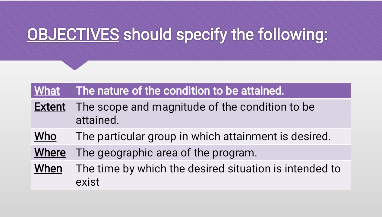
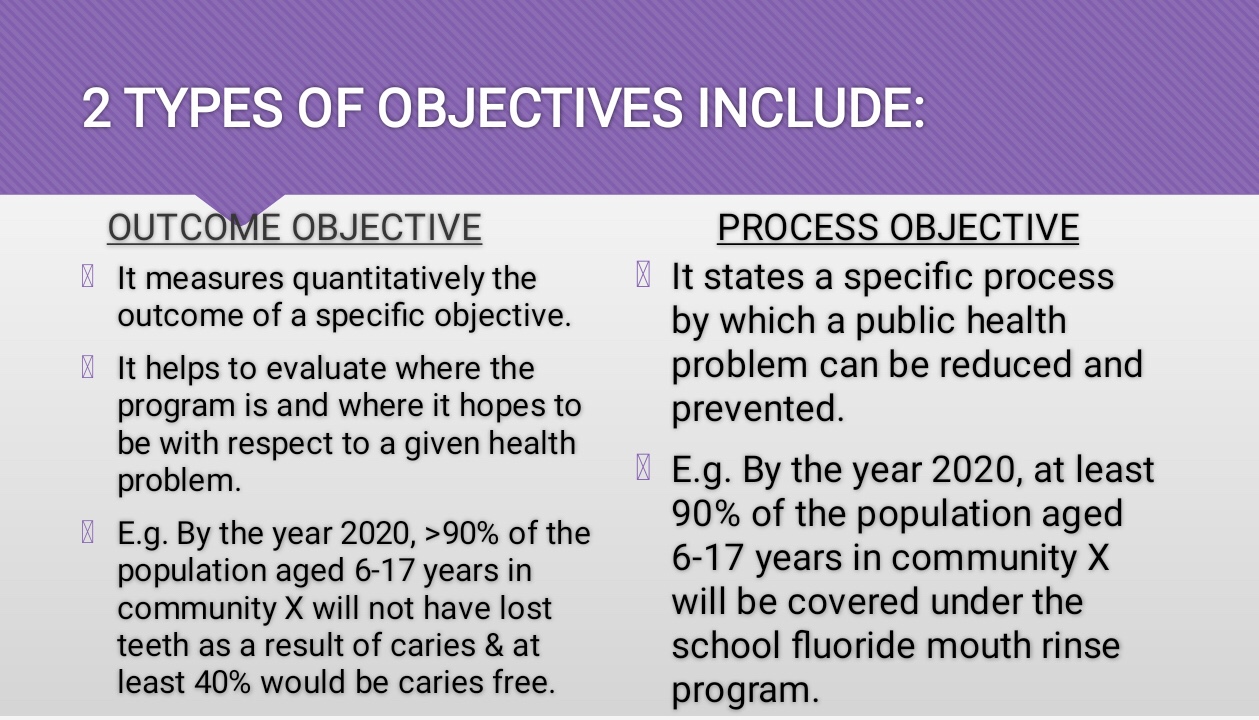


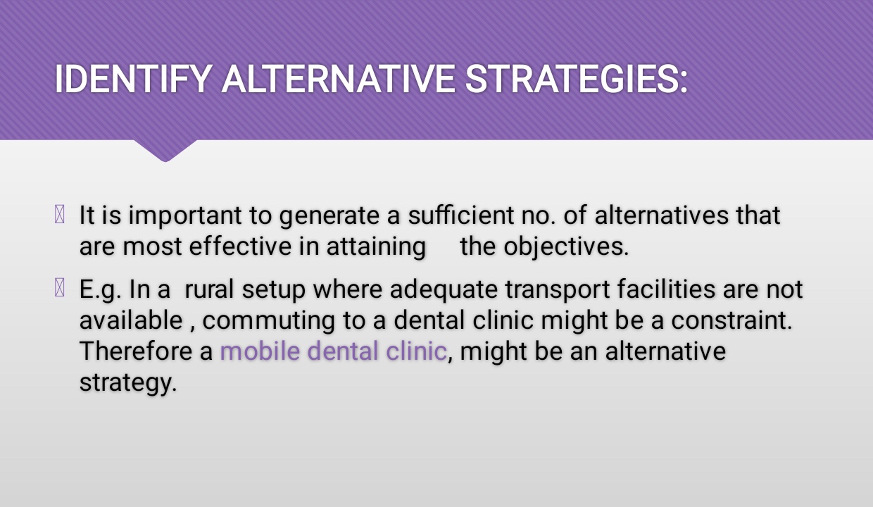

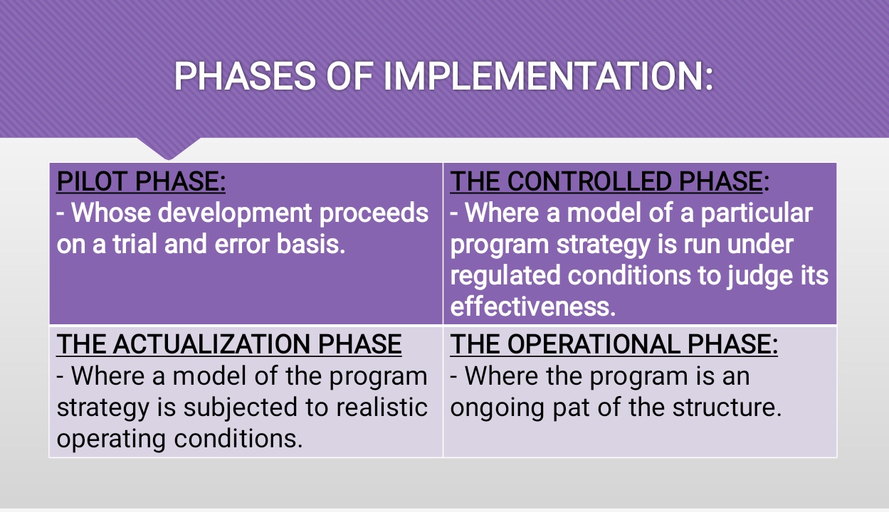

References: Textbook of Public Health Dentistry-Sober Peter(5th Edition)




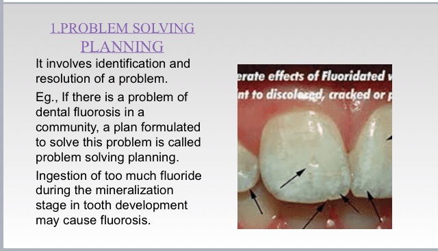
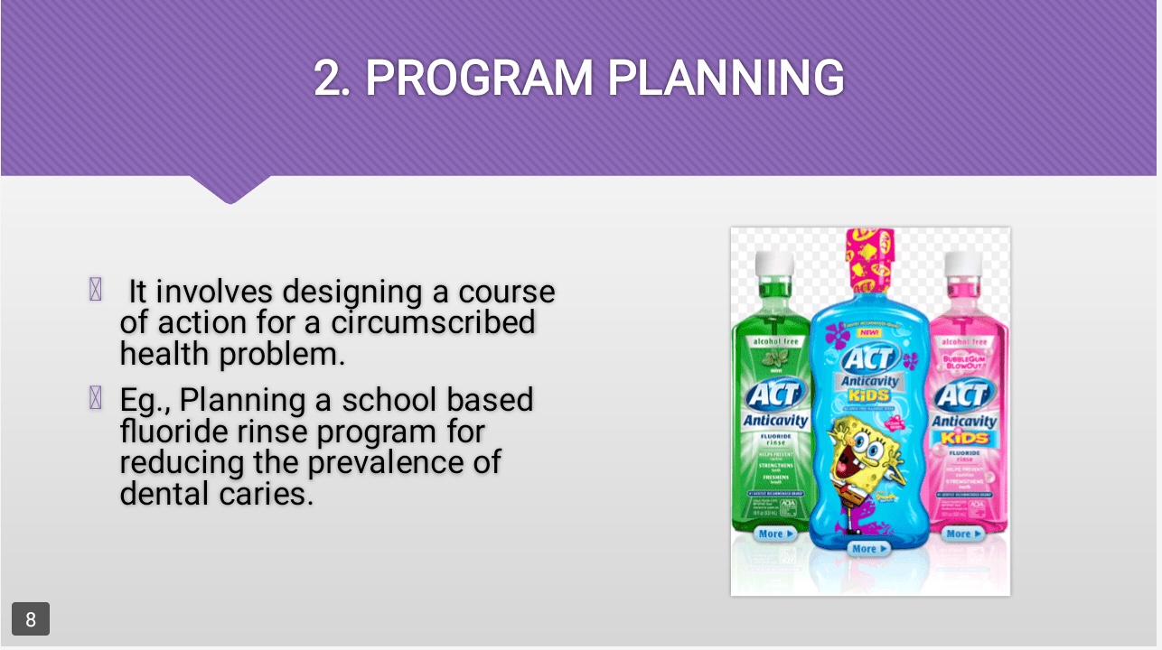

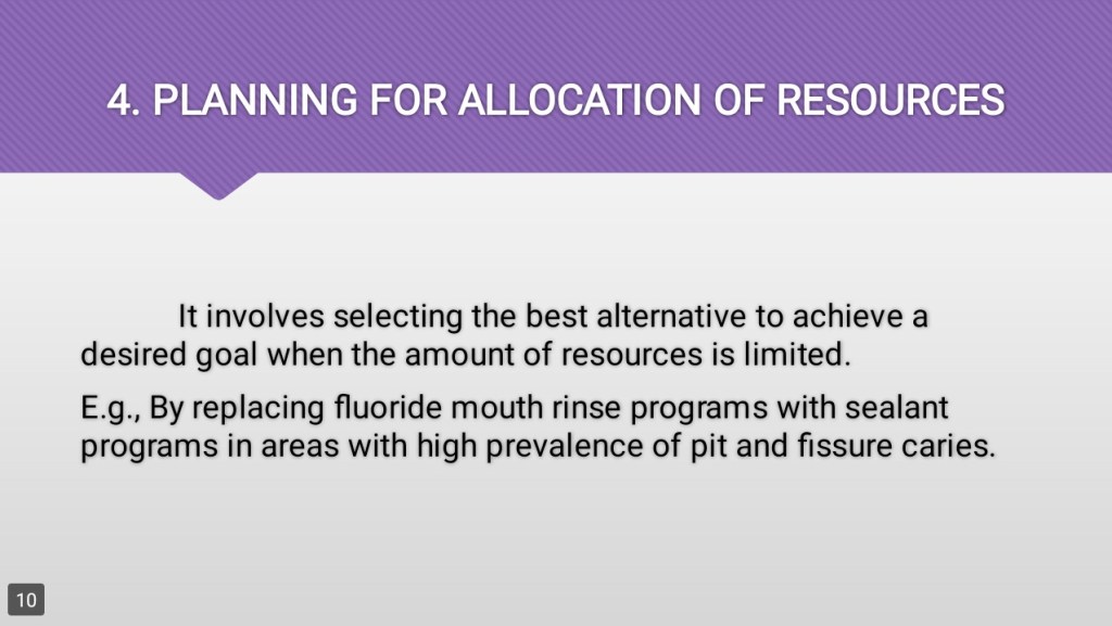
References: Textbook of Public Health Dentistry-Sober Peter(5th Edition); Random google images
Reference :Pharmacology
I) Trigeminal is the largest cranial nerve.
II) Vagus nerve is with vague or extensive distribution.
III) Olfactory is the smallest (in length) cranial nerve while trochlear is smallest in thickness
IV) Trochlear nerve shows largest intracranial course.
V) Buccal nerve is the only sensory branch of anterior division of mandibular nerve.
VI) Smallest branch of trigeminal nerve is opthalmic.
VII) Smallest branch of trigeminal nerve is opthalmic.
VIII) The branch of nasociliary that is frequently absent is posterior ethmoidal.
IX) Posterior superior alveolar nerve is a branch of maxillary nerve in pterygopalatine fossa.
X) Infraorbital nerve (middle & anterior superior alveolar nerves) is a branch of maxillary nerve in orbital groove. Middle superior alveolar nerve is present only in 28% of individuals.
XI) Mandibular nerve is largest of three branches of the trigeminal nerve.
XII) Inferior nerve is larger terminal branch of Mandibular nerve.
XIII) Greater Palatine nerve is also known as anterior Palatine nerve. Lesser palatine nerve induces middle and posterior Palatine branches.
XIV) Nervus intermedius is sensory branch of facial nerve.
XV) Nervus spinosus is from meningeal branch of main trunk of mandibular nerve.
XVI) Greater petrosal nerve is a branch of facial nerve and is parasympathetic to lacrimal glands, glands of nose and pharynx. The ganglion associated with greater petrosal nerve is pterygo Palatine ganglion.
XVII) Lesser petrosal nerve is a branch of GLOSSOPHARYNGEAL nerve and it is parasympathetic to parotid gland through auriculotemporal nerve. The ganglion associated with lesser petrsoal nerve is otic ganglion.
XVIII) The deep petrosal nerve is a branch of carotid plexus round the internal carotid artery. It joins the greater petrosal nerve to form nerve of pterygoid canal
XIX) External petrosal nerve is a branch of the sympathetic plexus round the middle meningeal artery.
Source: Dental Pulse MCQS for Dental PG Entrance Examinations. Volume 1,11th edition.

