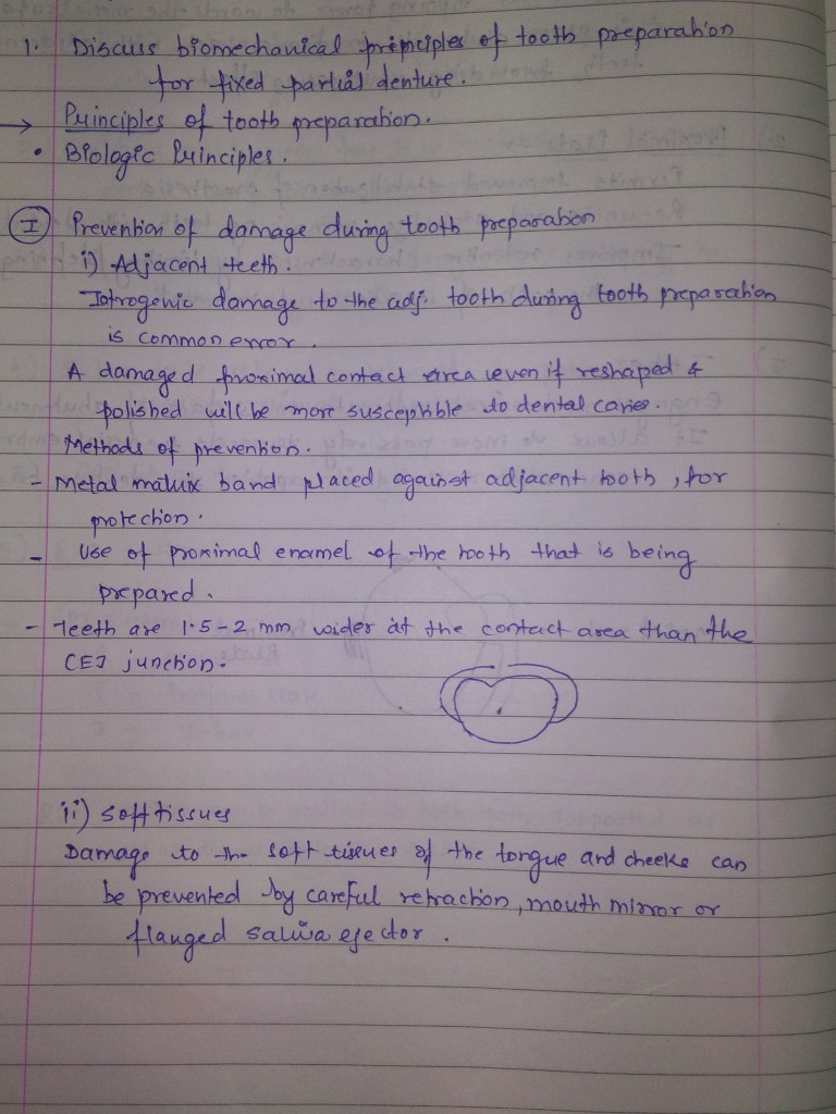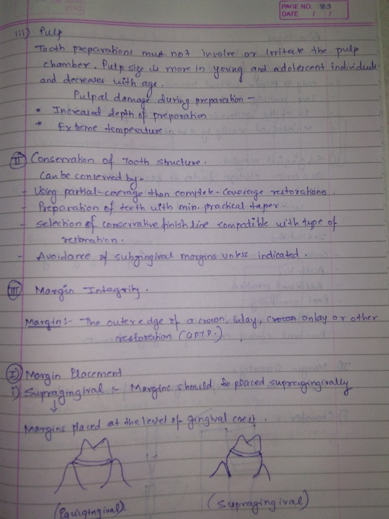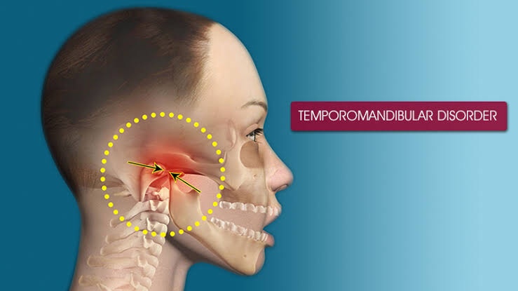
TEMPOROMANDIBULAR DISORERS(TMD)

TMD: They are the most common cause of facial pain after toothache.

🔹 Classification:

  1. TMD secondary to Myofacial pain dysfunction syndrome(MPDS)
  2. TMD secondary to true articular disease

🔹Etiology:

1) MPDS –

  • Malocclusion
  • Jaw clenching
  • Bruxism
  • Personality disorders
  • Stress & Anxiety
  • Increased pain sensitivity

2) Articular disease –

  • Disk displacement disorder
  • Degenerative joint disorders
  • Rheumatoid Arthritis, Ankylosis
  • Dislocation
  • Infection
  • Neoplasia

🔹Clinical Features:

➡️ The clinical manifestations in MPDS :

  • Pain without destructive changes of TMJ on radiograph
  • Tenderness
  • Spasm of Masticatory Muscles (Temporalis, Masseter, Lateral & Medial pterygoids)
  • Muscle hyperactivity & dysfunction due to malocclusion

➡️ Age: Young women (20-40 years); M:F – 1:4

➡️ 4 cardinal signs & symptoms of the syndrome:

  1. Pain (Periauricular, with chewing & radiate to head)
  2. Muscle tenderness
  3. Clicking/popping noise in TMJ
  4. Limitation of jaw motion

➡️ Pain: Dull type

  • unilateral/bilateral in MPDS
  • unilateral in articular origin except Rheumatoid Arthritis

➡️ Symptoms:

  • Otalgia
  • Neck pain
  • Shoulder pain
  • Dizziness

🔹Radiograph shows👇🏻

  • Erosion
  • Bony sclerosis
  • Osteophytes
  • Condylar-glenoid fossa remodelling

CT Scan – Material injected into joint cavity.

MRI – Articular pathology

Surgical – Traumatic TMD

Arthroscopy – Internal TMJ derangements

🔹Lab Diagnosis:

  1. Blood count to rule out any infection
  2. Rheumatoid Arthritis – Rheumatoid factor, ESR, Anti-nuclear antibody
  3. Gout – Uric acid levels

🔹Differential Diagnosis:

  1. Clustre/Migraine headache
  2. Temporal/Giant cell arteritis
  3. Post herpetic neuralgia
  4. Trigeminal Neuralgia
  5. Middle ear infections

🔹Treatment options for TMD:

• Self-care practices & conservative approach, surgical (last option)

• Basic treatments include –

  1. Apply moist heat or cold packs
  2. Eat soft foods, Do not chew gum
  3. Take medications – To avoid muscle pain and swelling; NSAID’s (Aspirin, Ibuprofen), Muscle relaxants (who clench teeth), Anti-anxiety drugs, Anti-depressants
  4. Use of night guards & splints
  5. Undergo corrective dental treatments
  6. Avoid extreme jaw movements

• Conservative –

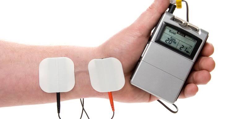
Transcutaneous electrical nerve stimulation (TENS) is a method of pain relief involving the use of a mild electrical current.
  • TENS
  • Ultrasound
  • Trigger point injections
  • Radiowave therapy

Dr. Mehnaz Memon🖊


References:

  1. https://www.nhs.uk/conditions/transcutaneous-electrical-nerve-stimulation-tens/
  2. https://www.epainassist.com/sports-injuries/head-and-face-injuries/tmd
  3. Image Source: Google
  4. Shafer’sTextbook Of Oral Pathology; Ghoms Oral Medicine

OEDEMA: PATHOGENESIS & TYPES

👉🏻Definition & Types:

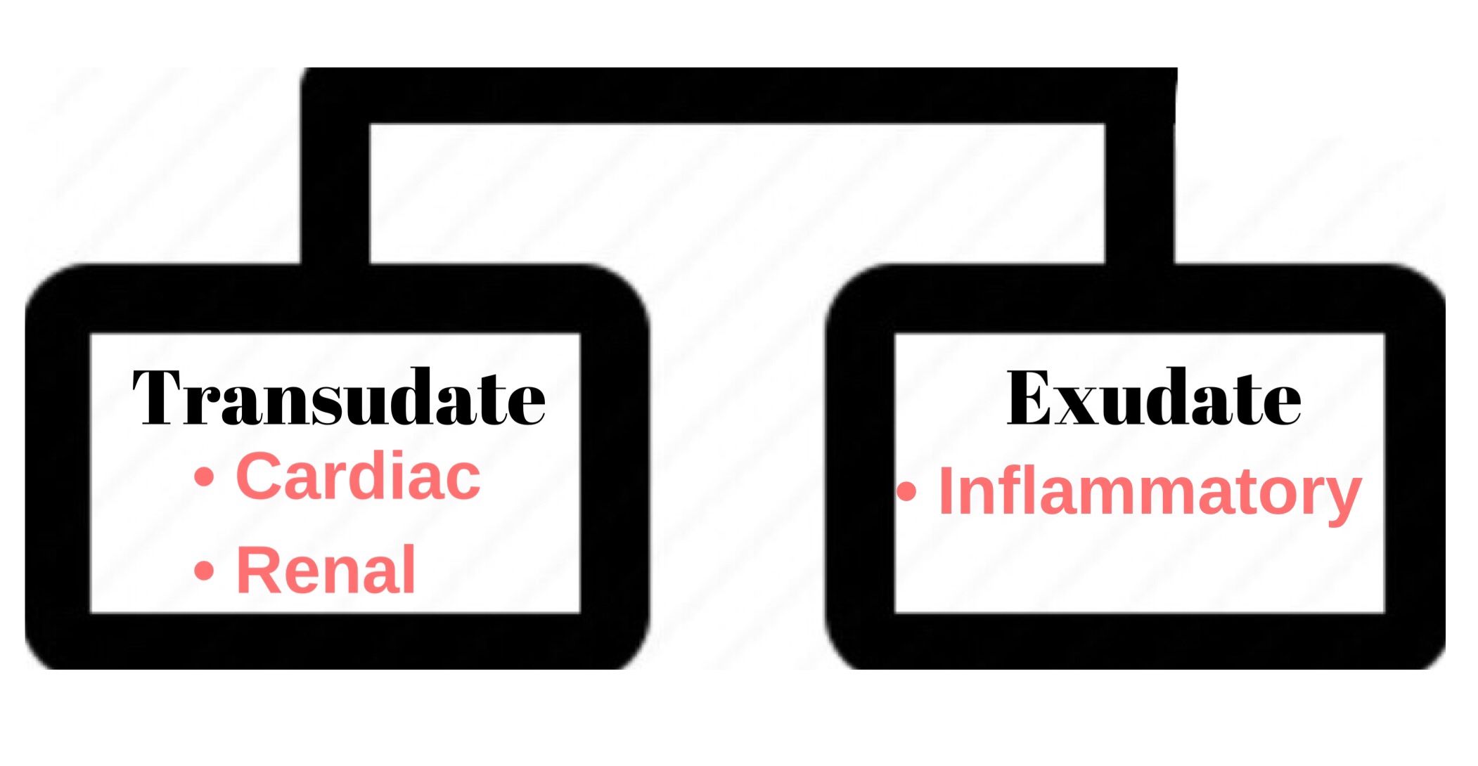
🔹OEDEMA〰️Swelling. Abnormal/excessive accumulation of free fluid in the interstitial tissue spaces & serous cavities.

🔹Accumulation of fluid (abnormally) inside the cell is intracellular edema/hydropic degeneration.

🔹Accumulation in body cavities:

  • Peritoneal Cavity – Ascitis
  • Pleural Cavity – Hydrothorax (T.B)
  • Pericardial cavity – Hydropericardium
TYPES OF OEDEMA👆🏻

🔹PITTING & NON-PITTING EDEMA:

Source: Healthline

➡️ When interstitial fluid volume increases, most of the fluid becomes free that is not bound to proteoglycan meshwork. This type of edema is called pitting edema because when this area is pressed with finger, fluid gets displaced producing depression or a pit as seen in SUBCUTANEOUS TISSUES.

➡️ Edema also develops due to swelling of the cells/clotting of interstitial fluid in the presence of fibrinogen. This is called Non-pitting/solid edema because its hard & a pit is not formed by pressing.
  • e.g. – Myxodema
  • – Elephantiasis

Oedema Fluid

🔹 Pathogenesis of Edema:

➡️ Causative mechanisms that produce edema are:

  1. ⬇️ Plasma oncotic pressure
  2. ⬆️ capillary hydrostatic pressure
  3. Lymphatic obstruction
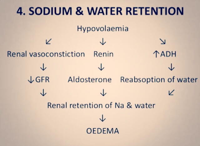
  4. Sodium & Water retention
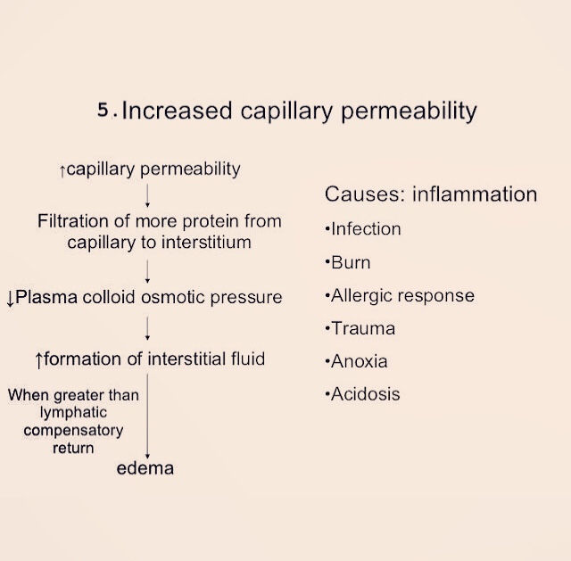
  5. ⬆️ capillary permeability
  6. Tissue factors

These mechanisms interfere with normal fluid balance of plasma, interstitial fluid & lymph flow.

  • It is exerted by total amount of plasma proteins that tend to draw fluid into the vessels normally.
  • A fall in the total plasma protein level (<5g/dl), results in lowering of plasma oncotic pressure in a way that it can no longer counteract the effect of hydrostatic pressure of blood.
  • Therefore, increased outward movement of fluid from capillary wall & decreased inward movement of fluid from the interstitial space causing edema.
Fluid pressure and colloid osmotic pressure forces operate at the capillary membrane, tending to move fluid either outward or inward through the membrane pores
Pathogenesis of oedema in hypoalbuminaemia. The normal balance of hydrostatic and oncotic pressures is such that there is net movement of fluid out of the capillaries at their arteriolar ends and net movement in at their venular ends (indicated here by arrows). Oedema can thus be due to an increase in capillary hydrostatic pressure, a decrease in plasma oncotic pressure or an increase in capillary permeability.

There are many possible causes of hypoalbuminaemia; a combination of which may be implicated in individual cases. For example, to list few of them👇🏻

  • Oedema of renal disease (Nephrotic syndrome, Acute glomerulonephritis)
  • Ascitis of liver disease e.g. in Cirrhosis.
  • Protein losing enteropathy
  • Malnutrition

➡️ Fluid from capillary wall to interstitial space by counteracting the force of plasma oncotic pressure.

➡️ Edema results when hydrostatic pressure at Venular end of capillary wall(12mmHg) becomes more than the plasma oncotic pressure, resulting in no reabsorption of fluid at the Venular end.

  • Example, edema of Cardiac disease – Congestive cardiac failure, constrictive pericarditis
  • Cirrhosis of liver
  • Passive congestion – e.g. mechanical obstruction due to thrombosis of veins of lower legs, varicosities, pressure by pregnant uterus, tumours etc.
  • Postural edema e.g. ⬆️ Venous pressure (Edema of feet & ankle)

➡️ Lymphoedema is caused due to obstruction of lymphatic channels causing localized edema.

  • e.g. removal of axillary lymph nodes in radical mastectomy for carcinoma of breast cancers.
  • Rupturing of main lymphatic channel causing chylothorax & chylous ascitis

– Inflammation of Lymphaties:

  • Filariasis (infection with Wuchereria Bancrofti) – Chronic lymphoedema of scrotum & legs

– Abnormal development of lymphatic channels:
  • Milroy’s disease/hereditary lymphoedema
  • Edema comfined to one or both lower limbs.
Source: Slideshare
Source: Slideshare

Forces acting in the interstitial space –

1. Oncotic Pressure

⬇️

⬆️ Vascular permeability & inadequate removal of proteins by lymphatics

⬇️

Elevation

2. Tissue Tension

⬇️

Lowered (e.g. loose subcutaneous tissues of eyelids & external genitalia)


Dr. Mehnaz Memon🖊

References: Textbook of Pathology, Harsh Mohan; Internet; https://basicmedicalkey.com/plasma-proteins-and-enzymes/