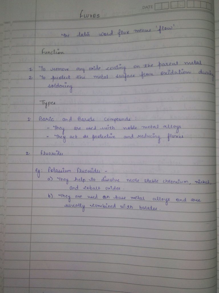
Source Gurkeeraat Singh textbook
Source Gurkeeraat Singh textbook
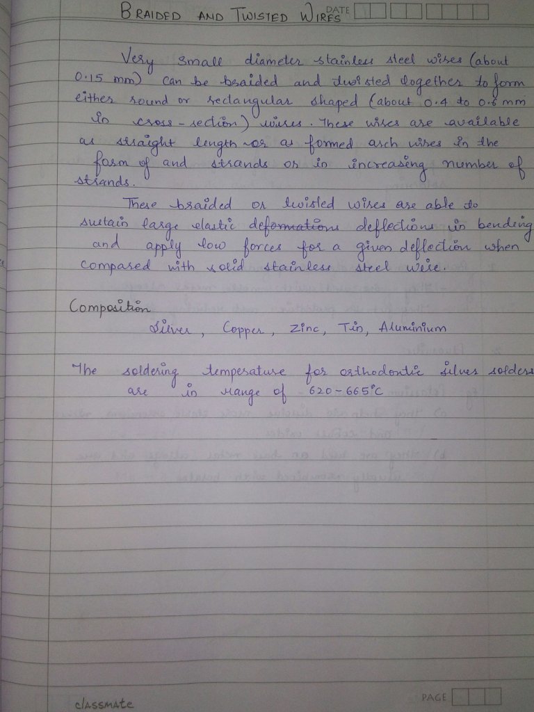
Source Gurkeeraat Singh textbook
After the registration of the maxillo-mandibular , the artificial teeth must be set in centric occlusal position.
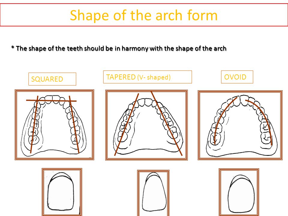
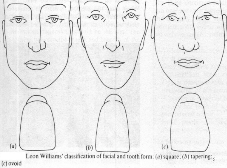
The facial form of the patient should be classified into-
–square
–ovoid
– tapering
-The form of the teeth should be in harmony with the form of the face, square teeth are used for those with a square face and so on.
-In addition, the patient’s age, gender and personality should be taken into consideration when trying to improve to improve the appearance.
-The color is also important, dark and opaque teeth should be selected for elderly patient, light and translucent teeth for the young patients.
-The size of the teeth must also be in harmony according to the size of the face and the gender of the patient.
to provide a comfortable and atraumatic occlusion
to assist in preparing food for deglutition
to impart a pleasing and natural appearance
to assist in speech
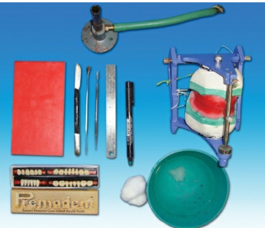
Armamentarium
•Maxillary occlusal rim with cast
• Mandibular occlusal rim with cast
• Mean value articulator
• Teeth shade
• Measurement scale
• Wax knife
• Roach carver
• Wax spatula
• Wax sheet
• Gas torch
• Cotton
• Rubber bowl with cold water
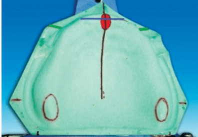
Guidelines for anterior teeth setting are marked on the cast and the land area of the cast.
I. The centre of the incisive papilla (marked in red)
II. A line is drawn along the midline of the cast through the centre of the incisive papilla (marked in black).
III. A perpendicular line is drawn to the above line through the centre ofincisive papilla till the land area (marked in blue; CPC line).
IV. During setting of maxillary canine, the cuspid tip of the canine should be placed in line with this point
The crest of the lower residual alveolar ridge is marked in red colour along the posterior aspect of the lower cast. A corresponding line is marked on the upper cast in blue
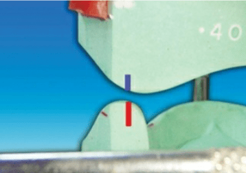
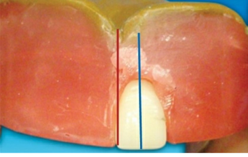
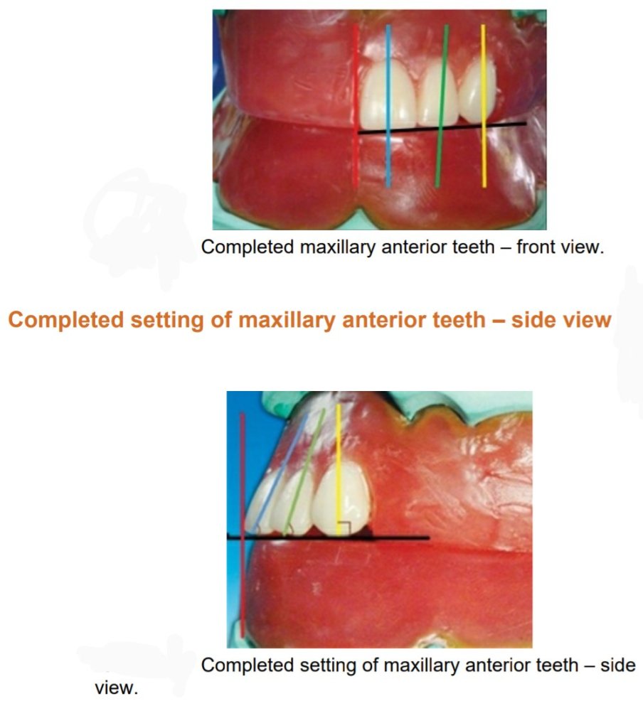
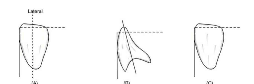
Placement of the maxillary central incisor in relation to all three planes.
🔻The long axis of the central incisor (the blue line) is positioned parallel to the
vertical axis (the red line) when viewed from the front
🔻The incisal edge of the maxillary central incisor is positioned in line with the
anterior occlusal plane . The central pin of the articulator will point the mesioincisal line edge of the central incisor.
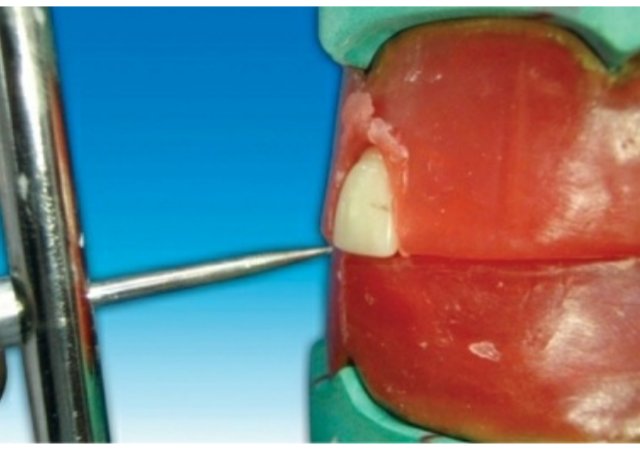
🔻The central pin of the articulator will
point the mesioincisal line edge of the central incisor.
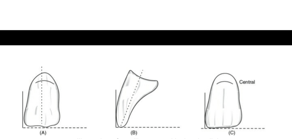
When viewed from:
Front: Long axis parallel towards the vertical axis.
Side: Slopes labially about 15 degrees.
Occlusal plane: Incisal edge is in contact with occlusal plane
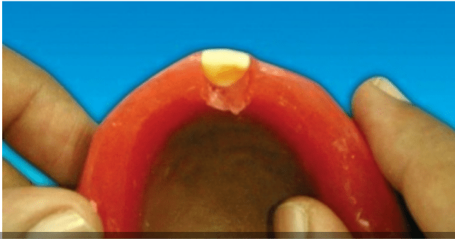
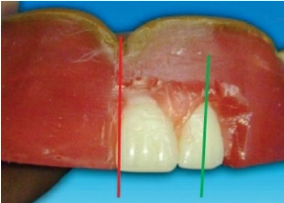
🔻The long axis of the lateral incisor (green line) is placed slightly inclined to
the vertical axis (red line).
🔻The incisal edge of the maxillary lateral incisor is placed slightly short of the
occlusal plane (marked as yellow line).
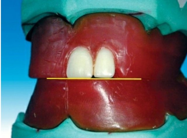
🔻The incisal edge of the maxillary lateral incisor should follow the outline of
the occlusal rim when viewed occlusally
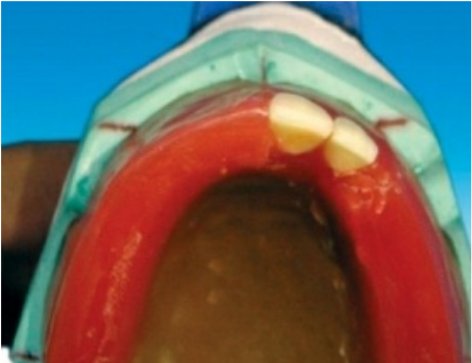
When viewed from:
Front: Long axis slopes towards midline at incisal edge.
Side: Slopes labially about 20 degrees.
Occlusal plane: Incisal edge is 1 mm short of occlusal plane
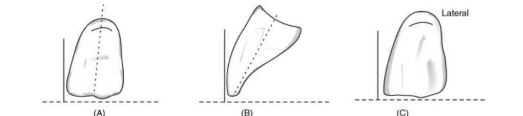
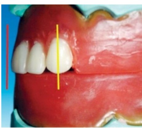
🔻The long axis of the maxillary canine (yellow line) should be parallel to the
vertical axis (red line) when viewed from the front.
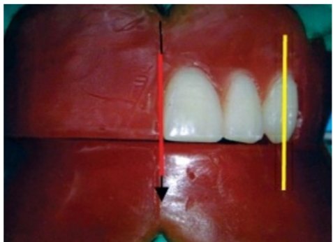
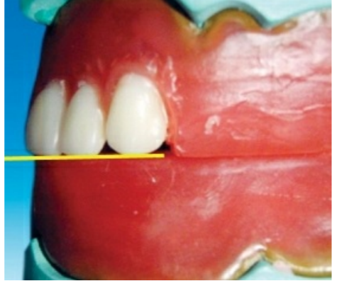
🔻The long axis of the maxillary canine (yellow line) should be parallel to the
vertical axis (red line) when viewed from the side .
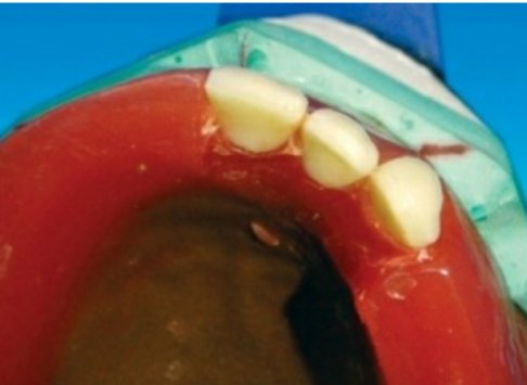
Maxillary canine follows the outline of occlusal rim, and the tip of the canine
corresponds to the canine papillary line
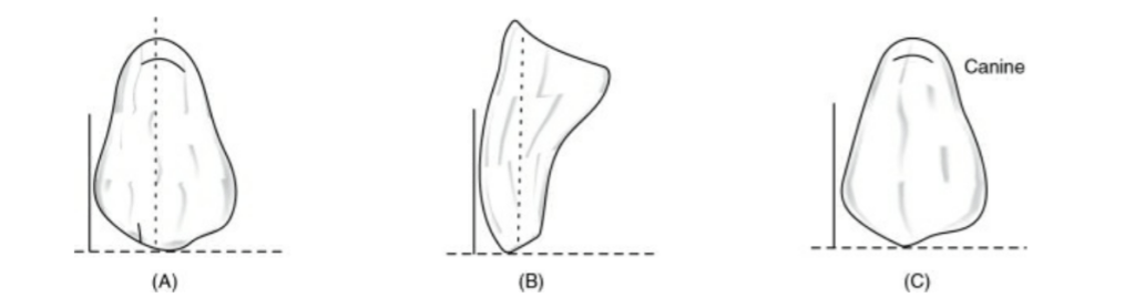
When viewed from:
Front: Long axis parallel towards the vertical axis.
Side: Long axis parallel towards the vertical axis.
Occlusal plane: Cuspid tip is in contact with occlusal plane
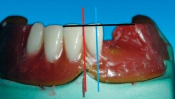
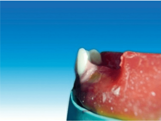
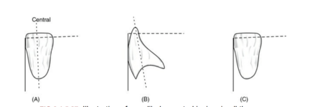
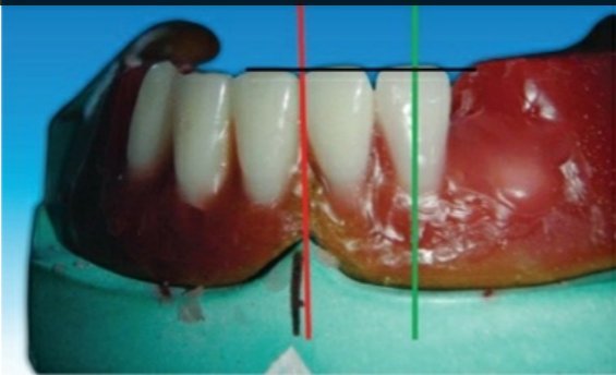
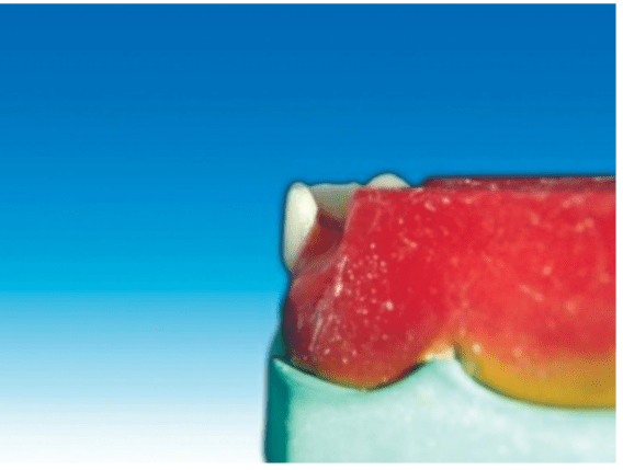
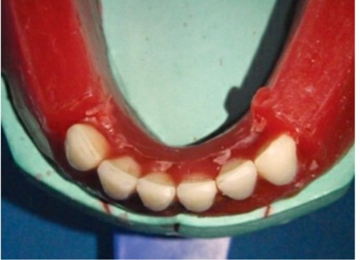
🔻The incisal edge of the mandibular central incisor should follow the outline
of the occlusal rim, and the incisal edge of the mandibular central incisor tooth
should not cross over the land area when viewed occlusally
When viewed from:
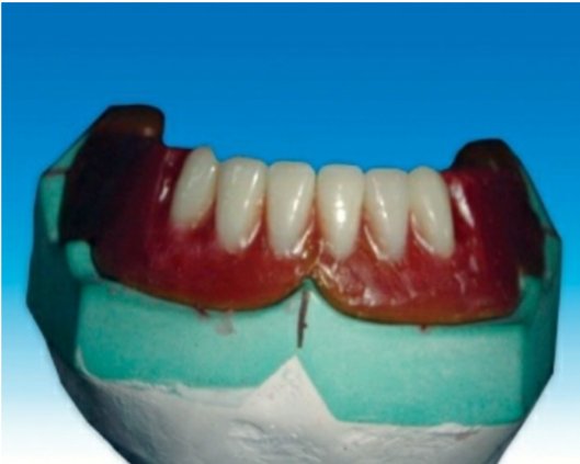
Front: Long axis inclines slightly towards the vertical axis.
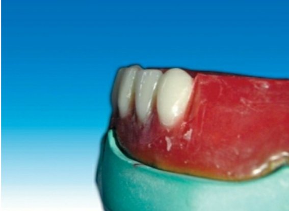
Side: Slopes labially.
Occlusal plane: Incisal edge is 0.5–1 mm above the occlusal
plane
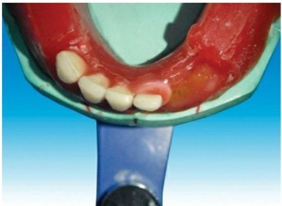
When viewed from:
Front: Long axis inclines towards the vertical axis.
Side: Slopes labially less than central incisor.
Occlusal plane: Incisal edge is 0.5–1 mm above the occlusal
plane
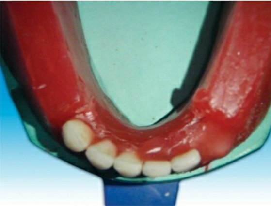
When viewed from:
Front: Long axis inclined towards midline.
Side: Slopes lingually.
Occlusal plane: Cuspid tip is 0.5–1 mm above the occlusal
plane
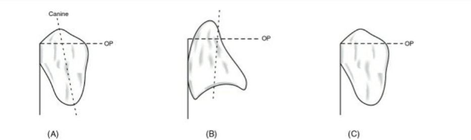
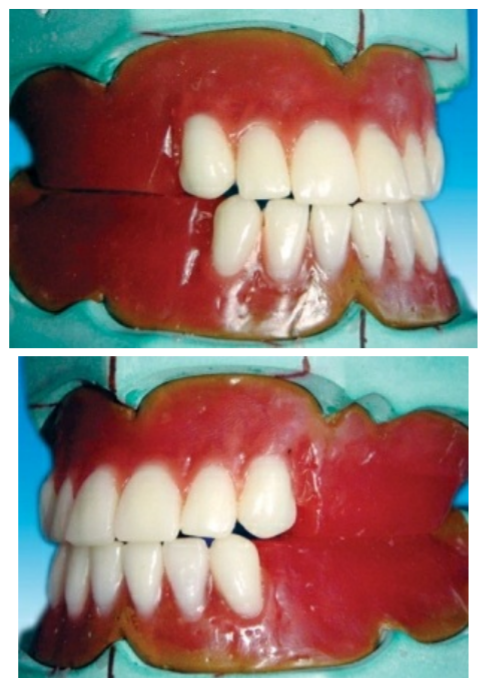
The mesial slope of the maxillary canine should overlap the distal slope of
the mandibular canine
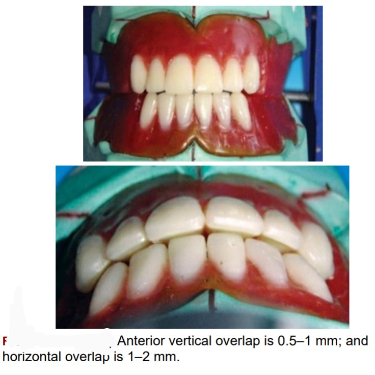
🛑The vertical overlap (over bite) between the maxillary and mandibular
incisal edge should be 0.5–1 mm, and the horizontal overlap (over jet) between
the maxillary and mandibular incisal edges should be 1–2 mm.
🛑The horizontal overlap should be uniform from left maxillary to right maxillary canine
Source – preclinical manual of prosthodontics S . Lakshmi
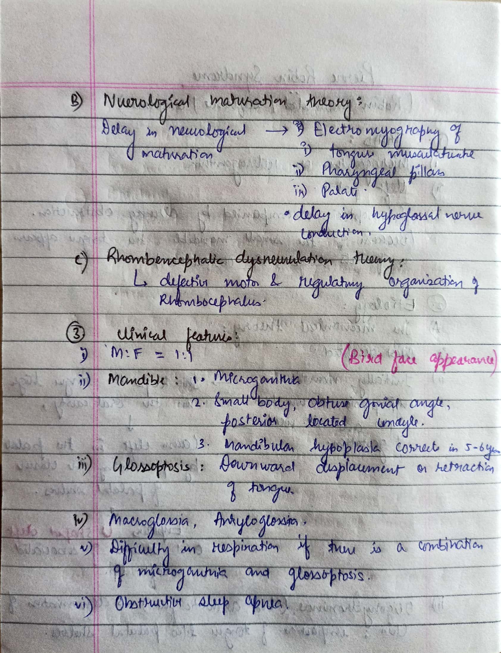
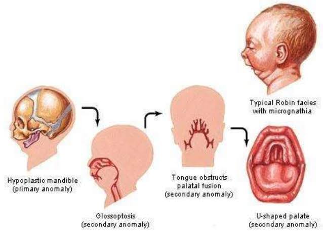

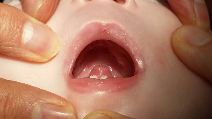
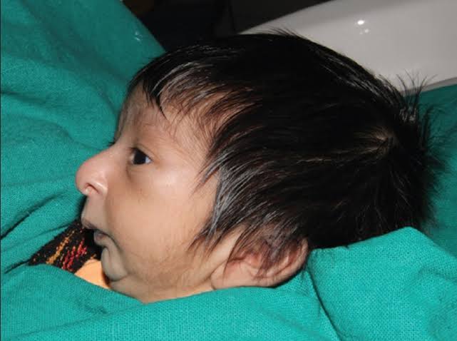
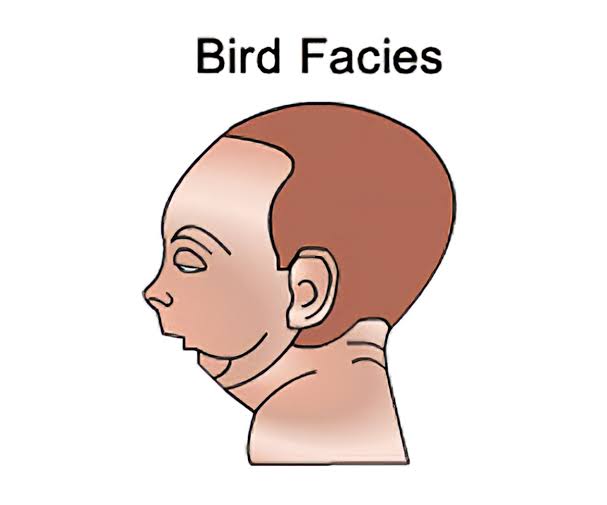
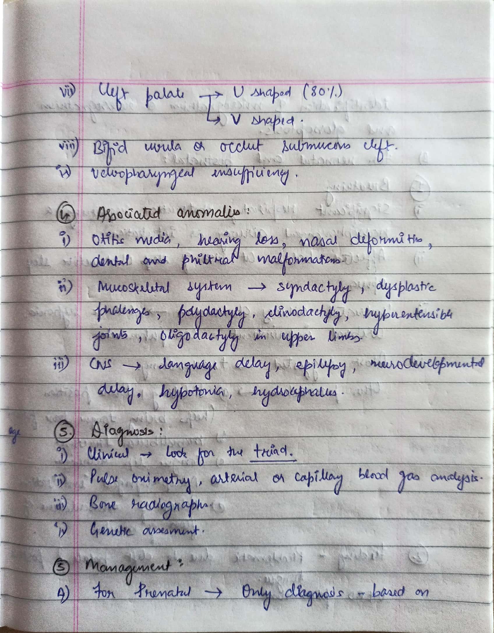

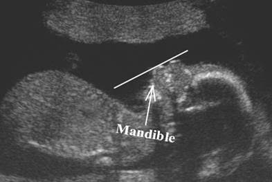


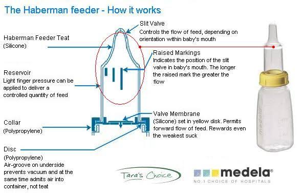
Sources – Shafer’s textbook of oral pathology , slideshare.com
Image source – Google images
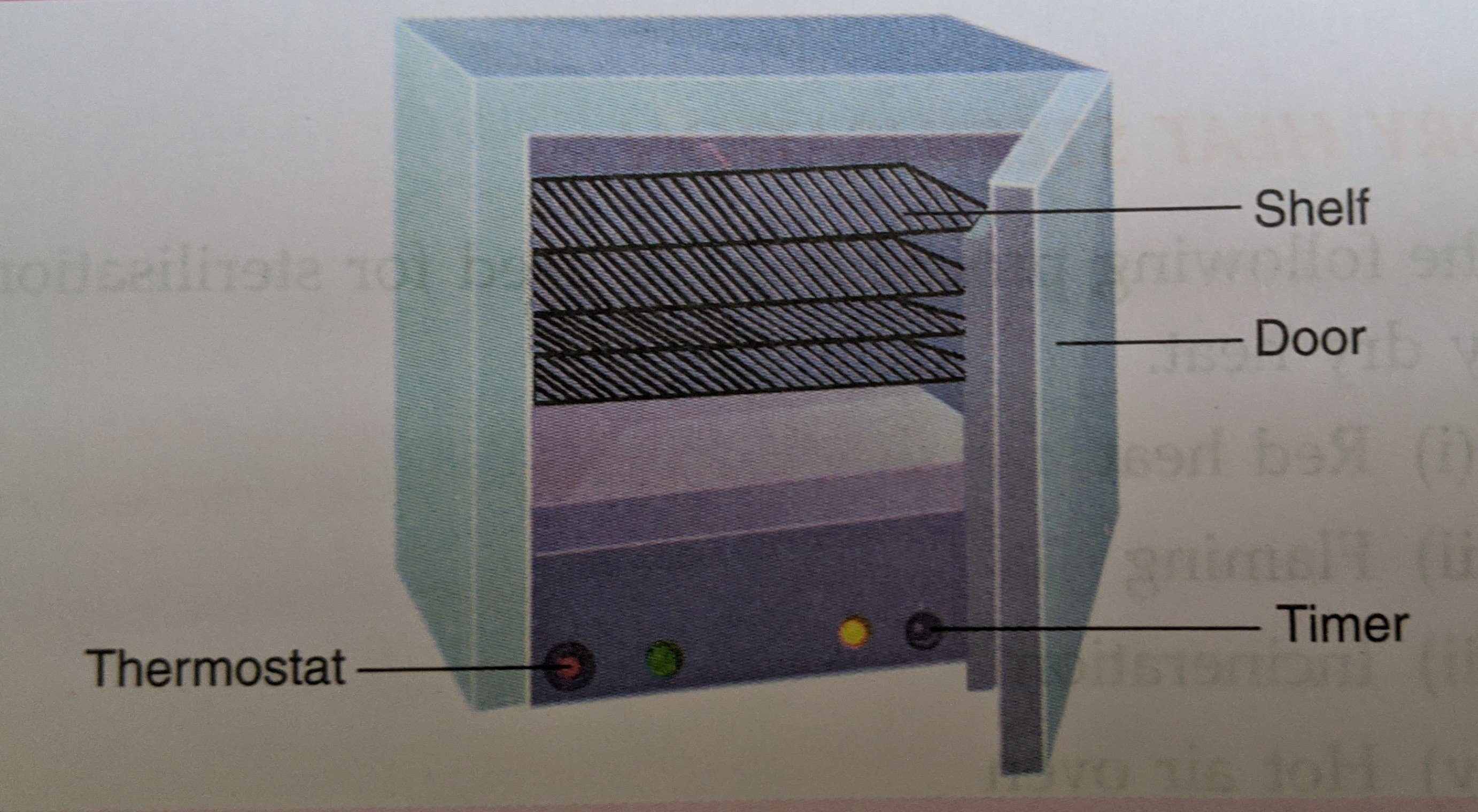
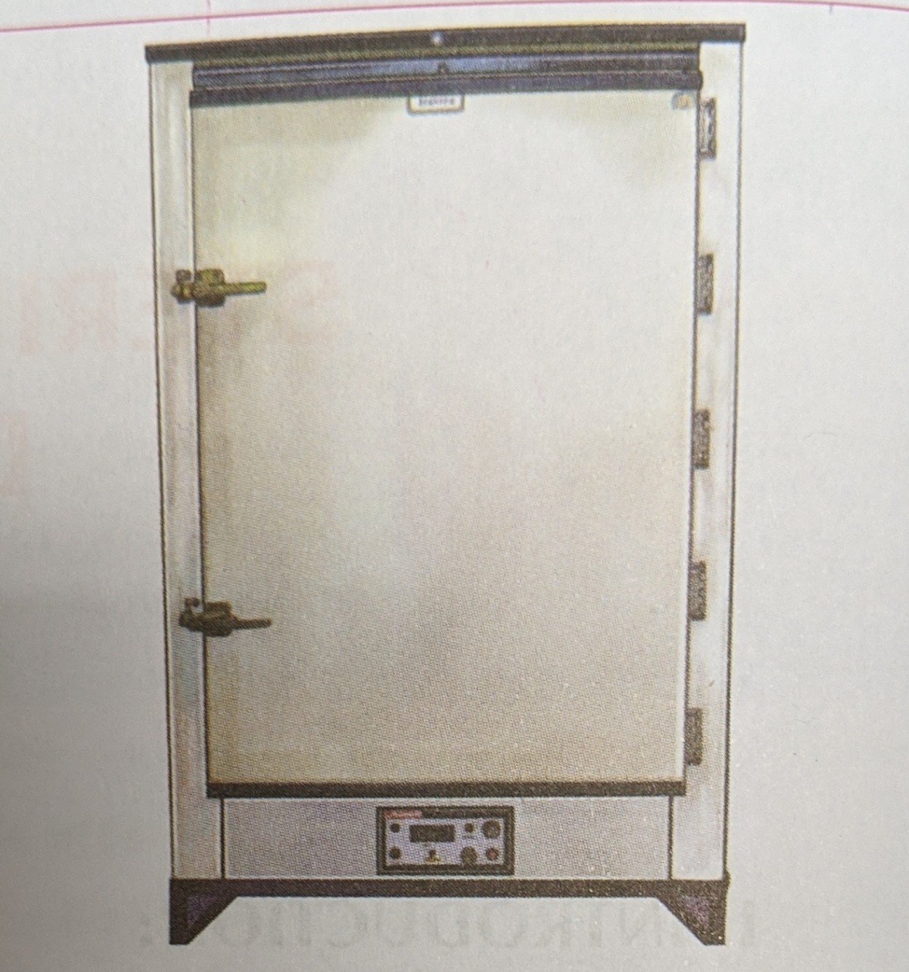
🔻It is the most widely used method of sterilisation by dry heat.
🔻 The oven is electrically heated and is fitted with a fan to ensure adequate and even distribution of hot air in the chamber
🔻It is also fitted with a thermostat that maintains the chamber air at a chosen temperature.
Temperature and time
160°C for two hours (holding time) is required for sterilisation.
Uses
It is used for sterilisation of
(i) Glassware like glass syringes, petri dishes, flasks, pipettes and test tubes.
(ii) Surgical instruments like scalpels, scissors, forceps etc.
(iii) Chemicals such as liquid paraffin, fats, sulphonamide powders etc.
(i) It should not be overloaded.
(ii) The material should be arranged in a manner which allows free circulation of air.
(iii) Material to be sterilised should be perfectly dry.
(iv) Test tubes, flasks etc. should be fitted with cotton plugs.
(v) Petri Dishes and pipettes should be wrapped in craft paper.
(vi) Rubber materials (except silicone rubber) or any inflammable material should not be kept inside the oven.
(vii) The oven must be allowed to cool for two hours before opening the doors, since the glasswares may crack by sudden cooling.
Sterilisation control
(i) The spores of nontoxigenic strain of Clostridium tetani are kept inside the oven. These spores be destroyed if the sterilisation is proper
(ii) Thermocouples may also be used.
(iii) Browne’s tube with green spot is available .
After proper sterilisation a green colour is produced (after two hour at 160°C).
Source – textbook of microbiology for dental students c p baveja
Source Gurkeeraat Singh textbook
Source Gurkeeraat Singh textbook
Source Gurkeerat Singh textbook
Source Gurkeerat Singh textbook
Immunoglobulin M (IgM)
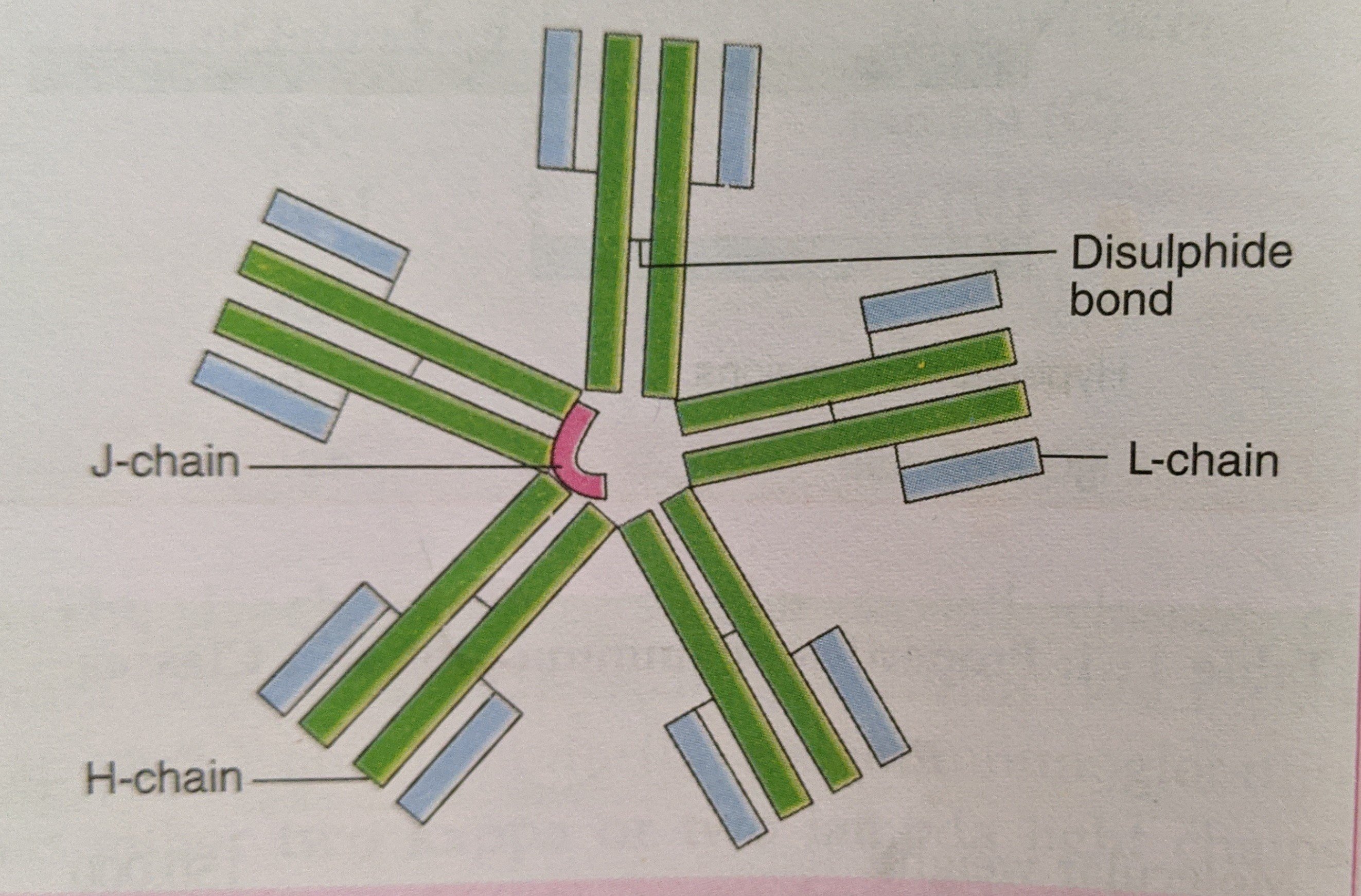
(i) IgM is a pentamer consisting of 5 immuno globulin subunits and one molecule of J chain, which joins the Fc region of the basic subunits.
(ii) It constitutes about 5-8 percent of total serum immunoglobulins. The normal level in serum is 0.5-2 mg/ml.
(iii) Half life is about five days.
(iv) It is heavy molecule (19S) with a molecular weight 900,000 to 1000000 hence also called the ‘millionaire molecule‘.
(v) IgM is mainly distributed intravascularly (80%).
(vi) It is the earliest synthesised immunoglobulin by foetus.
(vii) It appears early in response to infection before IgG.
IgM antibodies are short lived, and disappear earlier than IgG. Hence, its presence in serum indicates recent infection
(viii) It can not cross the placenta, presence of IgM antibody in serum of newborn indicates congenital infection.
(ix) It is very effective antibody in agglutination and complement fixation. It is more efficient than IgG in these reactions.
(x) IgM provides protection against blood invasion by microorganisms.
Source – textbook of microbiology for dental students c p baveja

