
Reference Pulse edition 14
Reference Pulse edition 14
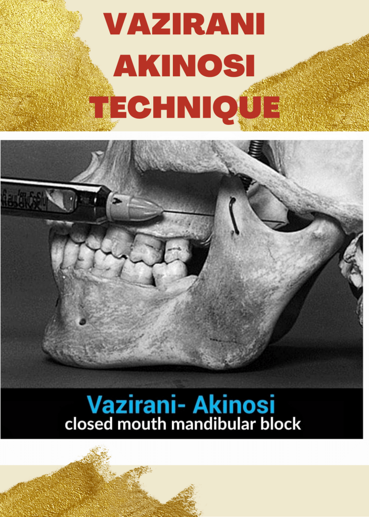
❇️ The Vazirani-Akinosi technique:
🔆“It is a specific method of nerve block in the mandibular region, carried out with the mouth closed.
❇️ The area of distribution of the anaesthesia includes
a) the corresponding dental arch,
b) the body of the jaw and the inferior ramus,
c) the gingiva/mucosa and vestibular periosteum, anterior to the mental foramen
d) the area of distribution of the lingual nerve: 2/3 of the anterior of the tongue and floor of the mouth, the gingiva/ mucosa and lingual periosteum.
🔆» The main indication, already anticipated, is trismus: classically this is contraction of the masticatory musculature which prevents the performance of effective inferior alveolar anaesthesia, for example, in cases of pulpitis or an abscess of a lower molar.
– By Dr. Sneha poeghal , Mallareddy institute of dental sciences , hyd .
References – Malamed


TRANSMISSION
Transmission occurs through three modes: inhalation, inoculation, or ingestion of spores
Mucormycosis infection in the clinical environment has been linked to use of adhesive bandages, wooden tongue depressors, hospital linens, negative pressure rooms, water leaks, poor air filtration, non-sterile medical devices, and building construction. This is a possible explanation for the recent rise in the number of mucormycosis cases during the covid outbreak.
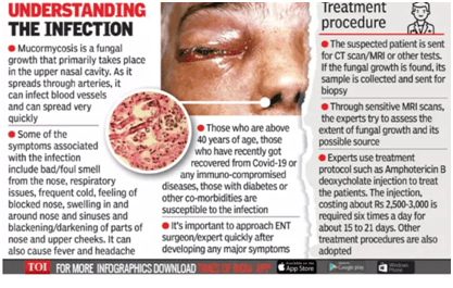
RISK FACTORS FOR MUCORMYCOSIS
Common predisposing factors include-
Depending on the location of fungal infection, mucormycosis is of 5 types-

Rhinocerebral form is the most common, accounting to nearly one-third of all the recorded cases of zygomycosis. This type of infection is primarily present in people with uncontrolled diabetes and those who had a kidney transplant.
Symptoms of Rhinocerebral Mucormycosis include-
Symptoms of pulmonary mucormycosis include-
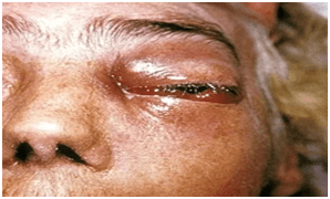
WHAT SHOULD THE DENTIST LOOK OUT FOR?
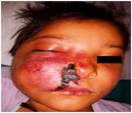

The commonly involved areas are the palate, face, eyes and the nasal passage. Some of the signs include-
DIAGNOSIS

Early diagnosis of mucormycosis infection is very crucial because of its rapid progression. Tools that help in making proper diagnosis are-
Radiographically, rhinocerebral mucormycosis shows nodular thickening of the sinus necrosis, sinus opacification without fluid level and spotty destruction of paranasal sinuses. CT scan with contrast/magnetic resonance imaging (MRI) can demonstrate erosion or destruction of bone and may help to determine the boundary of the disease.
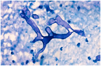
Histological examination of the biopsy specimen using KOH staining is useful in giving the final confirmation of the presence of fungal infection,which demonstrates long, broad, branching and nonseptate fungal hyphae. It is detected via a cotton swab sample of the nasal cavity, which is examined under a microscope.
COVID ASSOCIATED MUCORMYCOSIS
The mucormycosis cases reported after covid infection in a patient due to covid treatment related immunosuppression is called Covid-Associated Mucormycosis (CAM). Black Fungus is now commonly seen in patients who have recovered from covid-19 infection, who have been on oxygen support for a long time and diabetic patients. An infected mouth mask or an oxgen mask could also be the cause of the disease.
The fungus is normally present in the mucous lining of a healthy person, but a drop in the immune defense can trigger the growth of the fungus. After that it can reach to vital organs such as sinus, lungs and even brain. This is now happening more due to the heavy dose of steroids and anti-viral drugs which are used in the treatment of covid patients.
Diabetes further complicates the situation and promotes rapid fungal growth. The spread of the fungal infection is very aggressive and contagious in diabetic patients because of disruption of host defense mechanism and increased availability of nutrients such as free iron.
While steroids help in reducing lung inflammation to overcome the harmful effects of coronavirus, they also reduce immunity and cause a hike in blood sugar levels in both diabetics and non-diabetic Covid-19 patients. Thus, it is believed that this decline in immunity could be the driving force behind the rapidly increasing cases of mucormycosis.
TREATMENT OF MUCORMYCOSIS
The three main anti-fungal medicines employed in the treatment of Mucormycosis are-
In majority of the cases, surgical removal of the infected tissue is the only treatment option. Since there are high chances of reemergence of the fungal infection, it should be monitored carefully, with frequent follow-ups. Broadly speaking, the target organs of this fungal infection are eyes, lungs, skin and most recently, the oral cavity. This fungal infection is highly contagious as it spreads rapidly to other organs of the body, particularly to the brain.
Some simple oral hygiene practices to avoid the chances of getting mucormycosis after Covid infection are-

Maintaining good oral hygiene
Recent studies have proved that the anti-viral medicines and the steroids taken during the Covid infection period, lead to rapid growth of bacteria and fungus in the oral cavity, post recovery. This can cause a serious threat to the sinus, lungs, and even the brain. Routine brushing at least twice or thrice a day may help you to control the bacteria. Oral rising using a suitable mouthwash can also prove to be very beneficial.
New Toothbrush
It is advisable to the patients to change their toothbrush after recovery to avoid chances of reinfection from the virus present on the old toothbrush.
Disinfecting the toothbrush and tongue cleaner
Some experts suggest that a patient who has recovered from covid infection, should not keep their toothbrush and tongue cleaner in the same holder as of their other family members. This can help in spread of infection to other members of the family. It is also advised to clean the brush and tongue cleaner using an antiseptic mouthwash.
ROLE OF A DENTIST
Dentists can play a very important role in management of mucormycosis by diagnosing it early, carrying out proper investigations and referring to the appropriate specialists at the correct time. They can keep in mind the following clinical workflow for mucormycosis, to detect such cases coming in their clinics at their earliest-
History– A dentist should properly take history of previous Covid infection and hospitalization.
Symptoms and signs– A dentist should look out for the signs which have been mentioned previously
Investigations– Performing adequate investigations such as CT, MRI, Biopsy, etc.
Prescribe– A dentist can prescribe Analgesics and anti-inflammatory drugs, Antipyretics and Antibiotics to provide the patient relief for a shorter period, before referring to specialists.
Counselling– Counsel the patient to stay calm and follow the instructions.
Record– A dentist should keep a record of the clinical findings and the referrals.
Referral– A dentist should refer the mucormycosis patients to OMFS, ENT and Ophthalmologist for appropriate treatment.
Mucormycosis does not spread via contact transmission. Thus, the best way to keep oneself safe from the fungal infection is by self-care. One should always wear a mask before stepping outdoors even if their immunity is good. For health care professionals, it is very important to wear masks and gloves while doing bandages on the wounds and even while tending to fungal infected patients, to keep themselves safe.
DR. DEVYANI ALLEN
BDS, FRCD
Invisalign or clear aligners are orthodontic devices that are transparent, plastic form of dental braces used to adjust teeth.

They are designed as follows :
Each aligner is intended to be worn an optimal 2 hrs a day for one to two weeks. On average, the treatment process takes 13.5 months although it varies based on complexity of planned teeth movements.
Cost of clear aligners in India ranges from 1,50,000-3,50,000.
Pros of invisalign :
Cons of invisalign :
Quaffing and boozing have become common in almost all over the world. It does has a deleterious effect on liver but does it effect teeth too? Oh yes!!! it does has a role in effecting oral cavity in wide ways. Let us quickly go through it’s action.

How can we avoid such deleterious effects?? Well!! here are few tips :

References : wcdentalarts.com, deltadentalins.com, covingtondentalcenter.com


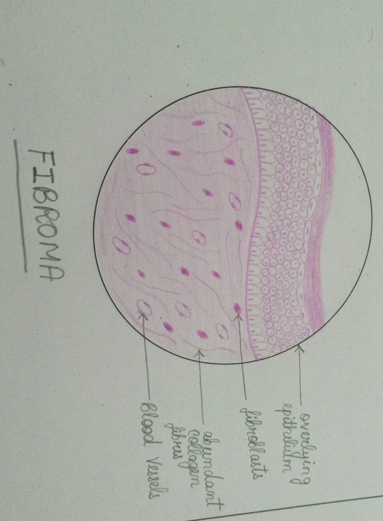
FIBROMA
LIPOMA

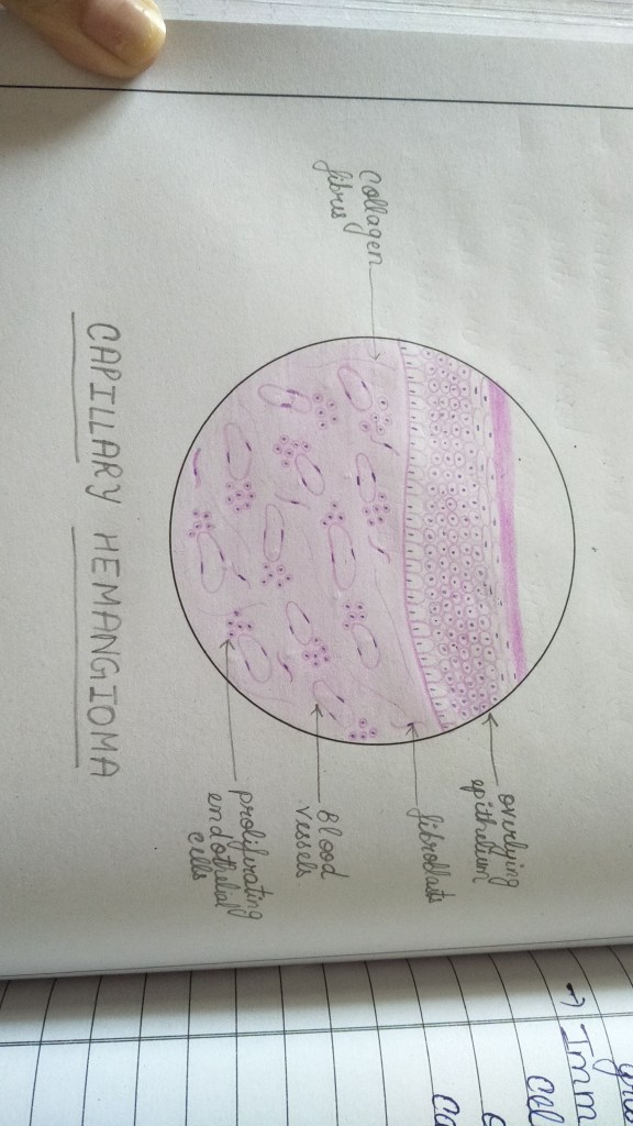
CAPILLARY HEMANGIOMA
CAVERNOUS HEMANGIOMA

LYMPHANGIOMA

NEUROFIBROMA

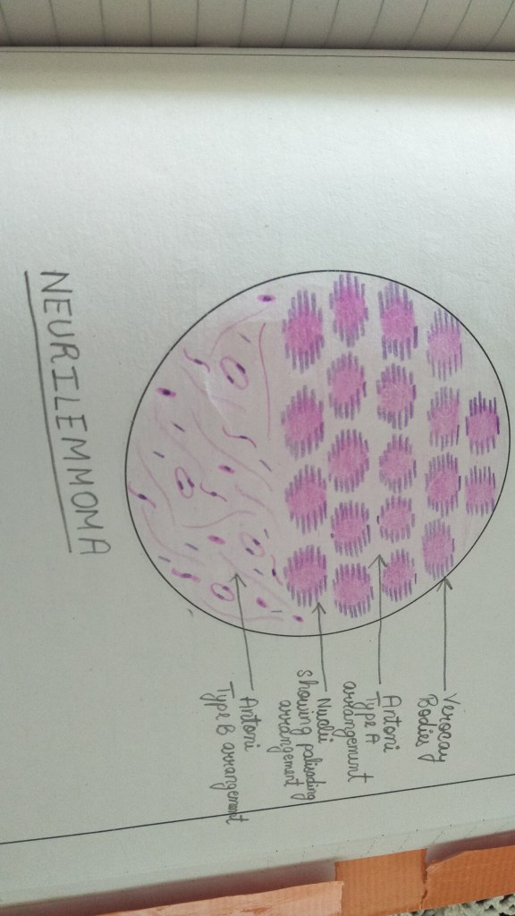
NEURILEMMOMA(SHWANNOMA)
OSTEOMA


REFERENCES- Shafer’s textbook of oral pathology 9th edition

We often come across few advices which are actually misconceptions among people. Let us see what are those myths and find out the actual facts to spread right knowledge about dentistry.
Myth 1 : Brushing harder cleans better
Fact : absolutely not!! It’s a misapprehension that plaque can be removed by brushing harder. Applying too much pressure may slowly erode enamel, which cannot repair itself once it suffers significant damage. One may experience increased sensitivity and a heightened risk of cavities due to such activity. Brushing too hard can cause the gum tissue to shrink back (gum recession). So brush in a soft and right way.

Myth 2 : White teeth are healthy teeth
Fact : one can have pearly white teeth and still have gum infections or cavities. Likewise one can have perfectly healthy teeth which are off white, yellowish or even brownish. Enamel is on the surface of every tooth and it has a natural hue of white. However, the underlying dentin layer has a slightly yellowish colour. This yellowish hue shows through the enamel in almost everyone.

Myth 3 : If one has no oral health concerns, there’s no need for an exam.
Fact : definitely not!! Here are six reasons as to why one must visit a dentist once in every six months.
Reason 1 : oral cancer is an extremely serious disease that manifests itself in various ways. Without knowing the signs of its early onset, oral cancer is often not diagnoses and can quickly progress and can become life threatening. Dentist is highly trained to recognise these signs and with regular checkup the likelihood of catching oral cancer in time is dramatically high.
Reason 2 : Even with the most diligent tooth brushes and flosses, there are small areas in the mouth that are missed by a regular brushing and flossing. Regular dental cleaning remove tartar from eroding teeth or cleaning holes in them, which is how cavities are created.
Reason 3 : Regular dental cleanings are essential in catching and addressing gingivitis before it gets out of hand. “Better to hold a tooth in mouth rather than replacing the lost ones by a veneer or implant”.
Reason 4 : There are many bad habits that can have a negative impact on our oral health, some of which one may not even realize are causing issues. Some of them include chewing ice, biting nails, clenching jaw, grinding teeth, eating particularly sticky or hard sweets, brushing too hard, drinking cofee and red wine and ofcourse smoking. A regular checkup can help in can identify oral damages caused by these which one may have not noticed.
Reason 5 : A crucial part of visiting dentist is getting one’s teeth and jaw bone x-rayed. X-ray images allow dental professionals to see what is happening beneath the surfaces of your mouth and can find, diagnose issues that may be invisible to naked eye. Like impacted tooth, bone decay, swelling, cysts or tumours.
Reason 6 : In addition to checking mouth, gums and tongue for signs of oral cancer, dentist will also check one’s neck, jaw and lymph nodes located just below jaw line for any swellings or lumps or other abnormalities.

Myth 4 : Teeth cleaning / scaling and polishing will abradd the enamel and cause sensitivity issues.
Fact : Cleaning safely remove the plaque and bacteria that builds up over time on the teeth and gums. They don’t damage enamel on the teeth. Infacf, if teeth are not cleaned regularly, inflammation can occur and this can lead to gum disease sue ro the bacteria residing in the plaques.

Myth 5 : Braces are only meant for the younger.
Fact : getting braces may be a little easier or go a little faster during adolescence but adults from all walks of life should know that age is just a number when it comes to receiving and benefiting from orthodontic treatment. Adults count for 20% of orthodontic patients according to AAO statistics. Although harder bone rissue can mean a longer, more involved treatment process for adults with braces, the right orthodontic treatment plan is usually all it takes to straighten teeth, improve bite alignment, make oral hygiene easier and create a perfect smile at any age.

Myth 6 : There is no need to wear retainers after orthodontic treatment.
Fact : oh yes!! You need to wear those retainers given by your dentists for atleast 9 months and then dropping down to nightly wear after that. Going a year without retainer means that your teeth will have continued to move back to their original position and may even be crooked. The solution may be to restart treatment with braces. Isn’t it better to have retainers in their place rather than spending again on braces?!!

Myth 7 : Dental treatment costs a fortune.
Fact : the only reason one has ro spend so much on a dental treatment is because he/she didn’t take care of their teeth as much as they should have. Neglect, or rathee, result of neglect is always costlier than the the routine dental appointment that pops up in one’s calender twice a year. Moreover, the dental equipment are costlier too because of which a normal dentist demands more which is actually normal compared to what he invests.
Myth 8 : A fast prosthodontist is a good prosthodontist.
Fact : you are mistaken. Fast is not always good. A prosthodontist offers specialized treatment that cannot be rushed. It’s important that they take their time to make sure the job is done right the first time. While dentists may want to keep their patients moving, a prosthodontist takes his time and does what is best for the patient.
Myth 9 : I’m better off with my natural teeth or no teeth.
Fact : ofcourse natural teeth are always preferable. However, if one has lost one or few teeth to injury, disease or decay, a dental implant is truly the next best thing. The tooth that opposes the site of missing tooth may start to grow out from its position because it no longer has the opposing tooth to resist it. One may experience increased sensitivity and other issues around this super-erupted tooth.

References : 123dentist.com, reeseortho.com

Flossing is an integral part of our daily oral hygiene routine. It helps in removing food particles from the areas where toothbrush cannot reach, such as the gum line between the teeth. If food particles and bacteria accumulate in such areas, it eventually leads to gum problems and early gum recession.
Majority of the people brush their teeth regularly but they don’t floss them. To prevent plaque accumulation over the teeth and it between them, it is important to follow a proper flossing technique.
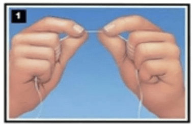
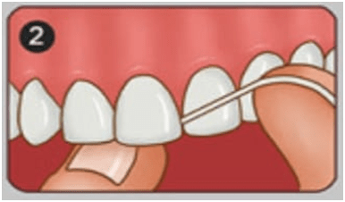
As per the ADA recommendations, the correct steps to be followed while flossing are as follows-
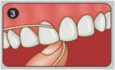
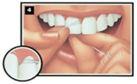
It is recommended that you floss first before brushing your teeth, as flossing loosens the debris and bacteria between the teeth, which can then be easily removed by brushing.
Take about 10-15 minutes daily to floss your teeth, for a healthy and naturally white smile.
DR. DEVYANI ALLEN
BDS, FRCD
Toothbrushing comprises the most important part of our daily oral hygiene regime. Thus, it becomes very important for us to choose a toothbrush which is comfortable to use as well as functionally efficient.
Nowadays, the market is flooded with different types of brushes, be it manual or electric. This evolvement in the variety of toothbrushes has made it difficult for us to decide upon which toothbrush is best for us.
Here are few practical tips which can help you make the correct choice for your healthy whites-
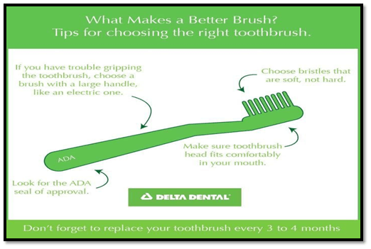
Irrespective of the brush you choose, it is important that you follow the correct brushing technique and brush for atleast 2 minutes to increase the lifespan of your whites.
If you are using a manual brush, it is important to replace it once every 3 months or whenever the brush shows any signs of wear and tear.
So, next time when you are in the supermarket checking out the vast variety of brushes, try to look out for these few things in your brush and you will be able to make the correct choice.
DR. DEVYANI ALLEN
BDS, FRCD
