Source: Shafer’s Textbook of Oral Pathology, 8th Edition
Source: Shafer’s Textbook of Oral Pathology, 8th Edition


References: Shafer’sTextbook Of Oral Pathology


Source: Shafer’s Textbook of Oral Pathology, 8th Edition

🌏 African Jaw Lymphoma
🌏 The endemic form is linked to malaria and to the Epstein-Barr virus (EBV), a common virus that also causes glandular fever.
🌏 A tumour peculiar to children of central Africa was reported by Denis Burkitt in 1950. It is a lymphoreticular cell malignancy.

▪️It is a high grade B-cell neoplasm & has 2 major forms:
1. Age – between 6 & 9 years
2. Sex – M:F – 2:1
3. Site: In African form (Endemic),
American form (Non-endemic) – Oral: only 1 quadrant involved. Other: Mainly involves Abdomen.
4. Onset & progress – fast growth with tumor doubling time of <24 hours.
5. Symptoms –
6. Sign – Lymphadenopathy (Non-tender)
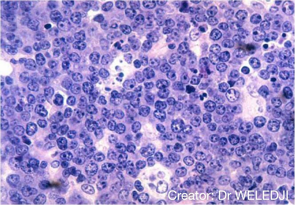
1. Monoclonal proliferation of B-lymphocytes characterized by small non-cleaved cells.
2. Burkitt cells are homogenous in size & shape with –
References: Shafer’sTextbook Of Oral Pathology
Dr. Mehnaz Memon🖊
Ann Arbor Staging System for Lymphoma: https://dentowesome.wordpress.com/2020/06/15/ann-arbor-staging/
Ghost cells are altered epithelial cells characterized by the loss of Nuclei with presence of basic cell outline.
References: Shafer’sTextbook Of Oral Pathology
Dr. Mehnaz Memon🖊
References: Shafer’sTextbook Of Oral Pathology


References: Shafer’sTextbook Of Oral Pathology


Dr. Mehnaz Memon🖊
References: Shafer’sTextbook Of Oral Pathology
By KRITI NAJA JAIN:-
REFERENCES:-

Source: Shafer’s Textbook of Oral Pathology, 8th Edition
