MALIGNANT MELANOMA

A neoplasm of epidermal melanocytes and third most common cancer of skin.

▪️Arises in preexisting mole. Appearance – Large, flat, spreading lesion; deeply pigmented Nodule

🔹Precursor Lesions:

(i) Congenital Nevi

https://dentowesome.wordpress.com/2020/05/18/oral-nevi/

(ii) Dysplastic Nevi (Atypical Mole)

(iii) Lentigo Maligna: Also called as Hutchinson’s freckle, is a tan or black on the skin that looks like a freckle.

  • Grows slowly
  • Dark, thick, nodular, mottled
  • Seen on one side of the face of an older adult who had a large amount of sun exposure.

🔹Etiology:

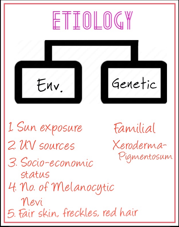
• Genes in the development of Melanoma:

• Phases in the growth of Melanoma:

  1. Radial
  2. Vertical

🔹Classification:

1. Superficial spreading Melanoma:

  • most common type (65%)
  • Radial growth phase – premalignant melanosis/pagetoid melanoma in situ
  • Vertical growth phase – Increase in size, color, nodularity/ulceration
  • Lesions are usually flat, scaly or crusty & 2 cm in diameter
  • Found in trunk & back of Men; Legs of women
  • Median age of occurrence – 50’s

2. Lentigo Maligna Melanoma:

  • Least serious form
  • More in women
  • Macular lesion on malar skin of middle-aged and elderly

3. Nodular Melanoma:

  • Exhibits only vertical growth phase
  • Sharply delineated nodule, may be pink/black
  • Occur in men on skin of head, neck & trunk
  • Looks like blood blister

4. Acral Lentiginous:

  • Also called muco-cutaneous Melanoma
  • Less common with fair skin
  • Palms of hands, soles of feet, mucous membrane, nail beds
  • Median age of occurrence – in 50’s & 60’s

Assessing the ABCDE’s of Moles

Image Source: IG|medical.docs

🔹Oral Manifestations:
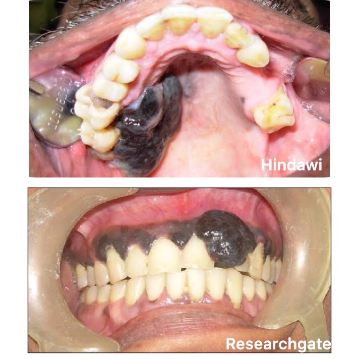
  • Age: 55 yrs
  • Sex: M>F
  • Site: Palate/Gingiva
  • Appearance: Deeply pigmented area; ulcerated/haemorrhagic; ⬆️ size
  • Amelanotic melanomas: 5-35% of oral cases

Melanoma stages 5 years survival rates:

  • Stage 0: Melanoma in situ ( Clark level I), 99.9% survival
  • Stage I/II: Invasive melanoma, 85-99% survival
  • Stage II: High risk Melanoma, 40-85% survival
  • Stage III: Regional Metastasis, 25-60% survival
  • Stage IV: Distant Metastasis, 9-15% survival

🔹Treatment depends on stage:

➡️ Metastases that cause symptoms but cannot be removed may be treated with radiation, immunotherapy, targeted therapy, or chemotherapy.

Dr. Mehnaz Memon🖊


References: Shafer’sTextbook Of Oral Pathology; Textbook Of Surgery by S.Das

PALATAL AND ALVEOLAR CYSTS OF NEWBORN

  • Dental lamina cyst of Newborn
  • Gingival cyst of Newborn
  • Epstein’s pearls, Bohn’s nodules

Dental lamina cyst of newborn are multiple, occasionally solitary, superficial raised nodules on edentulous alveolar ridges of infants that resolve without treatment; derived from rests of dental lamina & consisting of keratin producing epithelial lining.

Cystic keratin filled nodules; derived from epithelial remnants entrapped along line of fusion, usually seen at midpalatine raphe.

Keratin filled cysts scattered over the palate at the junction of hard and soft palate; derived from palatal salivary gland structures.

🔹Introduction:

  • A special form of odontogenic cyst – 80% of the infants.
  • Gingival cyst is M/E similar to epidermoid cyst.
  • It develops after 4 week in utero.
  • Palatal Cyst arises from epithelial remnants in stroma after fusion of palatal processes usually at posterior midline of hard palate.

🔹Clinical Features:

➡️ Gingival Cyst:

  1. They are multiple, superficial raised nodules on edentulous alveolar ridge of infants that resolve without treatment.
  2. They are localized in corium below the surface epithelium.
  3. Those found in anterior portion of jaw are displaced lingually & in posterior portion are found occlusal to crown of molars.
  4. They are asymptomatic and do not produce any discomfort to the infant.
  5. These are small discrete, white swellings, blanched from internal pressure.

➡️ Palatal Cyst:

  1. They are Multiple (<6), 1-4mm in size & yellow-white in color. Sessile mucosal papules of posterior hard palate.
  2. They are larger & less numerous than gingival cyst. Both are so superficial that several may be ruptured at the time of examination.

🔹 Histopathological Features:

  • Thin, stratified squamous epithelium – produce keratin.
  • Fibrovascular connective tissue stroma without inflammatory infilterate.
  • Cystic lumen filled with degenerated keratin, formed into concentric layers/onion rings.
  • Epithelium lacks Rete processes.
  • Dystrophic calcification & Hyaline bodies seen.

🔹Treatment:

Generally self limiting, (within 3 months) no treatment is required in most of the cases. If baby is having feeding difficulties, complete excision of the cyst can be done.

References: Shafer’s Textbook of Oral Pathology 7th Edition


Dr. Mehnaz Memon🖊

PAGET’S DISEASE

Introduction & Etiology👆🏻
Clinical Features & Phases of PDB..👆🏻
Clinical Features contd…👆🏻
O/M of PDB..👆🏻
Radiographic Features of PDB..👆🏻
Histological Features of PDB…👆🏻
Lab. Findings & Treatment of PDB..👆🏻

References: Shafer’s Textbook of Oral Pathology 7th Edition


Dr. Mehnaz Memon🖊

TRAUMATIC CYST/PSEUDO CYST

  • Solitary Bone Cyst
  • Hemorrhagic Cyst
  • Extravasation Cyst

🔷 Etiology: Trauma Hemorrhage Theory

Origin from traumatic injury

⬇️

Intramedullary Hemorrhage

⬇️

Clots break down and leave empty space within bone

🔷 Clinical Features:

  • Age: 18 years
  • Sex: Male predominance
  • Site: Posterior region of mandible ( More common). Also due to presence of hemopoietic marrow – incisor region also reported.
  • In majority of cases, pulp of involved teeth is vital.
  • Cavity contains Sero-sanguinous fluid, shreds of necrotic blood clot, fragments of Connective tissue.

🔷 Radiographic Features:

  • Smoothly outlined radiolucent area of variable size, sometimes with thin sclerotic borders. (D/D – Lingual salivary gland depression of mandible)
  • However the latter lesion is usually located below the mandibular canal, whereas Traumatic Cyst usually lies above it.

🔷 Histological Features:

  • Bone cyst lined by thin Connective tissue membrane.
  • On outer surface of cortical plate – Osteophytic Reaction

🔷 Treatment and prognosis:

➡️ Surgical exploration is often undertaken. The bony cavity is scraped to generate bleeding, which is considered the treatment of choice for this condition. Other methods of treatment have been tried, such as packing the curetted cavity with autologous blood, autologous bone, and hydroxyapatite. Exploration surgery usually leads to healing. Recurrence is rare.

References: Shafer’s Textbook of Oral Pathology 7th Edition, Internet


Dr. Mehnaz Memon🖊