PIGMENTS & SPOTS

Pigmentation is the process of deposition of pigments in the tissue.Mostly found in the mouth and could be attributed to oral manifestations of systemic diseases or malignancies.This could be due to :

  • Increased number of melanocytes
  • Augmentation of melanin production
  • Deposition of accidentally introduced exogenous materials

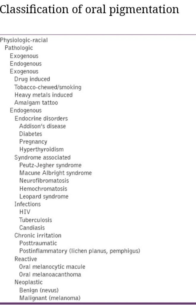
The various oral pigmentations can be in the form of:

1)Blue /Purple vascular lesions-

  • Hemangioma
  • Varix
  • Angiosarcoma
  • Kaposi’s sarcoma
  • Hereditary hemorrhagic telangiectasia

2)Brown melanotic lesions-

  • Ephelis & oral melanotic macule
  • Nevus
  • Malignant melanoma
  • Drug induced melanosis
  • Physiologic pigmentation
  • Cafè au lait pigmentation
  • Smoker’s melanosis
  • Endocrinopathic pigmentation
  • Peutz-jeghers syndrome
  • HIV oral melanosis

3)Brown heme associated lesions-

  • Ecchymosis
  • Petechia
  • Hemochromatosis
  • Hemorrhagic mucocele
  • Thrombosed varix

4)Gray/black pigmentations-

  • Silver amalgam tattoo
  • Graphite tattoo
  • Heavy metal ingestion-lead,mercury,bismuth
  • Hairy tongue

SPOTS

Appearance of peculiar spots in specific sites on the body are often diagnostic for major systemic disease or condition or an infection.Some of them are discussed here;

  • Koplik spots -measles (rubeola)
  • Pink spots on teeth – internal resorption
  • Roth spots – subacute endocarditis, typhoid fever
  • Bitot’s spots white plaque on conjunctiva of vitamin A deficient children
  • Herald spots -primary lesion seen in pitryiasis rosea
  • Sore spots -traumatic ulcers from denture irritation mostly
  • Cafè au lait spot-neurofibromatosis, Macune-Albright syndrome,Peutz-jeghers syndrome

Sources: Shafers textbook of oral pathology ,http://www.ncbi.nlm.nih.gov (oral pigmentation : A review),http://www.gibbleguts.com

Hunter syndrome

Features of Mucopolysaccharidosis Syndromes Hunter syndrome:-

  • Type- II
  • Eponym-Hunter.
  • Inheritance- X-Linked R.
  • Enzyme Defi ciency- Iduronate-2-sulfatase.Stored Substrate- HS heparan sulfate and DS DS, dermatan sulfate.
  • Clinical Features – Appears at 1 to 2 years of age; clear corneas, reduced intelligence, growth retardation, stiff joints
  • Differs from Hurler’s syndrome in –

  1. Mode of inheritance (X – linked).
  2. Absence of corneal clouding.
  3. Milder clinical course..Results from deficiency of iduronate – 2 – sulfatase (I2S).
  4. Without enough I2S, partially broken-down mucopolysaccharides accumulate in the organs and tissues of the body and become toxic.

Clinical features :-

  • Hunter syndrome is divided into two types.

I-   Type A is he severe form, which usually is diagnosed in children aged 18-36 months.

  • Considered the classic form.
  • Children with type A may survive into the second and third decades of life.
  • Symptoms in type A may include:

  1. coarse facial features and short stature.

  2. enlarged liver and spleen.

  3. progressive and profound mental retardation.

  4. ivory-colored skin lesions on the upper back and sides of the upper arms and thighs.skeletal changes, joint stiffness, short neck, broad chest, and too-large head.

  5. progressive deafness.

  6. atypical retinitis pigmentosa and visual impairment.

II.Type B Hunter syndrome is much milder than type A

  • May not be diagnosed until adulthood.
  • Individuals with type B may live into their 70s.
  • Their physical features are similar to those in type A.
  • Individuals with type B, however, usually have normal intelligence and do not have the severe skeletal problems of type A.

Diagnosis:-

  • In type A Hunter syndrome, the child’s appearance combined with other symptoms such as enlarged liver and spleen and the ivory-colored skin lesions can suggest the child has mucopolysaccharidosis.
  • Type B Hunter syndrome is much harder to identify, and might only be recognized when looking at the maternal relatives of a child with Hunter syndrome.
  • In either type, the diagnosis can be confirmed by a blood test for deficiency of I2S.

Treatment:-

  • Medical care is directed towards relieving the symptoms of Hunter syndrome.
  • Treatment with Elaprase (idursulfase) replaces I2S in the body and helps reduce symptoms and pain.

References:-

1.SHAFERS 8th edition

2.NEVILLE ‘S 3rd edition

Hurler’s syndrome

Features of Mucopolysaccharidosis Syndrome-HURLER’S SYNDROME(MPS I H, Gargoylism):

  • Type- I-H
  • Eponym-Hurler
  • Inheritance-Autosomal Ressesive
  • Enzyme Deficiency- α-L-Iduronidase
  • Stored Substrate- HS- heparan sulfate and DS-dermatan sulfate
  • Clinical Features – Appears in infancy; cloudy corneas, growth retardation, reduced intelligence, coronary artery disease; rarely live 10 years.
  • Chromosomal abnormality on chromosome arm 4p16.3

  • Characterized by increased levels of MPS in urine.

Clinical features:-

  • Becomes apparent in first 2 years of life, life expectancy of 6-10 yrs.
  • Head appears large; facial features quite typical.
  • Prominent forehead
  • Broad saddle nose & wide nostrils
  • Puffy eyelids with coarse bushy eyebrows
  • Hypertelorism
  • Thick lips
  • Large tongue with open mouth
  • Nasal congestion & noisy breathing.
  • Progressive corneal clouding classic manifestation.
  • Hepatosplenomegaly, results in protrudent abdomen
  • Short neck & spinal abnormalities typical.
  • Flexion contractures result in “claw hand”.
  • Dwarfed, mentally retarded individuals.
  • hhs

Histologic features:-

  • Excessive accumulation of intracellular mucopolysaccarides in many tissues & organs.
  • Accumulation of dermatan & heparan sulfate in cells of mononuclear – phogocyte system, fibroblasts & endothelial cells.
  • Affected cells are swollen, have clear cytoplasm resulting from accumulation of PAS +ve material in engorged, vacuolated lysosomes.
  • Involved fibroblasts assume appearance of ‘clear’ or ‘gargoyle’ cells.
  • ‘Hurler cells’ relatively large with metachromatically staining cytoplasm with crescent shaped nuclei.
  • Cells not identified with normal H/E stain but with toluidine blue or Alcian blue/ aldehyde fuchsin stains.
  • Should be differentiated from mast cells.

Oral Manifestations:-

hhss

  • Shortening and broadening of the mandible with prominent gonions.
  • Localized areas of bone destruction in the jaws.
  • Teeth may be small and widely spaced.
  • Gingival hyperplasia has been repeatedly described in
    patients with Hurler syndrome.
  • the tongue is also characteristically enlarged.

Laboratory findings:-

  • Elevated levels of mucopolysaccharides in urine.
  • Metachromatic granules or ‘Reily bodies’ often demonstrated in cytoplasm of circulating lymphocytes.

Treatment:-

  • Because of multisystemic involvement in patients with mucopolysaccharidosis type I (MPS I), treatment is multidisciplinary and encompasses both the curative and palliative elements.
  • Corrective surgery may be necessary for patients with mucopolysaccharidosis type I (MPS I) who have joint contractures or foot and hand deformities.

  • Corneal transplants may be required if vision problems become severe.

  • Given the numerous mutations at this genetic locus, identification of which allele or alleles are involved requires referral to medical geneticists for diagnosis and genetic counseling.

References :-

1.SHAFERS’s 8th edition

2.NEVIELLE’S 3rd edition

COMMON SITES

Correct and early diagnosis of a lesion / abnormality in the oral cavity is very crucial as these could be vital indicators to some of the underlying systemic diseases or other chronic conditions. Most of the oral conditions show a peculiar pattern of occurrence only on specific sites.Hence,knowledge of these common sites can help to some extent in proper identification, differentiation from other similar conditions and treatment planning.A few of such conditions and findings has been mentioned here.

Sources:Shafers textbook of oral pathology ,burkets oral medicine ,www.aurametrix.weebly.com