• Composed of proliferating odontogenic epithelium in a cellular ectomesenchyme resembling the dental papilla.
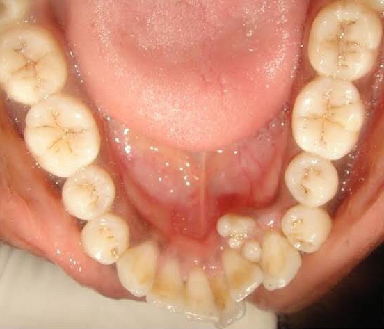
• Odontomas are composed of all mature components of dental hard & soft tissues: Enamel, dentin & pulp.
• Because of their limited slow growth and well differentiation – considered hamartomas rather than true neoplasms
Clinical Features:
1. Interfere with eruption of permanent teeth.
2. No sex predilection
3. Age: 2nd decades
4. Asymptomatic in nature
5. Associated with impacted, malpositioned teeth.
6. Cause malformation and displacement of adjacent teeth
- Early lesion: Radiolucent with smooth, well defined contours between the roots of teeth.
- Later stage: Radiopaque.
Types of Odontomas:
- Complex odontoma (less common)
- Compound Odontoma
1. Complex Odontoma:
• Location: Posterior region of either Jaw. (70% – Mandibular Molars)
• Composed of haphazardly arranged dental hard and soft tissue with no resemblance to normal tooth.
• Arises from normal tooth follicle.
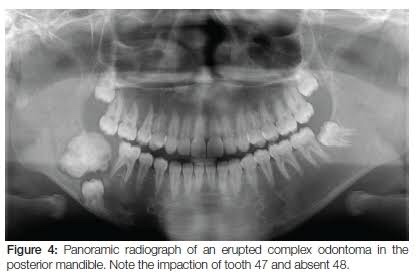
• Radiographically, Sunburst opacities with thin uniform radiolucent rim.
2. Compound Odontoma:
• Location: Anterior maxilla (62% – unerupted canine)

• Collection of small radio-opaque masses, some/all may be tooth like structures “denticles“
• Formed by exuberant growth of dental lamina or proliferation of enamel organ
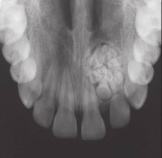
• Radiographically, cluster of multiple tiny toothlike structure within a fine radiolucent rim.
Histological features:

• Connective tissue capsule around odontoma is similar in all aspects to follicle surrounding normal tooth.
• Presence of ghost cells also found.
Differential Diagnosis:
- Periapical cemental dysplasia
- Cemento-ossifying fibroma
- Focal sclerosing osteitis
- Cementoblastoma
Treatment: Surgical Removal
References: Shafer’sTextbook Of Oral Pathology; Random google images


[…] https://dentowesome.wordpress.com/2020/04/09/odontomas/ […]
LikeLike