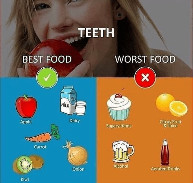
GROWTH HORMONE PHYSIOLOGY & PATHOLOGY

Overview

  • Growth hormone, aka, somatotropin, (somatotrophin) is secreted by anterior pituitary cells called somatotrophs.
  • Somatotrophs make up more than half of the anterior pituitary, and growth hormone is the most abundantly secreted anterior pituitary hormone.
  • Growth hormone is responsible for growth of almost all tissues, whether directly or indirectly via insulin-like growth factor 1 (IGF-1).
  • Growth hormone secretion is pulsatile, and is highest during sleep.
  • Growth hormone levels vary throughout life; they begin to fall after adolescence.
  • Negative feedback loops regulate growth hormone secretion.

Growth Hormone Physiology

  • First, we draw the hypothalamus and pituitary gland.
  • Neurosecretory cells originate in the arcuate nucleus of the hypothalamus, and their axons terminate on capillaries of the hypothalamic-pituitary portal system.
  • Within the anterior pituitary, there are various collections of endocrine cells;
    – We label the somatotrophs with an S, and show the nearby capillaries that deliver hormone products to the blood supply.

Growth Hormone Release

  • The hypothalamus secretes growth hormone releasing hormone (GHRH) into the neurosecretory cells.
  • When it reaches the anterior pituitary, GHRH stimulates somatotropin release of Growth Hormone (GH), which then travels in the blood to tissues throughout the body.

Growth Hormone Effects

  • Growth effects: it increases cell growth, proliferation, and differentiation throughout the body.
  • Direct effects on growth:
    – Increases bone length and muscle mass (growth hormone promotes protein deposition).
  • Indirect effects on growth:
    – Diabetogenic effects: growth hormone increases glycogen breakdown in the liver to increase blood glucose, which can be used as fuel by growing body tissues.
    – Growth hormone also increases insulin-like growth factor 1release from the liver, which in turn promotes growth and differentiation of various tissues.

Be aware that IGF-1 is sometimes referred to as somatomedin-C

– Glucose-sparing effects: it increases lipolysis, and decreases glucose uptake by skeletal muscle and adipose tissue, which frees up energy for growth of tissues.

Growth Hormone Regulation

  • Key regulators:
    – At the hypothalamus:
    Sleep, hypoglycemia, and stress trigger the release of growth hormone releasing hormone.
    Age and obesity are associated with reduced GHRH release, and, via negative feedback loops, glucose, IGF-1, and growth hormone also reduce GHRH release.
    – At the level of the anterior pituitary:
    Release of growth hormone is inhibited by growth hormone inhibitory hormone (aka, somatostatin, which is released by the hypothalamus), and via negative feedback signals from IGF-1 and Growth hormone (notice that IGF-1 and growth hormone provide negative feedback at both the hypothalamus and the anterior pituitary).
  • Additional factors that promote and inhibit growth hormone secretion:
    – Stimulates Secretion:
    Growth hormone releasing hormone
    Decreased blood glucose
    Decreased blood free fatty acids
    Increased blood arginine (an amino acid)
    Protein deficiency and starvation
    Stress and excitement (including a variety of experiences, including exercise and trauma)
    Testosterone and estrogen
    Deep sleep
    Ghrelin, which is a growth hormone secretagogue (GHS) that is produced mainly in the stomach.

– Inhibits Secretion:
Growth hormone inhibiting hormone (aka, somatostatin)
Increased blood glucose
Increased free fatty acids
Exogenous growth hormone
Insulin-like growth factor 1 (via negative feedback)
Aging
Obesity

Pause to recognize that low blood glucose, low levels of free fatty acids, and increased levels of amino acids in the blood stimulate growth hormone release, which ultimately reverses each of these states to maintain homeostasis.

Growth Hormone Pathologies

Growth Hormone Defects

Growth hormone deficiency

  • Characterized by low levels of growth hormone and IGF-1
    – If there is not enough growth hormone to trigger its production, IGF-1 will necessarily be low.
  • Isolated growth hormone deficiency is often due to defects in the GH1 gene, which is responsible for growth hormone production.
  • In other cases, growth hormone deficiency is an aspect of general hypopituitarism, in which other anterior pituitary hormones are also low.
  • Patients can be treated with recombinant human growth hormone.
    Growth hormone insensitivity is characterized by normal or high levels of growth hormone, and low levels of IGF-1.
  • This is the result of growth hormone receptor defects
    – May be hereditary, as in Laron Syndrome.
    – May acquired as a result of malnutrition, liver disease, diabetes, growth hormone receptor antibodies, or other pathologies.
  • Patients can be treated with recombinant IGF-1.
  • In Children:
    – Linear growth is slow (sometimes called pituitary dwarfism).
  • Children often have delayed puberty, with small gonads and genitalia (micropenis).
  • In Adults:
    – Body composition changes, including reduced muscle and bone mass, and increased fat mass.
    – Increased risk of cardiovascular disease (including left ventricular dysfunction and hypertension), and are more likely to develop insulin resistance.
    – Additionally, reduced exercise capacity, lethargy, and depression contribute to a lower quality of life in these patients.

Growth Hormone Excess

  • Excessive section of growth hormone leads to elevated growth hormone and IGF-1
    – Be aware that prolactin is also often elevated.
  • In both children and adults, the most common cause of growth hormone hypersecretion is an adenoma in the anterior pituitary.
    – Treatment often requires tumor removal.
  • In Children: Gigantism
    – Growth hormone hypersecretion occurs prior to fusion of the epiphyseal plates.
    – This causes accelerated linear growth, and children are taller than their age/sex-matched peers; delayed puberty is also likely.
  • In Adults: Acromegaly
    – Growth hormone hypersecretion occurs after epiphyseal plate fusion.
    – Thus, linear growth can’t occur, but bone deformation, soft tissue swelling, and enlargement of facial features, hands, and feet are common.
    – Patients are more likely to experience sleep apnea, diabetes, cardiovascular disease, arthropathy, carpal tunnel, headaches and visual disturbances due to tissue swelling and metabolic changes.
HYPOTHALAMIC & PITUITARY HORMONE OVERVIEW

Overview:

  • The hypothalamus collects information from throughout the body and uses it to regulate pituitary hormone secretion.
  • Hypothalamic neuroendocrine cell axons terminate in the median eminence and posterior pituitary, where they secrete various neurohormones.
    – 5 hypothalamic hormones act on the anterior pituitary lobe.
    – 2 hypothalamic hormones are released by the posterior pituitary lobe.
  • The anterior lobe, sometimes referred to as the adenohypophysis because of its gland-like components, comprises “-troph” cells that receive inhibitory and/or releasing signals from the hypothalamus via the hypothalamic-hypophyseal portal system.
    – The anterior lobe synthesizes and releases 6 peptide hormones that regulate growth, reproduction, and metabolism.
  • The posterior lobe, sometimes referred to as the neurohypophysis because it comprises nervous tissue, releases 2 peptide hormones that are synthesized in large-bodied neurons with cell bodies in the hypothalamus.
  • “Tropic” means that a hormone acts on other endocrine cells, as opposed to having direct actions on target organs; this will be an important function of several hypothalamic and anterior pituitary lobe hormones.

Hypothalamus and Pituitary Gland

  • First, we draw the hypothalamus and pituitary gland; indicate anterior and posterior lobes.
  • Within the anterior lobe reside the following clusters of endocrine cells:
    – Somatotrophs, Corticotrophs, Thyrotrophs, Lactotrophs, and Gonadotrophs.
    – Be aware that some authors end these cells with the suffix “tropes” instead of “trophs” (for example, corticotropes).
  • Hypothalamic hormones that act on the anterior lobe:
    – Growth-hormone Releasing hormone (GHRH)
    – Growth-hormone Inhibiting hormone (GHIH)
    – Corticotropin-releasing hormone (CRH)
    – Thyrotropin-releasing hormone (TRH)
    – Prolactin-Inhibiting hormone (PIH)
    – Gonadotropin-Releasing hormone (GnRH).

    Notice that there are 2 inhibitory hormones and 4 releasing hormones in this group.

Anterior Lobe Hormones

Growth-hormone releasing hormone stimulates somatotroph release of growth hormone (aka, somatotropic hormone, aka, somatotropin).

  • Growth hormone has multiple effects throughout the body: it facilitates bone and muscle growth and has diabetogenic effects on metabolism (it increases blood glucose).
  • Growth hormone inhibiting hormone, aka, somatostatin, inhibits somatotrophic cell release of growth hormone.

Corticotropin-releasing hormone triggers corticotrophrelease of adrenocorticotropic hormone (ACTH) (aka, corticotropin).

  • ACTH, in turn, stimulates the adrenal glands to release cortisol, which has a variety of effects, including reduction of inflammatory and immune responses and increases in gluconeogenesis, lipolysis, and proteolysis.
  • ACTH release is inhibited via short and long negative feedback loops (see the links in our notes for more details).

Thyrotropin-Releasing hormone stimulates thyroprophicrelease of thyroid-stimulating hormone (TSH).

  • Thyroid-stimulating hormone stimulates thyroid glandproduction and release of thyroid hormone, which promotes growth and maturation, increases metabolism, and stimulates beta-adrenergic receptors.

Prolactin-inhibiting hormone, which is dopamine, tonically inhibits lactotroph release of prolactin.

  • Dopamine’s effects ensure that prolactin is secreted in low levels in males and non-pregnant/non-breastfeeding females; be aware that prolactin receptors are located in various tissues, including the prostate and immune cells, but its exact effects are uncertain.
  • In pregnant and breastfeeding women, dopamine’s effects are inhibited, and lactotrophs release prolactin, which stimulates breast development and lactogenesis (milk formation).

Gonadotropin-releasing hormone (GnRH) promotes gonadotroph release of follicle-stimulating hormone (FSH) and luteinizing hormone (LH), which act on the gonads.

  • Follicle stimulating hormone works at the testes to support Sertoli cell growth sperm cell proliferation.
  • In the ovaries, FSH promotes granulosa cell growth, follicle maturation, and aromatase synthesis; aromatase is the enzyme that converts androgens to estrogens.
  • In the testes, Luteinizing hormone promotes Leydig cell secretion of testosterone.
  • In the ovaries, LH has multiple effects:
    – It promotes thecal cell secretion of androgens, which are then converted to estrogens by the nearby granulosa cells;
    – The LH surge induces ovulation;
    – Post-ovulation, LH promotes the development and functioning of the Corpus Luteum (notice that “luteinizing” hormone promotes the “luteum”).

Posterior Lobe Hormones

  • Neurons in the hypothalamus produce Oxytocin and Anti-diuretic hormone (aka, vasopressin), which are transported to the posterior pituitary, which releases them into the blood.

Oxytocin is responsible for myometrial contractionsduring childbirth to expel the fetus, and, during lactation, it promotes milk ejection.
– Recall that prolactin was necessary for milk production and secretion; oxytocin is required for its ejection.

Anti-diuretic hormone (ADH) regulates body water and blood pressure via the following mechanisms:
– In the kidney, ADH acts on the nephron collecting ducts to promote water reabsorption, thus increasing body water (and, therefore, blood volume, blood pressure, and cardiac output).
– Vasoconstriction is a secondary function of ADH, which binds to vascular smooth muscle receptors (hence its alternative name, vasopressin); in hypovolemic shock, increased endogenous production of ADH is an important compensatory mechanism.

CARDIOVASCULAR PHARMACOLOGY

Here we’ll learn which drugs are used to treat common cardiovascular disorders; because there are so many of them, antiarrhythmic drugs are discussed separately.

Hypertension

  • First, indicate that the goal of these drugs is to reduce blood volume, systemic vascular resistance, or cardiac output, so as to return the blood pressure to normal.
  • Many patients will require combination therapy to achieve these goals.

Vasodilation reduces systemic vascular resistance:

  • Angiotensin-converting-enzyme inhibitors (ACE inhibitors):
    – These drugs tend to end in “pril” (ex: Lisinopril, benazapril, captopril), and are considered first line therapy; additionally, they protect against diabetic kidney disease, which makes them particularly useful in patients with hypertension and diabetes mellitus.
    – However, they are not recommended as first line therapy in patients with asthma (because they can cause coughing), and that they are contraindicated in pregnancy and bilateral renal stenosis.
  • Angiotensin II receptor blockers (ARBs):
    – Often end in “sartan” (ex: candesartan, azilsartan, eprosartan, etc.).
    – As their name suggests, these drugs block the angiotensin II receptors on blood vessels, thereby prohibiting the vasoconstrictor effects of angiotensin II.
    – Like ACE inhibitors, these drugs should not be used in pregnancy or by patients with bilateral renal stenosis; however, they are suitable for patients with asthma, as they are not associated with respiratory side effects.
  • Calcium channel blockers:
    – Predominantly act on the vessels to produce vasodilation are the dihydropyridines; the specific drugs end in “pine” (ex: felodipine, nifedipine, amlodipine, etc.).
    Write that these are another example of first line therapy for hypertensive patients, and evidence suggests that they are particularly effective in elderly and African American individuals.
  • Hydralazine (brand name: Apresoline):
    – Sometimes used to treat severe hypertension, and, as we’ll see, hypertension during pregnancy.

Cardiac Output Reduction

  • Beta-blockers:
    – End in “lol” (ex: metoprolol, acebutolol, bioprolol, labetalol, etc.).
    – These drugs block norepinephrine and epinephrine receptors on blood vessels.
    – Because of their effects on the heart, beta blockers are contraindicated in patients with bradycardia and AV nodal block.
    – Be aware that, in patients with diabetes, beta blockers can mask signs of low blood sugar;
    – And, non-selective beta blockers should not be prescribed to patients with asthma or COPD, because they can induce bronchoconstriction.

Increase Urine Output

  • Thiazide diuretics:
    – Typically end in “ide” (ex: chlorothiazide, indapamide, etc.), and thiazide-like diuretics.
    – Thiazide diuretics are another example of first line therapy, but be aware that they should not be given to patients with sulfa allergies.
  • Aldosterone antagonists:
    – End in “one” (spironolactone and eplerenone) are used in resistant hypertension, and are contraindicated in pregnancy.

Hypertensive Emergency

  • Defined as systolic BP > 180 mmHg or diastolic BP > 120 mmHg with acute organ damage.
  • Nitrates (ex: nitroprusside and nitroglycerine)
  • Calcium channel blockers (ex: clevidipine and nicardipine)
  • Dopamine-1 – agonists (fenoldopam)
  • Adrenergic blockers (labetalol)

Hypertension during Pregnancy

  • Methyldopa
  • Labetalol
  • Nifedipine
  • Hydralazine

Hypotension

  • Hypotension medications work to increase cardiac output and/or systemic vascular resistance.
  • Vasoconstrictors include alpha-agonists; examples include methoxamine and phenylephrine.
  • Cardiostimulatory drugs used to increase cardiac output include beta-agonists, which increase heart rate and contractility; examples include norepinephrine and dopamine.

Heart Failure w/ Reduced Ejection Fraction

  • Treatment objectives are to improve cardiac functioning and relieve symptoms.
    Increase urine output to lower blood volume:
    – Diuretics
    – Aldosterone antagonists
    Vasodilators:
    – ACE inhibitors
    – ARBs
    Cardioinhibitory drugs
    – Beta-blockers (once the patient is stable)
    – Ivabradine, which is a sinus node inhibitor that is prescribed to patients with persistent heart rate above 70 beats per minute, despite treatment with beta blockers.
    Inotropic drugs
    – Digitalis (ex: digoxin), may be given to some patients to improve cardiac contractility and cardiac output.
    Digitalis should not be given to patients with hypokalemia, sinus or AV block, or reduced renal function.
  • Be aware that there are no proven medication strategies for treating heart failure with preserved ejection fraction.

Angina

  • The objective of treatment is to reduce the ratio of oxygen demand to supply in order to reduce the symptoms and improve prognosis.
    Vasodilator
    – Nitroglycerine is used to prevent and treat sudden attacks; nitroglycerine be administered sublingually, orally, or transdermally.
    Cardioinhibitory drugs reduce contractility and heart rate, and include:
    – Beta-blockers
    – Non-dihydropyridine calcium channel blockers, verapamil and diltiazem, which are more cardio-selective than are the dihydropyridine calcium channel blockers used to treat hypertension.
    Ranolazine
    – A late sodium current blocker, ranolazine, is also sometimes prescribed; this drug decreases heart wall tension and improves coronary blood flow.
    Anti-thrombotic drugs
    – Anti-platelets and anti-coagulants may be prescribed to reduce clotting and improve blood flow.

Myocardial infarction

  • Therapeutic goal is to reduce the ratio of oxygen demand to oxygen supply.
    Restore coronary blood flow
    – Thrombolytic and antiplatelet drugs
    Vasodilators
    – Nitroglycerine
    Reduce oxygen demand
    – Beta blockers
  • Other drugs may also be used to alleviate pain and MI complications, including morphine, anti-arrhythmics, and diuretics.

Be aware that the medications we’ve learned here may not be appropriate in all cases, and that other interventions, such as lifestyle changes and surgical procedures, may also be necessary. Furthermore, be aware that underlying disease states, such as dyslipidemia, may need to be addressed.

RESPIRATORY PHARMACOLOGY PART 2 (ASTHMA & COPD)

Overview

  • Asthma and COPD are obstructive respiratory diseases characterized by airflow obstruction, chronic inflammation, and airway remodeling.
  • Asthma is defined as intermittent, reversible obstruction and hyper-reactivity with excessive mucus production in the bronchi.
  • COPD is chronic, progressive, irreversible obstruction.
    – COPD is an umbrella term that includes chronic bronchitis, small airway disease, and emphysema.
  • Asthma-COPD overlap syndrome, which involves the airway hyper-reactivity associated with asthma plus elements of COPD.
  • Asthma and/or COPD have shared treatment goals:
    – Open their airways and reduce air trapping, which will relieve dyspnea, and, reduce airway remodeling and prevent exacerbations.

BRONCHODILATORS

  • Used to treat both asthma and COPD.

We draw a bronchial tube with thick layers of smooth muscle.

Beta-2-adrenoreceptor agonists (often shortened to simply “beta-2 agonists”) bind beta 2 receptors on the bronchial smooth muscle, which increases local cAMP and induces smooth muscle relaxation and bronchodilation.

  • Beta-2 agonist bronchodilators may cause adverse effects associated with sympathetic activation (tremors, irregular heartbeat).
  • Short-acting beta-2 agonist bronchodilators (SABAs) are used as “rescue” interventions for acute dyspnea due to bronchoconstriction and hyper-reactivity; they are effective within 1-5 minutes of administration and the effects last up to 4 hours.
    – Selective beta-2 agonists include: albuterol, terbutaline, pirbuterol, and metaproterenol (“ols”).
    – Non-selective beta agonists, such epinephrine, are associated with more adverse effects; fortunately, selective beta-2 agonists have replaced them.
  • Long-acting beta-2 agonist bronchodilators (LABAs) are administered once or twice daily as “maintenance” treatments to maintain open airways; onset is slower, but the effects last for 12-24 hours.
    – Some important examples of LABAs are salmeterol and formoterol (“ols”).

Muscarinic antagonists are also used to treat asthma and COPD. They block acetylcholine from binding muscarinic receptors in the bronchial smooth muscle, which prevents bronchoconstriction; these drugs also reduce vagal-mediated mucus secretion in the bronchi.

  • Muscarinic antagonist bronchodilators are associated with dry mouth, dizziness, gastrointestinal problems, and cough.
  • Short-acting muscarinic antagonist bronchodilators (SAMAs) are used as “rescue” treatments; they take effect within minutes of administration and last about 4 hours.
    – Ipratropium is a commonly used short-acting muscarinic antagonist.
  • Long-acting muscarinic antagonist bronchodilators (LAMAs) are used for maintenance therapy, as their effects last 12-24 hours.
    – Tiotropium, aclidinium, and umeclidinium are examples of these drugs (“iums”).
  • Dual Therapy: Beta-2-adrenoreceptor agonists and muscarinic antagonists work synergistically to open the airways, so they are often used together in dual therapy to maximize their effects.

Methylxanthines

  • Theophylline can be used to treat asthma, but that it is not a first-line therapy. Theophylline is a nonselective phosphodiesterase-4 inhibitor that also blocks adenosine receptors to induce smooth muscle relaxation.
    – The high dosages required to effectively open the airways produce systemic side effects, including convulsions and arrhythmias, as well as gastrointestinal problems and headache.
  • Rofumilast improves exercise tolerance in COPD patients. Rofumilast is thought to work via histone deacetylation, which produces anti-inflammatory effects.
    – Thus, it can be prescribed to enhance the anti-inflammatory effects of corticosteroid treatments.

ANTI-INFLAMMATORIES

Recall that inflammation is a key part of the pathology of asthma and COPD, and promotes infiltration of various immune cells, remodeling, and constriction of the airways. Thus, reducing inflammation is an important component of treating obstructive pulmonary diseases, especially asthma.

  • Corticosteroids, i.e., glucocorticoids, downregulate inflammatory genes to reduce inflammatory cytokines, chemokines, adhesion molecules, and other pro-inflammatory mediators; thus, they reduce the infiltration of inflammatory cells in the airways.
  • Inhaled corticosteroids such as beclomethasone, are first-line therapy for moderate to severe asthma.
    – Used alone, they are less effective in patients with COPD, but can be combined with long-acting beta-2-agonist bronchodilators.
    – Be aware that use of inhalers increases risk of infection in COPD patients.
    – Oropharyngeal candidiasis is a common adverse effect in patients using inhaled glucocorticoids.
  • Oral glucocorticoids, such as prednisone, produce systemic adverse effects, including increased risk of infection, hypertension, osteoporosis, and ocular disorders.
    – Thus, they are reserved for severe exacerbations and/or patients who are unresponsive to other therapies.

MONOTHERAPIES

Anti-IgE Antibodies such as omalizumab, bind free IgE and prevent it from binding to mast cell receptors. Thus, omalizumab is given to reduce exacerbations in severe allergic asthma.
– Recall that that IgE binding to mast cells triggers release of allergic mediators.

Anti-IL-5 drugs such as mepolizumab, reslizumab, and benralizumab.
– Interleukin 5 activates eosinophils and promotes airway inflammation.
– Anti-Interleukin-5 receptor blockers and antagonists prevent eosinophil activation and are therefore used as add-on therapies in severe eosinophilic asthma.

Cromolyn reduces mast cell release of inflammatory mediators; though it may be helpful to prevent asthma exacerbations in some patients, it is generally less effective than inhaled corticosteroids.

ANTI-LEUKOTRIENES

  • These drugs can be used to prevent aspirin- and exercise-induced asthma.
  • Leukotrienes aid in inflammatory cell migration, increase capillary permeability, and induce smooth muscle contraction.
  • Zileuton is a leukotriene synthesis inhibitor; montelukastand zafirlukast are leukotriene receptor blockers.

ADDITIONAL COPD THERAPIES

  • Smoking cessation is key to slowing progression of the disease.
  • Mucolytics help break up mucus and facilitate removal for easier breathing.
  • Oxygen therapy is recommended if oxygen saturation falls below 88%.
  • Infection prevention is important for reducing harmful exacerbations; patients may be prescribed antibiotics, such as azithromycin, to ward off bacterial infections, and influenza and pneumonia vaccines are recommended

RESPIRATORY PHARMACOLOGY PART 1 (RHINITIS & COUGH)

Here we’ll learn about medications used to treat rhinitis and cough; although we’ll single out specific drugs and their actions, keep in mind that many over-the-counter medications comprise various combinations of drugs.

RHINITIS

  • Rhinitis: the nasal passages are inflamed, causing a “runny” and/or “stuffy” nose, sneezing, and sinus congestion. * Rhinitis may be caused by allergies (IgE-mediated) or viral pathogens or irritants.

Decongestants are alpha-adrenoreceptor agonists that promote vessel constriction in the nasal mucosa, which reduces edema and swelling.

  • Epinephrine and oxymetazoline are nasal sprays.
    – Afrin is the name-brand version of oxymetazoline, which is also used in Visine for itchy, red eyes.
  • Phenylephrine is available in both sprays and tablet formulas.
  • Pseudoephedrine comes in tablet form.
    – Because it can be used to create methamphetamine, it has largely been replaced by phenylephrine in over-the-counter drugs.
  • Pseudoephedrine acts on both alpha and beta receptors,and is therefore associated with more systemic effects.
  • Adverse effects of alpha-adrenoreceptor decongestants are due to sympathetic activation, which particularly affects the central nervous and cardiovascular systems (again, this is especially true for pseudoephedrine).
    – If patients experience serious adverse effects, including irregular heart beat, shaking, or anxiety, they should stop using these medications.
    – Rhinitis medicamentosa: Although nasal sprays are better tolerated due to their localized effects, patients should not use them for more than 3-5 days at a time, as they can cause rhinitis medicamentosa, which is a form of rebound congestion.
  • Avoid or use caution in prescribing alpha-adrenoreceptor agonists to patients with heart disease, hypertension, thyroid disease, diabetes, enlarged prostate, pregnancy, or those who have used monoamine oxidase inhibitors in the last two weeks.

Glucocorticoids are available as nasal sprays; glucocorticoids bind to intracellular glucocorticoid receptors and downregulate the inflammatory response by up-regulate anti-inflammatory genes and suppressing pro-inflammatory genes.

  • Very effective against allergic rhinitis with discharge and blockage, and, that antihistamines are added to some formulas.

Cromolyn sodium blocks mast cell release of histamine and other inflammatory mediators that cause rhinitis; it is occasionally used in asthma, as well.
– However, because cromolyn has to be used multiple times a day and is generally less effective than other available drugs, it is not a first-line treatment.

Antihistamines are used to treat allergic rhinitis.

  • Histamines cause nasal vessel dilation and edema, which produces the increased watery discharged associated with a “runny” nose.
  • Anti-histamines block the action of histamines:
    – H1 antihistamines are inverse agonists that keep H1 receptors in their inactive states, which prevents mast cell degranulation and NF-kB (nuclear factor kappa B) activation.
    – Thus, these drugs reduce nasal discharge; antihistamines are also used to treat allergic inflammation and itching of the eyes and skin.
    – Additionally, some H1 antihistamine drugs also have other, non-histamine receptor effects (for example, some have anti-tussive effects).
  • Use caution when prescribing antihistamines to pregnant patients and those with cardiovascular disease, hypertension, urinary retention, and elevated ocular pressure.
  • First-generation antihistamines include diphenhydramine (aka, Benadryl), chlorpheniramine, brompheniramine, and hydroxyzine.
    – First-generation antihistamines readily cross the blood-brain barrier, and often cause sedation;
    headaches and weigh gain are also reported.
    – Additionally, these drugs have anticholinergic effects: blind as a bat, hot as a hare, mad as a hatter, dry as a bone, red as a beet.
    – First-generation antihistamines are also used as sleep aids and to prevent or treat motion sickness
  • Second-generation antihistamines* include loratadine (Claritin), cetirizine (Zyrtec), azelastine, and olopatadine (the last two are available in eye and nasal sprays).
    – Second generation formulas were designed to cause less sedation and have fewer anticholinergic side effects; they do not cross the blood-brain barrier as readily as the first-generation antihistamines.
  • Third generation antihistamines* include fexofadine, desloratadine, and levocetirizine.
    – These drugs are metabolites of the second generation H1 antihistamines, and, like those drugs, cause less sedation and fewer anticholinergic effects than first-generation antihistamines, and do not readily cross the blood-brain barrier.

Saline

Found in over-the-counter sprays and nasal irrigation systems.

  • Saline solutions flush away mucus and nasal irritants and sooth nasal membranes.

COUGH MEDICATIONS

Cough suppressants (aka, anti-tussives) target the medullary cough center.

  • They are NMDA receptor antagonists and Sigma-1 receptor agonists (NMDA stands for N-Methyl-D-aspartate).
  • These medications suppress cough caused by bronchial irritation due to flu and cold viruses, but are not used for chronic cough caused by smoking and lung disorders such as emphysema.
  • Dextromethorphan is a common over-the-counter anti-tussive drug.
    – Adverse effects are rare and include GI upset, drowsiness, and dizziness.
    – This should not be used by patients who have taken an MAOI in the last two weeks, nor should it be prescribed to children under four years old.
    – Furthermore, beware of dextromethorphan abuse;when taken at high dosages, it can cause euphoria, but it can also cause brain damage, seizures, and cardiovascular events.
  • Opioids (codeine, hydrocodone, etc.) also suppress the cough reflex.
    – Because of the potential for addiction and abuse, they are contraindicated in children and are not first-line therapy in adults.

Mucoactive agents are drugs that modify the production, secretion, or components of mucus, or that act on the respiratory cilia to facilitate easier mucus removal from the bronchi.

  • Guaifenesin (the main ingredient in Mucinex) is an expectorant – it increases the volume of watery mucus to facilitate productive coughing.
  • N-acetylcysteine is a mucolytic – it breaks down mucus components to reduce overall mucus viscosity.
    – It is sometimes used by patients with COPD.
    – In IV form, can be used to treat acetaminophen overdose.
  • Ambroxol is a mucokinetic drug – it increases the transportability of mucus by acting on the respiratory epithelium cilia.
  • Anticholinergic agents act as mucoregulators – they reduce mucus hypersecretion.

Nutrition, Diet & Dental Caries

  • Nutritional effects – systemically
  • Dietary effects – Locally

Vitamin D :-

➡️ Vit. D along with Parathyroid hormones & calcitonin play primary roles in regulating the concentration of Calcium & inorganic phosphate in plasma & ECF and in controlling mineralization of bones & teeth.

➡️ Quantitive defect in enamel tissue from metabolic injury to Ameloblasts – ENAMEL HYPOPLASIA

Source: Google
  • Clinical Significance: Roughened surface with discrete pitting / circumferential band like irregularities which post eruptively acquire a yellow brown stain.

Carbohydrate intolerance & Dental Caries:

➡️ Intolerance occurs because of deficiency of a specific enzyme involved in metabolism of sugar.

Hereditary fructose intolerance: (Froesch,1959)

  • Inborn error of fructose metabolism transmitted by an autosomal recessive gene.
  • Episodes of pallor, nausea, vomitting, coma & convulsions following ingestion of fruit containing fructose/cane sugar.
  • ⬇️ Dental caries.

Diet Modification:

➡️ Dietary sucrose has 2 effects on plaque:

  1. Frequent ingestion – S. Mutans colonization ⬆️
  2. Mature plaque exposed to sucrose metabolizes to organic acids – ⬇️ pH

Dietary Measures:

Source: mfine
  1. No. of meals + snacks as low as possible.
  2. Sugars – eliminated; Active chewing foods ➡️ desirable
  3. Fermentable Carbs.
  4. Flouride, Calcium, Phosphate, fats & proteins – in diet.
  5. Sugar substitutes – ⬇️ acid formation.

Adequacy of Diet:

Source: mfine
  1. Fats, oils, sweets – use sparingly.
  2. Milk, yogurt, cheese group: 2-3 servings
  3. Vegetable group: 3-5 servings
  4. Fruit group: 2-4 servings
  5. Meat, fish, eggs, nuts: 2-3 servings
  6. Bread, Rice, Pasta, Cereal: 6-11 servings.

Anti-cariogenic foods:

Source: Foodsmix
  1. Milk – least cariogenic
  2. Cheese – casein phosphatase
  3. Fibrous foods
  4. Tea
  5. Chewing gum – Salivary stimulant
  6. Xylitol👇🏻
  • Bacteriostatic
  • ⬆️ salivary flow
  • ⬆️ concentration of Amino acids & NH3 – Neutralize plaque acids.
  • Prevents S. Mutans binding to sucrose.

➡️ Dietary and nutrition education appropriate for dental settings are an essential component of guidelines or standards of practice that determine successful management of dental caries and the patient’s quality of life accross time.

Nutrition, Diet & Dental Caries:
Dr. Mehnaz Memon

References: Practical manual guide by CM Marya, Internet

Enzyme Naming Conventions

ENZYME NOMENCLATURE

  • Derived from substrate or chemical reaction the enzyme catalyzes
  • Most enzyme names have suffix “–ase”

Isozymes (isoenzymes)

  • Enzymes that catalyze same reaction but have different amino acid sequences (different proteins)

ENZYMES REQUIRING COFACTORS

Apoenzyme

  • Enzyme without the cofactor.

Holoenzyme

  • Enzyme and cofactor together

6 CLASSES OF ENZYMES

Oxidoreductases

  • Catalyze electron transfer reactions (oxidation/reduction reactions)
  • A- + B → A + B-

Transferases

  • Catalyze transfer of functional groups to a molecule
  • A-B + PO4 → A-B-PO4

Hydrolases

  • Catalyze breakage of bonds by hydrolysis (addition of water)
  • A-B-C + H2O → A-B + C

Lyases

  • Catalyze bond cleavage reactions that are not oxidation or hydrolysis reactions
  • A-B-C → A-B + C

Isomerases

  • Catalyze rearrangement reactions
  • A-B-C → A-C-B

Ligases

  • Catalyze reactions in which covalent bonds join two molecules
  • A-D + B-C→ A-D-B-C

Enzyme Active Site & Regulation

ENZYME ACTIVE SITE

  • 3-dimensional cleft where substrate can bind
  • Substrate specificity: depends on atom arrangement in cleft

Lock and key

  • Enzyme’s substrate fits perfectly into active site

Induced fit

  • Active site conforms to substrate’s 3D shape upon binding
  • Lock-and-key and the induced-fit are 2 models of substrate binding to active site

ENZYME REGULATION

Allostery

  • Effector molecules bind to enzyme and change substrate binding affinity

Transcription

  • Enzyme synthesis turned on or off to regulate production

Reversible covalent modification

  • Enzymes can be phosphorylated (activation or deactivation)

Isozymes

  • Different forms of enzymes can be used to affect reaction rates

Proteolytic activation

  • Some enzymes have part of their sequence cleaved to become active

ALLOSTERIC REGULATION

  • Effector molecule (inhibitor or activator) binds to effector site
  • Effector site: distinct from active site

Enzyme Inhibition

KEY VALUES

  • Vmax
    – Maximal rate of a reaction (every active site bound by substrate)
  • Km
    – Inversely proportional to binding affinity of enzyme to substrate

ENZYME INHIBITION

  • Occurs when a substance reduces activity of an enzyme

Types of inhibition

  • Competitive
    – Substrate & inhibitor compete for active site
    – Greater [S] overcomes inhibition
    – Increases the apparent Km but does NOT affect Vmax
  • Noncompetitive
    – Inhibitor reversibly binds to enzyme outside of active site to deactivate it.
    – Enzymes regain function when inhibitor removed from system
    – Does NOT change Km but lowers Vmax

NOT uncompetitive inhibition in which inhibitors bind enzyme-substrate complexes

  • Uncompetitive inhibition: requires preassembled enzyme-substrate complexes–>more effective when [S] is high
  • Irreversible
    – Inhibitor binds to and permanently deactivates enzyme
    – Only overcome by synthesis of new enzymes

CLINICAL CORRELATION

  • Heavy metals (mercury & lead)
    – Irreversible inhibitors: bind tightly to sulfur groups in enzymes
    – Permanently deactivate them
  • Ethylene glycol (antifreeze) metabolites
    – Toxic to human body
    – Ethanol (competitive inhibitor): used to inhibit alcohol dehydrogenase active site to prevent metabolism of ethylene glycol
  • Angiotension-converting enzyme (ACE) inhibitors
    – Blood pressure lowering agents: noncompetitively inhibit ACE
    – Prevent formation of angiotensin (acts on kidneys to inc. blood pressure)

Enzyme kinetics

Enzyme kinetics – Definition

  • Study of rates of catalytic reactions involving substrates and enzymes

ENZYMATIC REACTION VARIABLES

Substrate (S)

  • Substance that enzyme modifies
  • Typically the controlled variable

Velocity (V)

  • Rate of product formation; typically the measured variable

Maximum velocity (Vmax)

  • Reached when every enzyme’s active site is bound by a substrate
  • Determined experimentally (reactions do not typically reach Vmax)

Reaction constant (Km)

  • Substrate concentration corresponding to 1/2 of maximum velocity
  • Determined experimentally
  • Determines binding affinity of enzyme to substrate

MICHAELIS-MENTEN MODEL

  • Examines reaction of single substrate w/ single enzyme to create product.

Assumptions

  • No intermediates
  • No product inhibition
  • No allostericity or cooperativity
  • Total enzyme concentration is constant (and [S] >>> [E])
  • Pseudo-steady-state hypothesis: rate of [ES] formation = rate of [ES] breakdown
  • Initial velocity (rate measured as soon as substrate/enzyme are mixed)

First-order kinetics

  • Change in [S] changes velocity of rxn

Zero-order kinetics

  • Change in [S] does NOT change velocity of rxn

Vmax

  • Asymptotic: rxn rate approaches Vmax, never actually reaches it

Km

  • Estimates enzyme binding affinity
  • High affinity = requires a low [S] to reach 1/2 Vmax

LINEWEAVER-BURK MODELS

  • Uses inverse of variables in Michaelis-Menten model to represent graph as straight line

1/Vmax

  • Y-intercept: x = zero.

1/Km

  • X-intercept: y=0

FACTORS THAT AFFECT REACTION VELOCITY

  • Increasing temperature: increases reaction velocity until denaturation
  • Change in pH: can denature enzymes –> loss of function –> ionization of amino acids (changes active site conformation
  • [E] and [S] substrate affect reaction velocity