Bone Histology

HISTOLOGY OF BONE

GROSS STRUCTURE

  • The diaphysis is the shaft and notably comprises the marrow cavity.
  • The metaphyses comprises spongy bone.
  • The epiphyses, which are the ends – the sites of articulation.

The epiphyseal line (the ultimate regression of the growth plate) separates the epiphysis and metaphysis.

COLLAGENOUS STRUCTURES

  • Articular cartilage are derived from hyaline cartilage, at its ends.
  • Periosteum, along the shaft, is derived from a condensation of outer connective tissue. It comprises an external layer of collagens fibers and vasculature and an internal layer of osteoprogenitor cells.
  • Endosteum, centrally, is derived from derived from a condensation of inner connective tissue and helps separate the marrow cavity, internally, from the compact bony matrix that encapsulates it. It comprises an inner circumferential lamellae and osteoprogenitor cells.

MARROW CAVITY

  • Filled with hematopoetic marrow (red blood cell and white blood cell precursors) and fatty marrow (adipose tissue).

We learn about hematopoetic marrow further elsewhere. It comprises stem cells, which can self-renew, committed precursor cells, and cells that are in the process of maturation.

BONE TISSUES

  • The medullary cavity comprises spongy bone.
  • The outer layers of the diaphysis comprises compact bone.

MICROSCOPIC STRUCTURE OF BONE (ALONG THE DIAPHYSIS)

The periosteum.

  • Internal to it, lie columns of compact bone, called osteons.
  • Centrally, within the osteons, run the Haversian (aka central) canals, in parallel.
  • Volkman’s (aka perforating) canals run in perpendicular to them.
  • These canal systems form channels for the neurovasculature.
  • The osteon comprises concentric rings of lamellae – the bone connective tissue.
  • Osteocytes are a mature form of osteoblast (the bone-producing cells) within the bony matrix; they are concentrically arranged in between the circles of lamellae. They lie within lacunae (cavities) that interconnect via canaliculi (which are spindly, like spider legs) and through which the osteocytic cytoplasmic cell processes connect for the transportation of nutrients and waste.
  • The outer layer forms the cement line.
  • Interstitial lamella lie in between the osteons, which comprises remnants of partially resorbed osteons.
  • Sharpey’s fibers are collagenous fibers that anchor the periosteum to the outer lamellae.

Endosteum

  • Lies internal to the compact bone. It comprises:
    -An inner-circumferential lamellae
    -Osteoprogenitor cells
  • Spongy bone lies internal to the endosteum and comprises a network of lamellae that do NOT form the Haversian channels and osteons found in compact bone.

HISTOLOGICAL SLIDES

Compact bone

  • Osteon
  • Haversian (aka central) canal
  • Interstitial lamella
  • Osteocyte within a lacuna

Osteon (at higher resolution)

  • Concentric pattern of lamellae (the bony layer).
  • External to this lamella, show a lacuna.
  • Canaliculi radiate from it
  • Haversian canal
  • Edge of a Volkman’s canal, which perforates it perpendicularly.

Marrow cavity

  • Inner circumferential lamellae of the endosteum
  • Hematopoetic marrow
  • Fatty marrow, which increases with age
  • Vascular sinusoid

Bone Development

BONE DEVELOPMENT: OSTEOGENESIS (OSSIFICATION)

Endochondral ossification

  • An INDIRECT form of ossification, wherein a hyaline cartilaginous model (template) is replaced with bone, such as occurs with long bones (eg, the femur).

Intramembranous ossification

  • A DIRECT form of ossification mesenchymal cells directly differentiate to osteoblasts (no cartilaginous model is first formed), such as occurs with flat bones (the skull bones).

INTRAMEMBRANOUS OSSIFICATION

  • Embryologically, skeletal tissues typically derive from mesoderm: the midline (axial) skeleton derives from the somites and the appendicular (the limb) skeleton derives from the lateral plate.
  • Mesenchymal cells migrate to vascularized gelatinous extracellular collagen fiber matrix (a primary spongiosa).
  • They differentiate directly into osteoblasts.
  • Osteoblasts form bone in a loosely arranged (disorganized), immature initial form of bone, called woven bone.
  • Osteoblasts become trapped within their own bony matrix and become osteocytes; these bony matrices are referred to as trabeculae (aka fused spicules).
  • Woven bone later matures to form lamellar bone, a much tougher form of bone that constitutes both compact bone and spongy bone.

Compact Bone

  • The outer layer: the periosteum.
    -Comprises columns of compact bone, called osteons.
    -Centrally, within each osteon, lies a longitudinally-oriented canal, the Haversian (aka central) canal.
    -Each osteon comprises concentric rings of lamellae.
    -Osteocytes are a mature form of osteoblast (the bone-producing cells) within the bony matrix.
    -Internal to the compact bone, lies the endosteum, which comprises in inner circumferential lamellae and the osteoprogenitor cells, internal to it.

Spongy bone

Lies internal to the endosteum and comprises a network of lamellae that do NOT form the Haversian channels and osteons found in compact bone.

ENDOCHONDRAL OSSIFICATION

Origins

  • Mesenchymal cells migrate and differentiate to form a hyaline cartilage model, which comprises basophilic collagen and ground substance.
  • Chondrocytes are part of a cartilaginous model that are hyaline cartilage cells, which are purplish (basophilic).
    -Key histological features: their nucleus and lipid droplets.
    -Chondrocytes produce the structural components of cartilage: collagen, proteoglycans and glycosaminoglycans, and are usually found in clusters (isogenic groups) of recently divided cells.
    -Chondrocytes hypertrophy, which signals (via vascular endothelial cell growth factor) the sprouting of blood vessels, which we’ll draw next.

Bone Regions

  • The diaphysis (the shaft)
  • The epiphyses (the articulating ends of the long bone)
  • The metaphyses that separate them.

Periosteal Buds

  • Periosteal buds invade the center of the diaphysis. Vascularization occurs via the periosteal bud, which brings forth osteoprogenitor cells and forms the primary ossification center, which forms within the cavities that are created when the hypertrophic chondrocytes starve and apoptose (die).
  • Vasculature further invades the primary ossification center and the osteoprogenitor cells remodel bony matrix as the ossification center grows linearly (via interstitial growth).
  • The outer cartilage is perichondrium, which forms a periosteal bone collar of compact bone that grows in opposite orientation, increasing the bone thickness (via appositional growth) – here, the osteoblasts secrete bone matrix directly via intramembranous ossification. Periosteum distributes blood vessels to bone and is not found in synovial articulations or muscle attachment sites.

Medullary cavity

  • Forms as the primary ossification center degenerates cavitates and remodels via interstitial growth.
  • Endochondrium delineates it, which forms a layer of lamellar bone and osteoprogenitor cells.
  • The marrow cavity is filled with hematopoetic marrow (which comprises red and white blood cell precursors)
  • Vasculature invades the cavity to fill it with marrow.

Secondary ossification centers.

  • Unlike the primary ossification center, they never grow large enough to create marrow cavities but instead remain constituted with spongy bone.

Bone Growth

  • Further appositional growth (widening) of the periosteal bone collar occurs along the diaphysis (again which forms compact bone via intramembranous ossification).
  • There is an epiphyseal (growth) plate at the border of the metaphysis and the epiphysis: elsewhere we learn about the zones and processes of interstitial growth, but pay attention that Indian hedgehog (Ihh) was discovered to be important for the stimulation of chondrocyte growth with delay of chondrocyte hypertrophy (thus delaying a key step in endochondral ossification).

MAJOR BONES & THEIR DEVELOPMENT

Intramembranous Ossification

  • Cranial Vault
  • Maxilla/Mandible
  • Clavicles

Consider that the skull bones must ossify prior to delivery of the fetus, so the brain isn’t squashed during childbirth to help us remember that intramembranous ossification is a more direct form of ossification.

Endochondral ossification

  • Skull base
  • Vertebrae
  • Pelvis
  • Long Bones

They grow extensively throughout pediatric development and require an amount of pliability via their cartilaginous template prior to committing to ossification too soon. Consider that for a time-period, children fall often and thus must bounce and not break!

Amelogenesis Imperfecta

➡️ Represents a group of hereditary defects of enamel unassociated with any other generalized defects. It is entirely an ectodermal disturbance, since the mesodermal components of the teeth are basically normal.

➡️ Otherwise known as…

  • AI
  • Hereditary enamel dysplasia
  • Hereditary brown enamel
  • Hereditary brown opalescent teeth
AI can be inherited as an X-linked Autosomal Recessive or Autosomal Dominant condition

Prevalence: 1 in 700 to 1 in 15,000

Etiology:

  • Dental enamel is a highly mineralized tissue with over 95% of the volume occupied by unusually large, organized structures called the hydroxyapatite crystals.
  • The formation of these is controlled in Ameloblasts through the interaction of a no. of organic matrix molecules that include –
MMP20 (Matrix Metallopeptidase 20)
DSPP (Dentin sialophosphoprotein)

Develoment of Enamel:

➡️ 3 stages:

  1. Formative – deposition of organic matrix
  2. Calcification – Matrix mineralization
  3. Maturation – Crystallites enlarge & mature

Types of AI classification (Witkop and Sauk)

Based on clinical, histological & genetic criteria:

🔹 TYPE I HYPOPLASTIC

  • Pitted Autosomal dominant
  • Local Autosomal dominant
  • Local Autosomal Recessive
  • Smooth Autosomal dominant
  • Smooth, X-linked dominant
  • Rough Autosomal dominant
  • Enamel agenesis, Autosomal Recessive

🔹 TYPE II HYPOMATURATION

  • Diffuse Pigmented, Autosomal Recessive
  • Hypomaturation
  • Snow-capped teeth, X-linked
  • Autosomal Dominant

🔹 TYPE III HYPOCALCIFICATION

  • Diffuse Autosomal dominant
  • Diffuse Autosomal Recessive

🔹 TYPE IV COMBINATION TYPE

  • Hypomaturation – Hypoplastic with taurodontism
  • Hypomaturation – Hypoplastic with taurodontism, Autosomal Dominant
  • Hypoplastic – Hypomaturation with taurodontism, Autosomal Dominant

Clinical Features:

1) Hypoplastic – Enamel not formed to full normal thickness.

2) Hypomaturation –

  • The enamel can be pierced by an explorer point under firm pressure.
  • Can be lost by chipping away from the underlying normal appearing dentin.
  • Teeth normal in shape but exhibit a mottled, opaque white, brown-yellow discoloration.
  • Snow capped pattern – Zone of white opaque enamel on the incisal or occlusal third of crown.

3) Hypocalcified –

  • The enamel is so soft that it can be removed by a prophylaxis instrument.
  • Yellow, brown or orange on eruption. Stained brown to black with time.
  • Rapid calculus apposition.
  • Coronal enamel lost with function except for the cervical portion which is mineralized better.
  • Autosomal Recessive – More severe.

Other Features:

  • Both dentition affected
  • Crown – Yellow to dark brown
  • Enamel might have numerous parallel vertical wrinkles or grooves.
  • Open Contact points.
  • Occlusal & incisal edges frequently abraded.

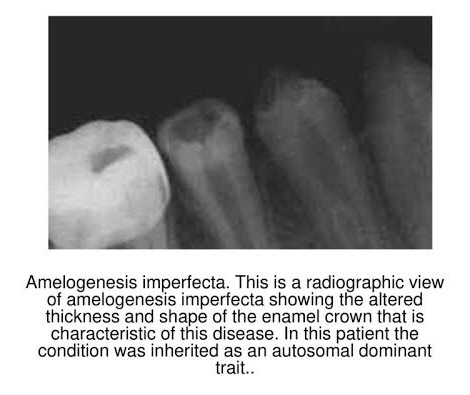
Radiographic Features:

Source: SlidePlayer
  • The enamel may appear totally absent.
  • When present may appear as a thin layer, chiefly over tip of cusps & on inter-proximal surfaces.
  • In some cases, calcification is so much affected that enamel and dentin seem to have the same radio density, making differentiation b/w the two difficult.

Histological Features:

  1. Hypoplastic: Disturbance in the differentiation/viability of Ameloblasts. Defect in matrix formation.
  2. Hypomaturation: Alteration in enamel rod & rod sheath structures.
  3. Hypocalcified: Defects of matrix structure & of mineral deposition.

Management:

  • Sealants/bonding
  • Prosthetic reconstruction

References: Shafer’sTextbook Of Oral Pathology; Internet

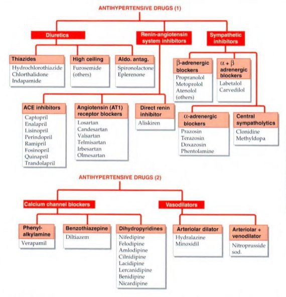
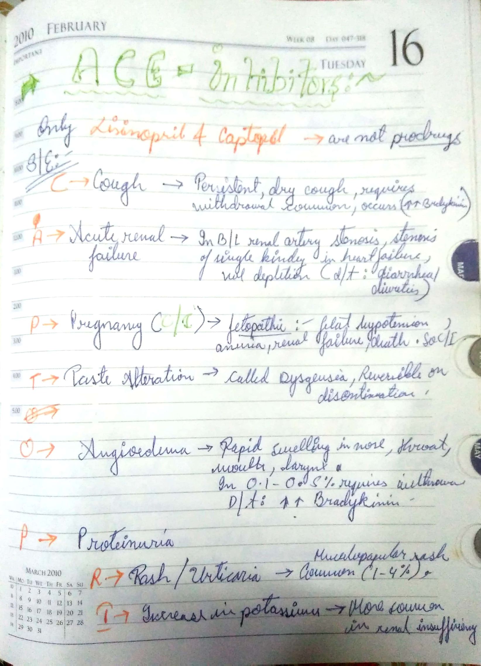
SIDE EFFECTS OF ANTI HYPERTENSIVE DRUGS

The objective of antihypertensive therapy is to reduce the incidence of adverse cardiovascular events, particularly CAD stroke and heart failures.

Here , we discuss some side effects of the Major drugs which is mainly prescribed for the treatment:-

The choice of antihypertensive therapy is initially indicated by the patient’s age and ethnic background.

Comorbid conditions also have an influence on initial drug selection,for example:-

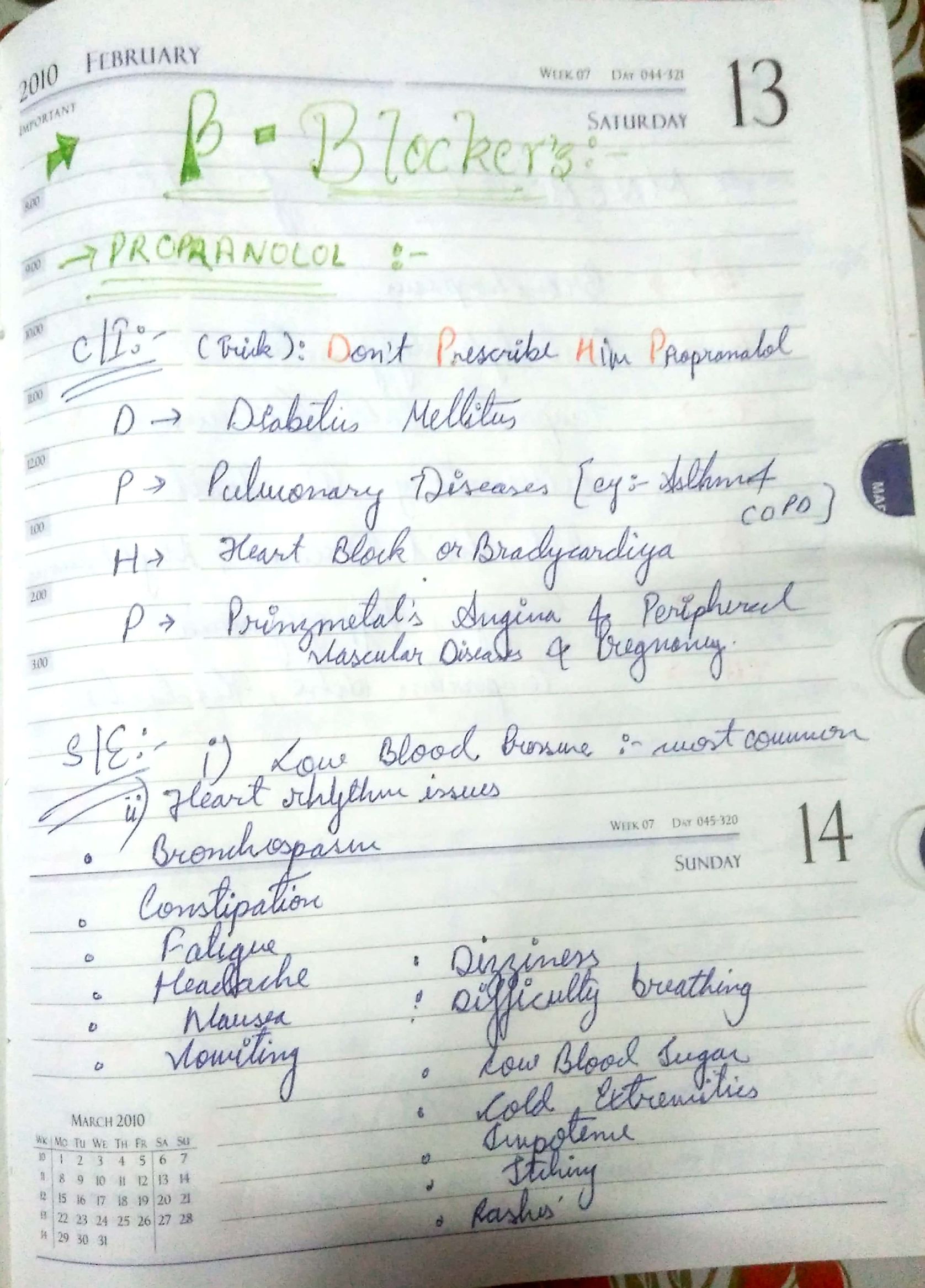
•beta blocker might be the most appropriate treatment for a pateint with angina.

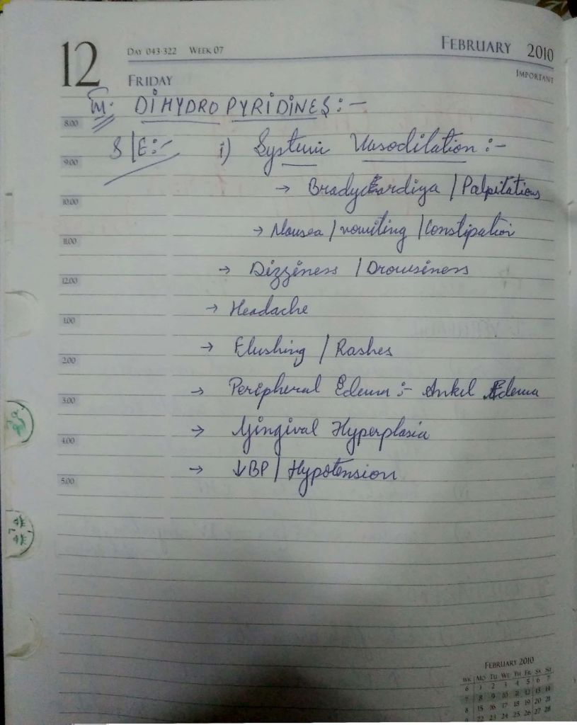
•Thiazides diuretics and dihydropyridines calcium channel blockers antagonists are the most suitable drugs for treatment in older people.

Thank you,

Regards,

KRITI Naja Jain 🙂

REference:-

  1. KDT 8th e
  2. Davidson 23rd e
  3. YouTube video

Generalised Seizure

Seizure

  • Denote that a seizure is an acute, transient neurological event (typically less than 5 minutes in duration) caused¬ by abnormal (excessive or synchronous) electrical discharges within the brain.

Epilepsy

  • Denote that epilepsy is the syndrome of recurrent, unprovoked seizures.
  • As we’ll discuss elsewhere, provoked seizures can occur from conditions like intracranial hemorrhage or metabolic causes; whereas; unprovoked seizures do not have a triggering cause.

Status Epilepticus

  • Denote that status epilepticus refers to seizure activity that fails to terminate within the anticipated time period (variably defined as anywhere from 5 – 30 minutes) OR it refers to a series of consecutive seizures without intervening recovery.
  • Indicate that status epilepticus has a mortality of 15 – 20%; however, it is the underlying etiology for the seizures that is the major contributor to this poor prognosis.
    • Thus, as clinicians, we should never “give-up” on a patient no matter how long the duration of the status epilepticus, as the seizures can persist for weeks and patients can still have a good outcome if the underlying etiology resolves or is treated.

Common Causes of Provoked Seizures

  • Denote that the following entities are considered the most common etiologies of seizures:
    • Stroke
    • Brain tumor
    • Brain injury
    • CNS infection

Most Common Cause of Epilepsy, Worldwide

  • Denote that neurocysticercosis (taenia solium) is the most common cause of epilepsy, worldwide.
    • The seizures come from the accumulation of cysts within the brain, which occurs when patients swallow the eggs found in the feces of a person who has an intestinal tapeworm.

GENERALIZED SEIZURES

Semiology

  • Define the tonic phase as a tonic as stiffening and define the clonic phase as rhythmic jerking.

Mimicker

  • Indicate that a key mimicker of tonic-clonic seizure is convulsive syncope, which are convulsions brought on by syncope, a loss of adequate cerebral perfusion, rather than by abnormal electrical activity in the brain.
    • Bear in mind that one of the most common mimickers of generalized seizures, and most seizure types, is actually non-epileptic spells (aka psychogenic seizures).

Syndromes

  • Indicate that generalized tonic-clonic seizures can occur in a wide-variety of epilepsy syndromes.

Detailed Semiology

  • To best understand the clinical semiology of a tonic-clonic seizure, let’s diagram what happens to a person at each phase of the seizure.
    • We’ll include the corresponding EEG (electroencephalogram), which can be illuminating, at the end.

Pre-ictal

  • Note that there is often a pre-ictal sensory prodrome that can last minutes to hours.
    • The ictal phase refers to the seizure, itself: the event.

Tonic Phase

  • Show that there is tonic stiffening: show that the back and neck are arched. The patient is lying down because there is a loss of consciousness.

Clonic Phase

  • Show that it’s characterized by rhythmic jerking (convulsions) of the face, arms, and legs.

Apnea

  • During the ictal phase, there is often apnea with frothing at the mouth, choking sounds, and cyanosis (a blue appearance to the skin), which can mimic a cardiac arrest.

Post-ictal Relaxation

  • Then, show that after the event, post-ictal, there is post-ictal relaxation, which involves a stupor with possible bladder or bowel incontinence and deep, slow respirations.

EEG Correlation

  • See: Tonic-Clonic Seizure
  • Now, let’s address the EEG rhythm, which even if we don’t read EEG, can help reinforce our understanding of the seizure presentation, itself.
  • Draw a small strip of EEG – we’ll just show a couple of tracings; normally, there would be at least 18 tracings to a page.
  • For reference, show the region between the vertical lines is one second.
  • The number of wave cycles within that one second is the frequency, which is measured in Hertz (Hz).
    • A single wave cycle in one second would be 1 Hz.

Tonic Phase

  • Indicate that at the initiation of the seizure, during the tonic phase, the EEG rhythm is a 10 Hz tonic (fast frequency), low amplitude waveform.

Clonic Phase

  • Now, indicate that during the clonic phase, the corresponding EEG rhythm is 4 Hz (slower frequency) spike-and-wave activity.

Post-ictal Slowing

  • Finally, indicate that after the event, there is post-ictal slowing, with only a few wave cycles per second.

MYOCLONIC – JUVENILE MYOCLONIC EPILEPSY (JME)

Semiology

  • Next, indicate that myoclonic seizures, manifest with brief, shock-like muscle jerks.

Mimicker

  • Indicate that they are often mistaken for a movement disorder.

Epilepsy

  • Write that myoclonic seizures are an important component of juvenile myoclonic epilepsy (JME), which begins in adolescence (12 – 18 years old).
    • It is one of the photosensitive epilepsies; seizures can be triggered by flashing or flickering lights.

Detailed Semiology

  • For juvenile, myoclonic epilepsy, draw a bed and a sunrise, because the events characteristically cluster upon awakening in the morning.
  • And draw a bolt of lightning because the jerks are described as “lightning-like”.
  • Draw our person lying in bed (because they tend to occur in the morning).
  • Show that they manifest with symmetric, irregular, shock-like, jerks of the shoulders and arms, most notably, which can cause the person to drop items, but can also affect the legs, which can cause falls.

EEG Correlation

  • Indicate that the EEG demonstrates polyspikes, which correlate with the myoclonic jerks, and characteristic disorganized, 4 – 5 Hz polyspike and wave discharges.
    • See: Polyspike-and-Wave Complexes
    • See: Status Myoclonus
  • As mentioned, these discharges have a strong photoparoxysmal response, so flashing lights are used during EEG to elicit these discharges.

ATONIC – LENNOX-GASTAUT

Semiology

  • Next, indicate that atonic seizures cause as loss of tone (drop attacks).
  • They manifest with brief loss of muscle tone in the postural muscles or head.

Mimicker

  • Indicate that they can be hard to distinguish from syncope, which also involves a sudden loss of tone.

Epilepsy

  • Write that they are an important feature of Lennox-Gastaut syndrome, which involves multiple seizure types, including atonic seizures and cognitive dysfunction. It peaks at ~ 4 years of age.

EEG Correlate

  • We’ll skip showing the EEG correlate of Lennox-Gastaut, but for reference, there are slow (1.5 – 2 Hz ) spike-and-wave discharges.

ABSENCE – CHILDHOOD ABSENCE EPILEPSY (CAE)

  • Finally, for the major non-motor, generalized seizure, we’ll address absence seizures.

Semiology

  • Indicate that absence seizures (aka petit mal seizures) manifest with a blank stare; patients appear to be daydreaming or zoning out.
  • These patients may exhibit rhythmic facial movements or motor automatisms.
  • Notably, there is no postictal confusion; patients can pick right back up where they left off with an activity.

Mimicker

  • Indicate that it is essential to distinguish absence seizures from an attentional disorder, as these patients can be mistakenly diagnosed with a learning disability.

Epilepsy

  • Indicate that they are the major seizure manifestation in childhood absence epilepsy (CAE), which typically occurs between 4 to 8 years of age, affects girls more than boys, and can involve 100s of seizures in a day.
  • Show that children with absence seizure appear to be “daydreaming” or “staring off” in school.

EEG Correlation

  • Indicate that the EEG demonstrates runs of well-organized 3 Hz generalized high-voltage rhythmic spike-and-wave discharges.
    • See: 3 Hz Generalized Spike-Wave Discharges

JUVENILE ABSENCE EPILEPSY (JAE)

  • For reference, juvenile absence epilepsy (JAE) is another generalized epilepsy syndrome, which we can think of (albeit a simplification) as a mixture of childhood absence epilepsy and juvenile myoclonic epilepsy:
    • it occurs at 9 – 13 years of age
    • it involves absence and myoclonic seizures that tend to occur shortly after awakening.

Focal Seizures

  • Note that the primary focal epilepsy syndrome is temporal lobe epilepsy, which is why in our diagram we show the seizure emanating from the medial temporal lobe, but focal seizures can occur from any cerebral lobe.
  • To localize the origination of the seizure, we can use both lateralizing and localizing signs.
  • On EEG, we look for focal epileptiform discharges.
    • See: Focal Epileptiform Discharge

LATERALIZING SIGNS

  • In regards to seizure laterality, indicate the following important signs:

Versive Movements

  • Indicate versive motor movements, which refers to contralateral turn of the head and/or eyes (away from the seizure).
  • Draw a brain and show a seizure emanating from the right hemisphere.
  • Then draw a pair of eyes and show that they exhibit forced eye deviation to the left: the side opposite (contralateral) to the side of seizure.

Todd’s Paralysis

  • Next, indicate that Todd’s paralysis refers to a post-ictal weakness in the side of the body opposite to the seizure.
  • Consider the post-itcal slowing we drew following a tonic-clonic seizure: the brain is slow and suppressed, so naturally the corresponding side of the body is limp and weak.

Stroke Mimickers

  • Naturally, then, these signs can be important mimickers of stroke. In stroke:
    • The eyes can drift toward the side of the stroke: they look at the healthy side of the body (this is the opposite direction from in seizure wherein they look away from the seizure).
    • There is weakness on the side opposite of the stroke, similar to a Todd’s paralysis.

Additional Signs

Some commonly discussed, albeit less reliable lateralizing signs, include the:

  • Figure 4 Sign
    • The arm contralateral to the seizure is extended at the elbow with the wrist in flexion and the fist is clenched.
    • The ipsilateral limb is in elbow flexion.
  • Fencing Posture
    • The arm contralateral to the seizure is raised and semi-extended above the head, as if holding a fencing foil.
    • The head is turned toward the raised arm while the ipsilateral arm is semi-flexed at at the patient’s side.

LOCALIZATION

  • Now, in regards to lobar localization, let’s address a few common seizure localities (note that seizures emanate supratentorially, above the brainstem and cerebellum).
  • Draw a medial face of a cerebral hemisphere; we do this, because as mentioned, the most common locality for focal seizures is the medial temporal lobe.
  • Divide it into the temporal, frontal, parietal, and occipital lobes.

Temporal Lobe

  • Indicate that temporal lobe seizures often manifest with sensory auras, automatisms, or speech arrest (or another form of cognitive impairment).
  • Sensory Auras:
    • Epigastric rising
    • Inappropriate fear
    • Olfactory hallucinations
    • Deja Vu
  • Autonomic features
    • Wide variety of sympathomimetic, gastrointestinal, and respiratory symptoms

Frontal Lobe

  • Indicate that frontal lobe seizures tend to be stereotyped and nocturnal. They are easily confused for psychogenic seizures or a movement disorder (especially because there is often NO loss of awareness or postictal phase).
  • Additional Features:
    • Manifestations of frontal lobe seizures range from emotionally-driven, fearful hallucinations to motor activity: tonic-clonic movements and the more complicated positions described previously (figure 4 sign, fencing posturing, etc…).

Parietal Lobe

  • Indicate that parietal lobe seizures tend to cause somatosensory auras, which we could predict given the role of the parietal lobe in sensory processing.
  • Note, however, that parietal lobe seizures are notoriously poorly localizing and they will commonly propagate to more regions with more readily obvious manifestations, such as the frontal lobe or the occipital lobe, before they are recognized.

Occipital Lobe

  • And finally, indicate that occipital lobe seizures often produce elemental visual phenomena, such as flashing lights or geometric shapes, much like migraine auras.

Insular Cortex

  • Characteristically manifest with symptoms involve the GI system and throat, such as vomiting, hypersalivation, dysarthria or strange thoracoabdominal sensations.

Muscle Stretch Reflex

Synonyms

  • Monosynaptic reflex, myotactic reflex, deep tendon reflex, tendon jerk

Definition

  • It is an automatic, monosynaptic reflex that involves a muscle and tendon, and produces a jerk.

Most commonly tested

  • Biceps (C5, C6)
    • Elbow flexion
  • Triceps (C7,C8)
    • Elbow extension
  • Patella (L2 – L4)
    • Knee extension
  • Achilles (S1,S2)
    • Foot plantarflexion

KEY MEDIATORS

  • Muscle spindles, which activate via muscle stretch.
  • Spinal neurons, which receive sensory input and generate motor output.
  • Muscle fibers, which contract.
  • Interneurons, which modulate neuronal firing.
  • Golgi tendon organs, which activate via muscle contraction to terminate the reflex.

ACTIVATION

  • When the patellar tendon is activated,
  • the muscle spindle sends an excitatory volley along the Type 1a sensory afferent,
  • which excites the extensor motor neuron.
  • It activates the muscle extensors, which extend the knee.

INTERNEURONAL INHIBITION

  • Renshaw cells are interneurons that lie in the anterior horn of the gray matter of the spinal cord.
  • When Renshaw cells are activated, they inhibit flexor motor neurons using the inhibitory neurotransmitter glycine.

TERMINATION

  • Golgi tendon organs are situated where the quadriceps tendon inserts into the patella.
  • Type 1b fibers project from the Golgi tendon organs to the Renshaw interneurons.
  • Inhibitory fibers project from the the Renshaw interneurons to the extensor motor neurons.
  • The Type 1a and 1b fibers fire at the same rate, but the muscle spindle fibers have a much lower threshold to fire than Golgi tendon organs, thus, the muscle spindle fibers fire first, and then later the Golgi tendon organs fire, which terminates the muscle stretch reflex.
  • Neurobiological influences, such as myosin ATPase and calcium re-accumulation into the endoplasmic reticulum aid in muscle contraction.

CLINICAL CORRELATION

  • In comatose patients, presence of the triple flexor reflex to plantar stimulations a sign of disinhibition, similar to the Babinski sign.

Myofibrils

Thick filaments.

  • Form from myosin
  • The A band refers to the length of the thick filaments, “think “A” for d-a-rk – they are aniosotropic (or birefringent) in polarized light.
  • H Zone is a zone of only thick filaments.
  • M line bisects the A band.

Thin filaments

  • Form from actin
  • The I band is the region along the thin filaments (between the thick filaments).
  • Think “I” for L-i-ght – they are “isotropic” (do not alter polarized light).

Z disks

  • Transverse bands at the ends of the thin filaments.

Sarcomere

  • The contractile unit of the myofibril.
  • Comprises the area between the Z-disks.

THIN MYOFILAMENT: DETAILS

  • The thin filament slides towards the H zone.

Actin

  • Spherical molecules joined in pairs of strands (like beads on a string). It is referred to as F-actin for filamentous actin, and comprises a polymer of G-actin monomers that are arranged in a double helix.

Tropomyosin

  • Threadlike strands

Troponin

  • Protein complexes that bind tropomyosin, actin, and also calcium (show their calcium-binding sites).

Cap Z

  • Binds the Z disk to the thin filaments.
  • This is (+) end stabilizer.

Tropomodulin

  • The (-) end stabilizer.

Troponin subunits

  • TnT binds to tropomyosin
  • TnI, which inhibits actin interaction with myosin.
  • TnC, which binds calcium ions.

Nebulin

  • Attaches to the Z disk, passes along the thin filament and binds to tropomodulin to help stabilize the thin filament and determines its length.
  • It is commonly stated that two nebulin molecules wrap around each thin filament to anchor it to the Z disk.

THICK FILAMENTS: DETAILS

  • Comprise myosin molecules (technically myson II), which form a golfclub shape, and comprise two heavy chains and two light chains.
  • The head forms from the heavy chain.
  • Unlike the thin filaments, the thick filaments are not directly connected to the Z disc. Instead, show that they are anchored to the ends of the sarcomere via titin molecules, which extend from the Z disks to the M line, and regulates the sarcomere’s elasticity, in addition to managing assembly of myosin.

MUSCLE CELL IN A CYLINDRICAL, 3-DIMENSIONAL VIEW

Here, we draw a muscle cell in a cylindrical, 3-dimensional view.

Sarcoplasmic reticulum (SR)

  • Form web-like rows.
  • Store calcium.
  • Are a key component to coupling muscle cell excitation to myofibril contraction.

Terminal cisternae (aka lateral cisternae) of the sarcoplasmic reticulum.

  • Flank the transverse tubules (T-Tubules)

Transverse tubules (T-Tubules)

  • Are tubular invaginations of sarcolemma.
  • Elsewhere, we see that T-tubules and terminal cisternae connect the terminal synapse firing (its depolarization) to the sarcoplasmic reticulum, again, ultimately coupling muscle cell excitation and myofibril contraction.

Triads of terminal cisternae

  • Triads of terminal cisternae with intervening T-tubules occur at the A-I junctions.

Desmin

  • Filaments encircle the Z disks.
  • Desmin-related myopathy (DRM) is an inherited disease in which, and results in disorganized and weak skeletal muscle fibers. DRM can be fatal, as it also affects cardiac and smooth muscles.

Plectin

  • Links the desmin filaments.

Alpha-beta-crystallin

  • A heat shock protein, which protects desmin from stress-induced damange.

Thus, together, desmin, plectin, and alpha-beta-crystallin constitute a Z-disk protection network.

Alpha-actinin

  • A Z disk component that binds to actin.

Costameres

  • Specialized regions of the sarcolemma, which bind to key protein complexes.

Dystrophin-associated glycoprotein complex (DAGC), which comprises:

  • The dystroglycan subcomplex, which links dystrophin (discussed soon) to laminin, a key external lamina protein (called laminin-2 in skeletal muscle).
  • The sarcoglycan subcomplex, which, when defective can cause sarcoglycanopathies – a similar manifestation of weakness as those from dystrophinopathies – and are a common cause of limb-girdle muscular dystrophy.
  • Dystrophin, which stabilizes the sarcolemma during muscle contraction.

DUCHENNE MUSCULAR DYSTROPHY (DMD)

Pathological slide

  • Muscle from an individual with Duchenne muscular dystrophy (DMD).
  • X-linked form of muscular dystrophy affects boys and occurs from a genetic mutation that prevents the synthesis of dystrophin.
  • We see a sparse group of muscle cells and see that much of the muscle is replaced with fatty and fibrous connective tissue, which presents with pseudohypertrophic muscles: muscles that are enlarged from fat and connect tissue (not muscle).
  • Eventually patients with this illness succumb to their weakness.

Syntrophins

  • Are recruited to the sarcolemma and manage the assembly of other proteins.

Dystrobrevins

  • Link desmin to dystrophin and syntrophin.