Posterior Abdominal Wall

Key Points

  • The posterior abdominal wall muscles contribute to respiration and movement of the torso and thigh.
  • The posterior abdominal wall is bound superiorly by the diaphragm, and, inferiorly, by the ilia of the pelvis.

Quadratus lumborum:

  • Originates on the iliac crest and the transverse process of L5.
  • Inserts on the transverse processes of L1-L4, and along the inferior border of rib 12 (intertextual variation regarding the specific origins and insertions of quadratus lumborum exists).
  • It depresses and stabilizes rib 12, and laterally flexes the torso.

Diaphragm:

  • Originates on the internal surface of the sternum, costal cartilages of ribs 6-12, and lumbar vertebrae L1-L3
    Inserts into the central tendon.
  • Contraction of the diaphragm expands the thoracic cavity, which decreases intrapulmonary pressure and induces inspiration (inhalation).

Iliacus:

  • Originates on the iliac fossa and lateral sacrum.
  • Inserts on the lesser trochanter of the femur.
  • Flexes the thigh at the hip.

Psoas major:

  • Originates on the lateral aspects and intervertebral discs of vertebrae T12-L5 Inserts, with iliacus, on the lesser trochanter.
  • Flexes the thigh at the hip.

Psoas minor:

  • Originates on the lateral aspects and intervertebral discs of T12-L1
  • Inserts on the pectineal line on the pelvis.
  • Weak trunk flexor; it does not travel to the thigh with psoas major, and, therefore, does not act on the thigh.

Anterior Abdominal Wall

Key Point:

  • The muscles of the anterior abdominal wall comprise thin sheets that compress and protect the abdominal contents, and, therefore, are involved in a range of actions, including defecation and micturition, parturition, and forceful expiration of air (as in heavy breathing, coughing, or yelling).

External oblique:

  • Originates from the external surfaces of ribs 5-12
  • Inserts on the ilium (the anterior ½ of the iliac crest and the anterior superior iliac spine), the pubic tubercle, and, via its aponeurosis, the linea alba.
  • The inferior border of this aponeurosis forms the inguinal ligament, which is a strong cord of connective tissue to which other muscles attach.
  • The fibers of the external oblique extend inferomedially from their posterior origins.
  • The external obliques compress the abdomen and flexes and rotates the trunk.

Internal oblique:

  • Originates on the lateral 2/3rds of the inguinal ligament, the iliac crest, and the thoracolumbar fascia.
  • It inserts at the linea alba, pubic crest and pectineal line, and external surfaces of ribs 9-12; it gives rise to an aponeurosis, which inserts at the midline.
  • Its fibers run superomedially at an oblique angle.
  • The internal oblique works with the external oblique to compress the abdomen and flex and rotate the trunk.

Rectus abdominis:

  • Originates on the pubic crest and pubic symphysis of the pelvis.
  • Inserts into the xiphoid process of the sternum and on the external surfaces of the costal cartilages of ribs 5-7.
  • Rectus abdominis depresses the ribs and flexes the trunk.
  • Rectus sheath covers anterior and posterior surfaces of rectus abdominis; it is formed by the aponeuroses of the other abdominal muscles.
  • Its fibers run vertically in four segments that are separated by horizontal tendinous intersections.

Transversus abdominis:

  • Originates on the lateral 1/3rd of the inguinal ligament, the iliac crest, the thoracolumbar fascia, and the external surfaces of the costal cartilages of ribs 7-12.
  • Inserts along the linea alba, pubic crest, and pectineal line.
  • Transversus abdominis compresses the abdomen and depresses the ribs.

Muscles of Respiration

Anterior scalene

  • Originates on the transverse processes of C3-C7
  • Inserts on the superior surface of the first rib.
  • Elevates the first rib during forced inhalation, or, when the rib is fixed, flexes the neck (moves the head closer to the chest or shoulder).

Middle scalene

  • Originates on the transverse processes of C2-C7.
  • Inserts with the anterior scalene on the first rib.
  • Elevates the first rib and flexes the neck.

Posterior scalene

  • Originates on the transverse processes of C4-C6.
  • Inserts on the second rib.
  • Elevates rib 2; when this rib is fixed, it flexes the neck.

Diaphragm

  • Originates on the inferior internal surface of the sternum, costal cartilages of ribs 6-12, and the lumbar vertebrae L1-L3.
  • Inserts into the central tendon.
  • Expands the thoracic cavity to decrease intrapulmonary pressure and induce inspiration (inhalation).

Three layers of intercostal muscles:

External intercostals

  • Originate on the inferior border of the ribs.
  • Insert into the superior border of the ribs below.
  • Elevate the ribs during inhalation.

Internal and innermost intercostals

  • Originate on the superior border of the ribs.
  • Insert into the inferior border of the ribs above.
  • Depress ribs during forced exhalation (normal exhalation requires no active effort).

Transversus thoracis

  • Originates on the posterior surface of the sternum (body and xiphoid process).
  • Inserts into the internal surfaces of ribs 2-6.
  • Depresses ribs during exhalation.

Vertebral Column

Vertebral column (aka, spinal column, aka, spine)

  • It encloses and protects the spinal cord.
  • It provides attachment sites for muscles of the head, neck, and trunk.
  • The individual vertebrae are named according to their region and vertical order. For example, T1 is the first vertebra of the thoracic region.
  • The S- shaped curve of the vertebral column facilitate its flexibility and resilience.

Regions of the Vertebral Column:

Cervical

  • 7 vertebrae within the neck.
  • C1 and C2 have unique anatomical features to support the skull.
  • Small bodies.
  • Transverse foramen for vertebral arteries.
  • Spinous processes of cervical vertebrae 2-6 are bifid (aka, bifurcated).

Thoracic

  • 12 vertebrae that articulate with ribs to form posterior wall of thoracic cage.
  • Bodies have costal facets for articulations with the ribs.
  • Spinous processes are typically longer and angled inferiorly.
  • Transverse processes have costal facets for articulation with ribs.

Lumbar

  • 5 vertebrae of the lower back.
  • Large bodies.
  • Spinous processes are typically short, and more horizontally oriented.
  • Short transverse processes.

Sacral/Coccygeal

  • 5 fused sacral bones.
  • 3-4 fused coccygeal bones.
  • Contribute to posterior wall of the pelvis.

General Features of Vertebrae:

Vertebral body (aka, centrum)

  • The disc-shaped weight-bearing portion of the vertebra.

Vertebral (aka, neural) arch

  • Pedicles
  • Laminae
  • Transverse processes
  • Spinous process
  • Superior and inferior articular processes for articulation with vertebrae above and below

Vertebral foramen

  • Space between body and arch
  • When stacked, vertebral foramina create vertebral canal, through which spinal cord passes

Intervertebral foramen

  • Where spinal nerves pass through vertebral column laterally

Intervertebral disc

  • Absorbs shock and acts as a cushion between the vertebrae, and allows for spinal flexibility.
  • It comprises:
    • The nucleus pulposus, which is an inner gelatinous substance
    • The anulus fibrosus is the outer ring of collagen and cartilage that encloses the nucleus pulposus

Clinical correlations:

  • Distorted spinal curvatures can be caused by poor posture, aging, disease, and other factors.
  • Scoliosis, which is a common curvature abnormality, is characterized by a lateral curvature of the vertebral column.
  • Herniated intervertebral discs are common; they occur when a fissure in the annulus fibrosus allows the nucleus pulposus to bulge and compress nearby nerves, which can cause deficits and pain.
Vertebral Column & Back Musculature: Intermediate Back

Deeper layer =

Erector spinae, from medial to lateral:

  • Spinalis, Longissimus, and Iliocostalis.

Spinalis:

  • Originates on the thoracic vertebrae and the first two lumbar vertebrae (specifically¬, from their spinous processes).
  • Inserts along the vertebral column as superiorly as the 2nd and 3rd cervical vertebrae. It sometimes has a cervical portion that inserts on the base of the skull.

Longissimus:

  • Originates on the transverse processes of the lumbar, thoracic, and cervical vertebrae
  • Inserts along the ribs and vertebral column (as superiorly as C2).
  • It also sends a portion of muscle to attach to the mastoid processes on the skull.

Iliocostalis:

  • Originates on the sacrum and ilia.
  • Inserts along the ribs and vertebral column as superiorly as cervical vertebrae 4-7.

Additional information about the erector spinae:

  • These muscles comprise many short segments of muscle fibers.
  • The erector spinae muscles are prone to injury as a result of improper heavy lifting.
  • When contracted bilaterally, the erector spinae muscles extend the back;
    Unilateral contraction produces lateral flexion of the back (moves the shoulder towards the lateral hip of the same side).
  • The erector spinae muscles can be further subdivided according to vertical region:
    For example, the portion of iliocostalis that lies within the lumbar region is referred to as iliocostalis lumborum; the portion of spinalis that lies with the thoracic region is referred to as spinalis thoracis, and so on.

Superficial layer =

Serratus posterior superior:

  • Originates on vertebrae C7-T3 (specifically, from their spinous processes and associated connective tissues), extends inferolaterally
  • Inserts as thin strips of muscle, on ribs 2-5, lateral to the erector spinae.
  • Elevates ribs 2-5 during forced inspiration.

Serratus posterior inferior:

  • Originates on vertebrae T12-L3
  • Inserts superolaterally on ribs 9-12 (specifically, to their inferior borders).
  • Depresses ribs 9-12 during forced expiration.
  • Serratus posterior superior and inferior muscles are mirror images of each other.
  • The thoracolumbar fascia separates the erector spinae and serratus posterior superior and inferior muscles.

Vertebral Column & Back Musculature: Superficial Back

Key points:

  • The superficial muscles of the back stabilize and also move the pectoral girdle and humerus.
  • The muscles of the superficial back are symmetrically paired, and trapezius lies superficially over the others.

Trapezius:

  • Originates on the skull (the external occipital protuberance, superior nuchal line) and the spines and connective tissues of C7-T12.
  • Inserts on the lateral 1/3rd of the clavicle, and on the spine and acromion of the scapula.
  • Because the fibers of trapezius are convergent, trapezius adducts, rotates, elevates, or depresses the scapula, depending on which fibers contract.

Levator scapulae:

  • Originates on the transverse processes of vertebrae C1-C4.
  • Inserts on the medial border of the scapula.
  • It elevates and rotates the scapula.

Rhomboid minor:

  • Originates on the spines of vertebrae C7-T1.
  • Inserts on the scapula at the medial border, adjacent to its spine.
  • It adducts the scapula.

Rhomboid major

  • Originates on the spines of vertebrae T2-T5
  • Inserts on the medial border of the scapula.
  • It adducts the scapula.

Latissimus dorsi

  • Originates on the spines of vertebrae T7-T12, the thoracolumbar fascia (which overlies the lumbar vertebrae and sacrum), ribs 9-12, and the iliac crest (of the ilium).
  • Inserts on the floor of the bicipital groove of the humerus.
  • Adducts, extends, and medially rotates the humerus, and also depresses the scapula.

Mastication

Muscles of mastication are involved in chewing.

Temporalis:

  • Originates on the temporal fossa of the temporal bone and inserts on the coronoid process and anterior ramus of the mandible; it elevates and retracts the mandible.

Masseter:

  • Originates on the zygomatic bone and arch and inserts on the angle and lateral ramus of the mandible, which it elevates.

Medial pterygoid has two heads:

  • The deep head originates on the medial aspect of the lateral pterygoid process of the sphenoid and inserts on the medial surface of the angle and ramus of the mandible.
  • The superficial head originates on the tuberosity of the maxilla and inserts with the deep head on the mandibular angle.
  • The medial pterygoid elevates the mandible and moves it laterally.

The lateral pterygoid also has two heads:

  • The upper head originates from the infratemporal fossa of the sphenoid and inserts on the temporomandibular joint capsule and mandible.
  • The lower head originates from the lateral surface of the lateral pterygoid plate and inserts with the upper head.
  • The lateral pterygoid protrudes the mandible and moves it laterally.

Movements of the mandible at the temporomandibular joint:

  • Protrusion moves the mandible anteriorly; the lateral pterygoid muscles produce this movement.
  • Retraction moves the mandible posteriorly; the masseter produces this movement.
  • Elevation moves the mandible superiorly towards the upper teeth; temporalis, masseter, and the medial pterygoid produce this movement.
  • Depression moves the mandible inferiorly.

Cheeks and Mouth

Orbicularis oris:

  • Originates on the skin and other muscles around the mouth and inserts into the skin of the lips; it closes the lips.

Zygomaticus major:

  • Originates on the lateral side of the zygomatic bone and inserts into the angle of the mouth; it draws the corner (aka, angle) of the lips laterally and superiorly.

Zygomaticus minor:

  • Originates on the anterior aspect of the zygomatic bone and inserts into the skin of the upper lip, which it elevates.

Levator labii superioris:

  • Originates on the maxilla (specifically, from the infraorbital margin), and inserts into the skin of the upper lip, which it elevates; it also dilates (flares) the nostrils.

Buccinator:

  • Originates on the surfaces of the maxilla and mandible, and inserts into the lips; it compresses the cheeks.

Risorius:

  • Originates on connective tissues on the lateral sides of the face and the skin of the cheeks and inserts into the angle of the mouth; it retracts the lips at the angle of the mouth.

Mentalis:

  • Originates on the body of the mandible and inserts into the skin of the chin and lower lip, which it elevates and protrudes.

Depressor labii inferioris:

  • Originates on the mandible (specifically, from the anterolateral body) and inserts into the skin of the lower lip, which it depresses.

Additional Information:

  • These muscles blend into one another
  • Cranial nerve VII innervates them.

Scalp, Eyes, Nose

Nasalis:

  • Originates on the maxilla and inserts on the nasal cartilage at the midline; it flares the nostrils.

Corrugator supercilii muscles:

  • Originates on the frontal bone (specifically, from the medial supraorbital margin) and insert on the medial half of the eyebrows and laterally on the supraorbital margin on the frontal bone; they depress the eyebrows (and pull them medially).

Orbicularis oculi:

  • Originates on the medial margin of the orbit (specifically, from the maxillary, frontal, and lacrimal bones), and inserts into the rim and skin of the orbit and eyelids; it moves the eyelids and surrounding skin to squint and close the eyes.

Frontalis:

  • Originates on the frontal bone and skin of the eyebrows, and inserts into the galea aponeurotica (aka, the epicranial aponeurosis); it elevates the eyebrows, which wrinkles the forehead horizontally.

Additional Information:

  • Cranial nerve VII (the facial nerve) innervates these muscles.
  • Bell’s palsy occurs when CN VII is damaged; Belly’s palsy results in asymmetrical paralysis of the muscles of facial expression.

Skull Foramina

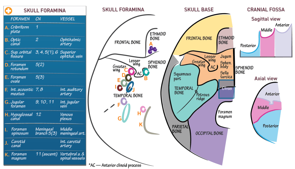
VASCULATURE AND CRANIAL NERVES OF THE MAJOR SKULL FORAMINA

The skull foramina are the entry/exit sites of the cranial nerves through the skull base and the entry/exit sites of the major cranial vasculature through the skull base.

  • Within the ethmoid bone, lie the foramina of the cribriform plate.
    -They contain CN 1, the olfactory nerve bundles.
  • Next, medial to the anterior clinoid process, lies the optic canal and lateral to it, lies the superior orbital fissure.
    -CN 2 traverses the optic canal.
    -CNs 3, 4, and 6, and the first division of CN 5 pass through the superior orbital fissure.
  • Posterior to the superior orbital fissure, within the greater wing of the sphenoid bone, lies foramen rotundum, and posterior to it, lies foramen ovale.
    -CN 5 (2) traverses foramen rotundum (think of R2D2 from “Star Wars”).
    -CN 5 (3) traverses foramen ovale.
  • Along the petrous apex of the temporal bone, lies the internal acoustic meatus.
    -Both CNs 7 and 8 pass through it.

Clinical Correlation – 8th Nerve Palsy

  • Posterior to the internal acoustic meatus, lies the jugular foramen.
    -CNs 9, 10, and 11 pass through it.
  • Medial to the internal acoustic meatus, lies the hypoglossal canal.
    -CN 12 passes through it.

MAJOR VESSELS THAT PASS THROUGH THE SKULL FORAMINA

  • The ophthalmic artery traverses the optic canal.
  • The superior ophthalmic vein passes through the superior orbital fissure.
  • The internal auditory artery (aka labyrinthine artery) passes through the internal acoustic meatus.
  • The internal jugular vein passes through the jugular foramen.
  • The hypoglossal canal contains a venous plexus.
  • Posterior to foramen ovale, lies foramen spinosum.
    -The meningeal branch of CN 5(3) passes through foramen spinosum and the middle meningeal artery passes through it, as well.

Middle meningeal artery rupture is the major cause of epidural hematoma.

  • The carotid canal lies along the petrous ridge.
    -The internal carotid artery runs within it.
  • Foramen magnum lies in midline.
    -CN 11 passes up through foramen magnum (before it passes out of the cranium through the jugular foramen) and the vertebral arteries and spinal vessels traverse foramen magnum, as well.

THE CRANIAL FOSSAE

The three cranial fossa are the: anterior, middle, and posterior fossae.

The anterior cranial fossa

  • Lies anterior to the posterior aspect of the lesser wing of the sphenoid bone.
  • The basal portions of the frontal lobes lie within this fossa.

The middle cranial fossa

  • Lies between the anterior cranial fossa and the petrous ridge of the temporal bone.
  • The basal portions of the temporal lobes lie within this fossa.

The posterior cranial fossa

  • Lies posterior to the petrous ridge.
  • The cerebellum and brainstem lie within this fossa.

The anterior depression is the anterior cranial fossa, the middle depression is the middle cranial fossa, and the posterior depression is the posterior cranial fossa.

BONES OF THE SKULL BASE

  • The anterior one-third is the frontal bone.
  • The ethmoid bone, which comprises the steeply peaked crista galli and the surrounding cribriform plate, lies in midline.
  • Posterior to the frontal bone, lies the sphenoid bone.
  • The midline portion of the sphenoid bone is the sphenoid body.
    -It subdivides into the jugum sphenoidale, anteriorly, and the sella turcica, posteriorly.
    -The lesser wing of the sphenoid bone lies anteriorly, and the greater wing lies posteriorly.
    -Topographically, the lesser sphenoid wing angles up over the greater sphenoid wing, which rolls downward.
    -The protuberance lies along the posteromedial ridge of the lesser wing as the anterior clinoid process.
  • The temporal bone lies posterior to the greater wing of the sphenoid bone.
  • We see the petrous ridge and the squamous part of the temporal bone lies laterally.
    -The squamous part makes up the bulk of the external surface of the temporal bone, whereas the petrous part makes up the bulk of the internal surface.
  • Posteromedial to the temporal bone, lies the occipital bone; it extends back to the occiput.
  • In the anterior one-third of the occipital bone, lies the foramen magnum, which is the entry zone of the brainstem.
    -The combined anterior occipital bone and posterior sphenoid bone form the clivus, which is steeply sloped, and is a clinically important anatomical region.
  • Along the lateral edge of the skull base, lies the parietal bone.
    -The parietal bones make up much of the lateral and superior surfaces of the skull.