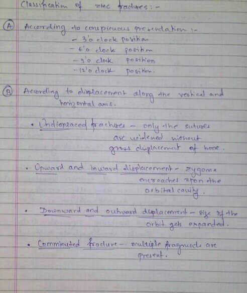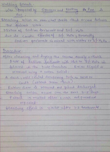
SOURCE- RAMYA RAGHU TEXTBOOK
SOURCE- RAMYA RAGHU TEXTBOOK
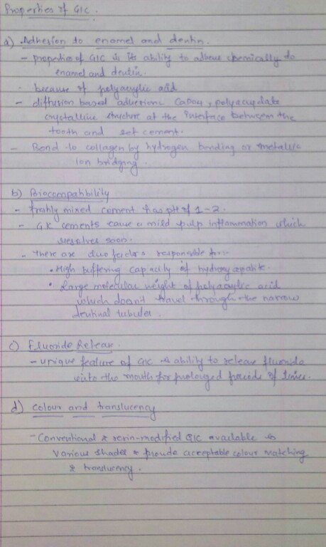
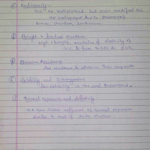
SOURCE- RAMYA RAGHU AND STUDERVANT TEXTBOOK
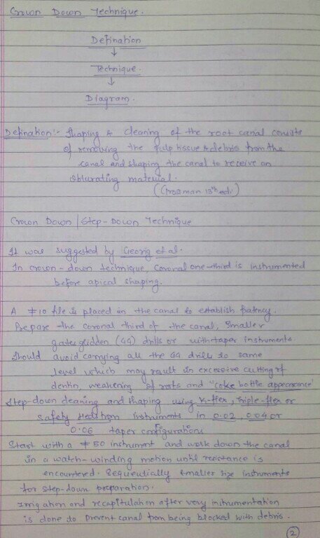
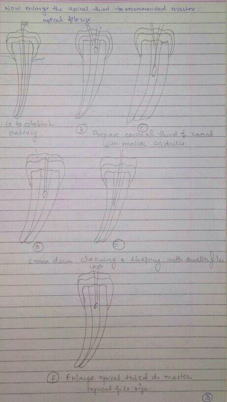
SOURCE- NISHA GARG AND GROSSMAN TEXTBOOK
• Decrease Thyroid hormones
Classification
• primary hypothyroidism – due to cause within the thyroid gland itself.
• secondary hypothyroidism – due to failure of TSH production following pituitary or hypothalamic disease.
Causes
• spontaneous atropic thyroidism
• iodine deficiency
• iodine therapy
• thyroidectomy
• hashimoto’s thyroiditis
• dyshormonogenesis
• drug-induced ( lithium , amiodrone etc)
• radiation therapy
clinical features
A. Thyroid gland enlargement
B. Gastrointestinal
• weight gain
• decrease in appetite
• constipation
• acsite
C. Cardiorespiratory
• angina
• bradycardia
• hypertension
• pericardial effusion
• Pleural effusion
• cardiac failure
D. Neuromuscular
•ache and pain
•muscle stiffness
•carpal tunnel syndrome
•delayed relaxation of tendon reflex • •depression
•psychosis
•cerebral ataxia
•myotonia
•deafness
E. Dermatological
• mycoderma(non- pitting edema of skin of hands , feet and eyelid)
• dry flaky skin and hair
• Alopecia
• purplish lips
• malar flush
• xanthelasma
• carotenaemia
F. Reproduction
• menorrhagia
• infertility
• galacoma
• impotence
G. Haemtological & miscellaneous
• macrocytosis
• anaemia
• cold intolerance
• tiredness
• harseness of voice
• low pitched sound
Slurred speech
Investigation
• serum T4 decrease; TSH increase ( > 20mU/l)
• serum cholesterol & triglycerides increase
• serum LDH, Ck increase
• serum Na+ decrease
• ECG – sinus bradycardia, low voltage QRS complex, ST – T wave abnormalities
• chest radiograph – Enlargement of cardiac shadow
Treatment
D.o.c – Thyroxine – 50 microgram/day for 3 weeks
then
100microgram/day for 3weeks
then
150microgram/day for maintenance dose
• pt with heart disease angina may worsen with thyroxine therapy -treated with beta block and vasodilators or CABG
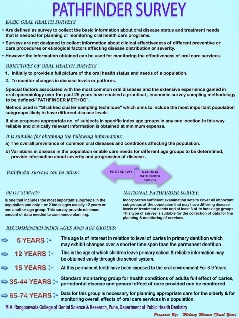
References: Textbook of Public Health Dentistry-Sober Peter(5th Edition)
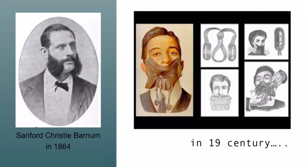
It’s hard to believe that a sheet of rubber can make you feel more comfortable during a dental treatment and allow dentists to do better, but a “RUBBER DAM” can . S.C. BARNUM
It is a direct method of isolation. It is a safeguard against the bacterial contamination from saliva and accidental swallowing of instruments.
HISTORY :
ADVANTAGES:
INDICATIONS:
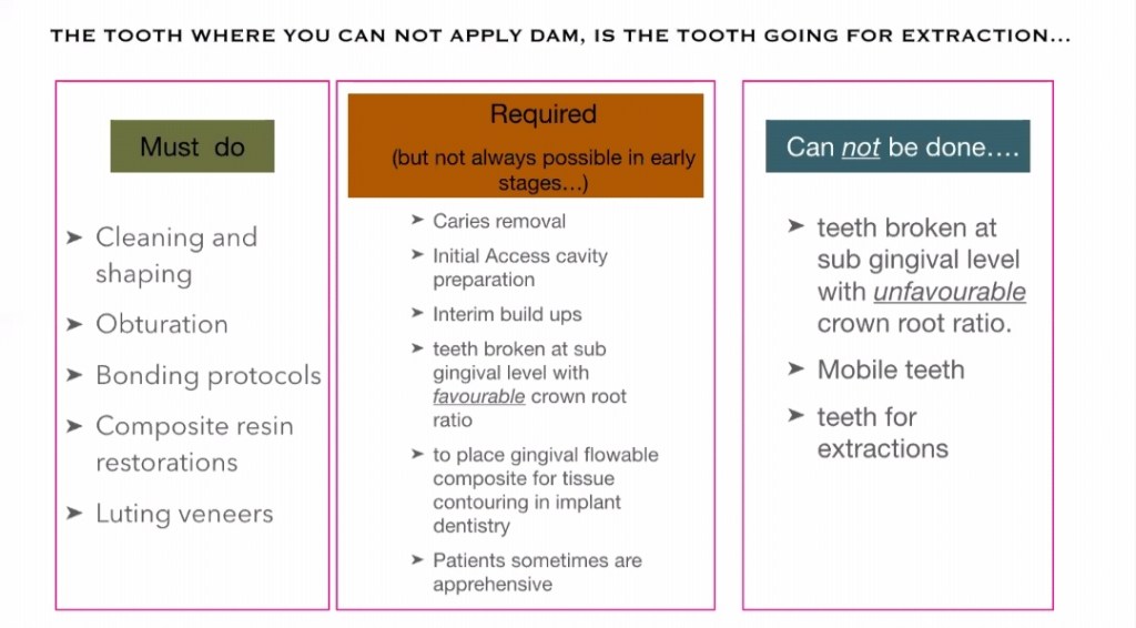
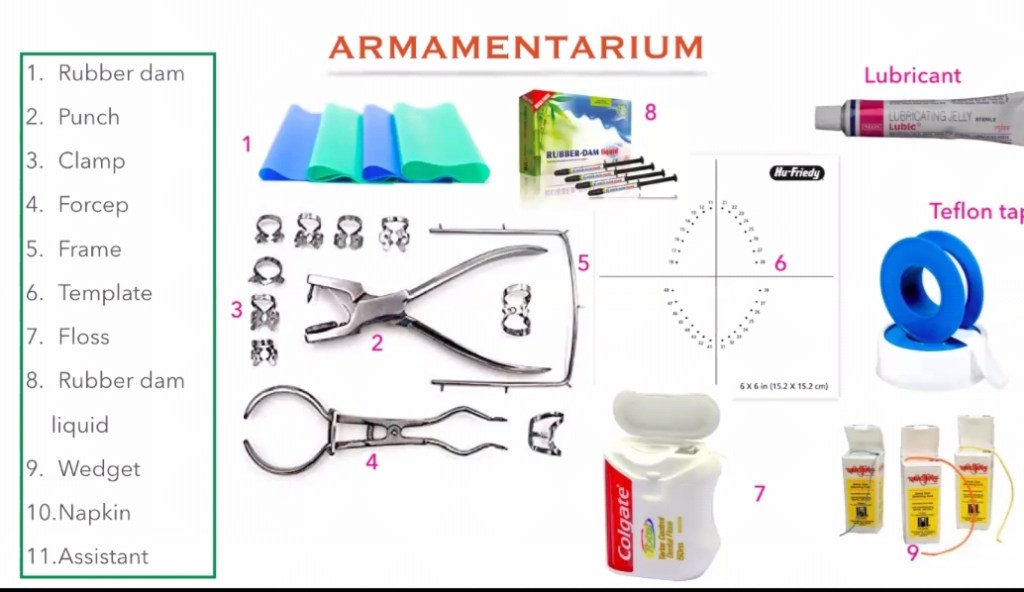
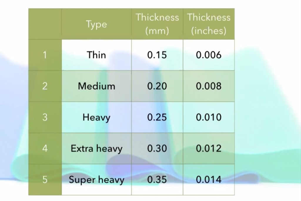
ARMAMENTARIUM:
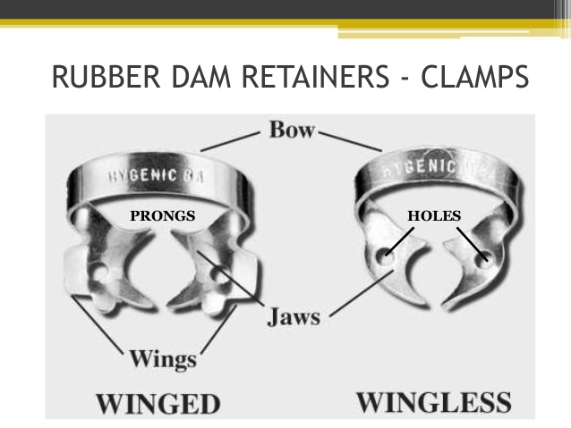
2, RUBBER DAM CLAMPS:
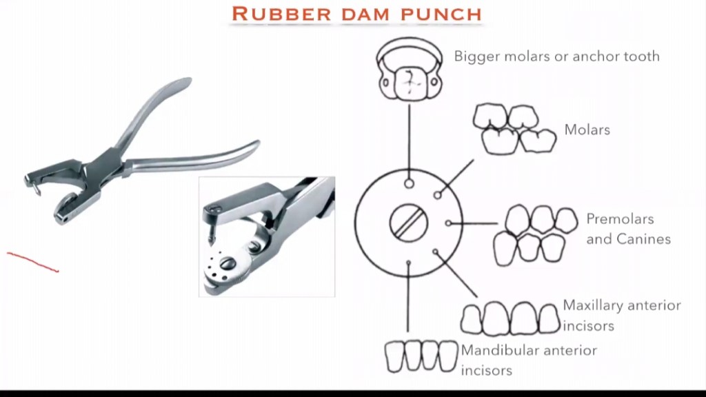
3. RUBBER DAM PUNCH :
It is an instrument which have a metal table and a tapered , sharp, pointed plunger to produce clear cuts holes on the rubber dam sheet so that the teeth can be isolated. It is of 2 types: Single hole punch and multi hole punch.
4. RUBBER DAM FRAME:
5. RUBBER DAM FORCEPS:
6. RUBBER DAM TEMPLATE:
7. RUBBER DAM NAPKIN :
8. WEDGET CORDS:
9. FLOSS:
REFERENCE: GROSS-MAN’S ENDODONTIC PRACTICE 13TH EDITION .
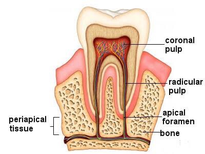
➡️ For all operative procedures, the shape of pulp chamber & its extensions into the cusps, pulpal horns is important.
➡️ In some developmental disturbances the pulpal horn projects high into the cusps & exposure of pulp can occur when it is least anticipated.
➡️ Pulpitis is the response of the traumatized pulp with trauma being the result of a bacterial infection as in dental caries or physical trauma to tooth structure.
➡️ Pulpitis in milder form could result in focal reversible pulpitis & may progress if left unchecked to acute & chronic forms of pulpitis.
➡️ Well vascularized pulpal tissue may at times in carious molar teeth of young adults & children with open apex exhibit a form of hyperplasia seen clinically in exposed pulp chamber as a protruding red mass of granulation tissue called pulp polyp or chronic hyperplastic pulpitis.
➡️ Inflammation within pulp may also sometimes result in a condition called pulp polyp, internal resorption or pink tooth.
➡️ Pulp infection can spread apically into PDL causing granuloma, abscess, cysts.
➡️ Pulp stones lying at the opening of the root canal may cause difficulty to locate the root canals.
➡️ A necrotic pulp can cause spread of disease to periodontium through an accessory canal.
➡️ Pulp capping is successful in non-infected or minimally infected accidentally exposed pulp in individuals of any age.
Source: Internet, Grossman’s Textbook of Endodontics
Dentowesome 2020 @ dr.mehnaz
➡️ The periapical expressions of pathologic Amelogenesis are hypoplasia, which is manifested by pitting, furrowing or even total absence of enamel & hypocalcification in the form of opaque or chalky areas on normally contoured enamel surfaces.
➡️ It is caused by systemic, local or hereditary factors
➡️ An example of systemic type of calcification of the enamel is so called Mottled Enamel
➡️ High flouride content in water – deficiency to calcify
➡️ The discoloration of the teeth from administration of tetracyclins during childhood is very common clinical problem
➡️ Dentin sensitivity of pain is exlained by hydrodynamic theory, the alteration of the fluid & cellular contents ending in contact with these cells.
➡️ Erosion of peritubular dentin & smear plug removal accounts for dentin hypersensitivity caused by agents like acidic soft drinks.
➡️ The rapid penetration & spread of caries in the dentin is the result of the tubule system in the dentin
➡️ Electron microscope of carious dentin show regions of massive bacterial invasion of dentinal tubules.
➡️ Smear layer consist of cut dentin surface which occludes the tubules & reduces permeability. Also prevents adhesion of restorative materials to dentin. Therefore this layer is removed by etching.
➡️ Cementum is more resistant to resorption than bone. It is for this reason orthodontic tooth movement is made possible.
➡️ Cementum resorption can occur after trauma or excessive occlusal forces. After resorption has ceased the damage usually is repaired either by formation of acellular/cellular cementum or alternate formation of both.
➡️ In most cases of repair there is tendency to re-establish the former outline of the root surface. This is called anatomic repair.
➡️ It is only a thin layer of cementum is deposited on the surface of resorption, the root outline is not reconstructed & a baylike recess remains. This is termed functional repair.
➡️ Hypercementosis – secondary to periapical infammation or extensive occlusal stress. Extraction of such tooth may necessitate the removal of bone.
Source: Internet
Dentowesome 2020 @dr.mehnaz

3 major types:
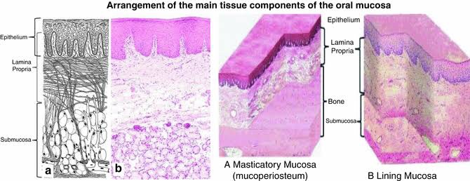
➡️ Contains 4 layers starting from the bottom:
▪️Stratum Basale:
▪️Stratum Spinosum:
▪️Stratum Granulosum:
▪️Stratum Corneum:
🔹Keratinized Areas:
🔹Non-Keratinized Areas:
References: Orban’s Oral Histology
Dentowesome 2020 @dr.mehnaz