Extraoral Examination of Temporomandibular Joint

Written by : Dr.Urusa I Inamdar

The importance is to determine deviation of jaw from the midline during the opening and closing of the jaws.

Causes of jaw deviation:

  • Traumatic injuries of the joint
  • Infection of the jaw
  • Fractures of the jaw
  • Muscular hypertrophy and hypotrophy

The lateral mandibular range of motion or movement is assessed ” Normal 8 to 10 mm ” by having the patient to occlude the teeth and then slide the jaw in both directions. The range of movement from midline and any pain, location and severity is recorded.

Maximum interincisal opening: As a general guide, mobility is considered to be reduced if the subject is unable to open his or her jaw to the width of two fingers ( <30 mm ).
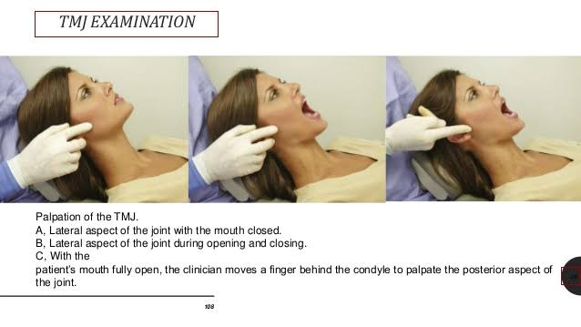
Palpation

  • Bimanual and bidigital Palpation or extra-auricular or intra-auricular.
  • Palpation may reveal pain and irregularities during condylar movement , described as clicking or crepitus. Clicking reveals the internal derangement of TMJ.
  • The lateral pole of condyle is most accessible for palpation during mandibular movements.
  • Palpation just anterior and posterior to the lateral pole detects pain associated with TMJ capsular ligament.
  • The comparison between both condyles must be assessed by palpation.

TMJ muscles :

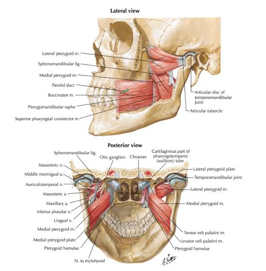
Gravity muscles ( Depression of mandible )

  • Geniohyoid
  • Digastric
  • Mylohyoid

Anti-gravity muscles ( Elevation of mandible )

  • Medial , oblique , anterior , vertical of temporalis.
  • Medial Pterygoid , masseter .

Protrusion of mandible

  • Medial pterygoid.
  • Lateral pterygoid.

TMJ disorders :

Developmental

  • Hypoplasia/aplasia of condyle
  • Hyperplasia of condyle
  • Bifid condyle

Traumatic

  • Dislocation of condyle
  • Fracture of condyle
  • Injury to articular disc

Inflammatory

  • Osteoarthritis
  • Rheumatoid arthritis
  • Septic arthritis

Neoplastic

References:

  • A practical manual of Public Health Dentistry – C M Marya
  • Slide share – Diagnosis of Temporomandibular disorder- Kelly
  • TMJ Anatomy – Geeky medics

LYMPH NODES EXAMINATION

Examination of neck nodes is important, particularly in head and neck Malignancies and a systematic approach should be followed.Neck nodes are better palpated while standing at the back of the patient. Neck is slightly flexed to achieve relaxation of muscles.

Lymph nodes of head and neck region are classified according to their position level:

  • Level I includes :
  1. IA Submental nodes , which lie in the Submental triangle, i.e between right and left anterior bellies of diagastric muscles and the hyoid bone.
  2. IB Submandibular nodes , lying between anterior and posterior bellies of diagastric muscle and the body of mandible.
  • Level II Upper jugular nodes :

They are located along the upper third of jugular vein, i.e between the skull base above and the level of hyoid bone ( or bifurcation of carotid artery ) below.

  • Level III Middle jugular nodes :

They are located along the middle third of jugular vein , from the level of hyoid bone above, to the upper border of cricoid cartilage.

  • Level IV Lower jugular nodes :

They are located along the lower third of jugular vein , from upper border of cricoid cartilage to the clavicle.

  • Level V Posterior cervical group :

They are located in the posterior triangle ,i.e between posterior border of sternocleidomastoid ( anteriorly ) , anterior border of trapezius ( posteriorly ) , and the clavicle below. They include lymph nodes of spinal accessory chain , transverse cervical nodes and supraclavicular nodes.

  • Level VI Anterior compartment nodes :

They are located between the medial borders of sternocleidomastoid muscles ( or carotid sheath ) on each side, hyoid bone above and suprasternal notch below. They include prelaryngeal, pretracheal , paratracheal nodes .

  • Level VII :

They are located below the suprasternal notch and include nodes of the upper mediastinum.

Examination of various lymph nodes:

  1. Submental nodes : Roll the fingers below the chin with patients head tilted forward.
  2. Submandibular nodes : Roll your fingers against inner surface of mandible with patients head gently tilted on one side.
  3. Parotid ( preauricular ) nodes : Roll your fingers in front of the ear , against the maxilla .
  4. Postauricular ( mastoid ) nodes : Roll the fingers behind the ears.
  5. Internal jugular chain : Examine the upper , middle and lower groups. Many of them lie deep to sternomastoid muscle which may need to be displaced posteriorly.
  6. Transverse cervical nodes
  7. Supraclavicular ( scalene ) nodes : Roll your fingers gently behind the clavicles.

A normal lymph node cannot be felt. If a node is palpable, it must be abnormal.

Area of lymphatic drainage of face :

The face has 3 lymphatic territories :

  1. The upper territory : Including the greater part of the forehead, the lateral halves of the eyelids, the conjunctiva, the lateral part of the cheek and the parotid area, drains into the preauricular (parotid) nodes.
  2. The middle territory : Including a strip over the median part of the forehead, the external nose, the upper lip, the lateral part of the lower lip, the medial halves of the eyelids, the medial part of the cheek and the greater part of the lower jaw, drains into the Submandibular nodes.
  3. The lower territory : Including the central part of the lip and chin, drains into the Submental nodes.

References:

A practical manual of Public Health Dentistry by CM Marya.

http://www.dentalcare.com

Written by - Dr.Urusa I Inamdar

Minimal Invasive Dentistry

current approach in dentistry in Covid state

Amidst the pandemic state with the high transmissibility of the disease through air & droplets and considering that routine dental procedures usually generate aerosols; alterations to dental treatment is of prime concern to maintain a healthy environment for patient & dental team.Here is where the approach of performing minimally invasive dental treatment becomes crucial.

Risks of infection – Human-to-human transmission

What is minimal intervention dentistry?

Minimal intervention dentistry( MID) is a conservative philosophy of professional care concerned with first occurrence,early detection & earliest possible cure of caries at a micro level ; followed by minimally invasive and patient friendly treatment to repair irreversible damage caused by dental caries.

Goals & Principles:

.Early diagnosis of dental caries

.Assessment of individual caries risk

.Disease control by remineralisation of incipient carious lesions.

.Repair rather than replacement of defective restorations

.Minimal invasive treatment

.Periodic follow up.

Caries diagnosis:

Includes early diagnosis & caries risk assessment

Early diagnostic aids
Factors relevant in caries risk assessment

Procedures:

Non invasive procedures: Biological approach

Remineralising agents

Minimal invasive treatments:

1)Air abrasion

Indications-

.for abrading the surface of old composites prior to new restoration ; minimal class I & class II preparations for composites ; for abrading ceramic or cast restorations for bonding ; for widening pits & fissures for sealants.

2) Sono Abrasion

Indications-

.opening pits & fissures for sealant restorations ; minimal preparation of incipient class II cavities

3) Chemicomechanical Caries Removal (CMC)

Carisolv – 2 syringe system ,one containing NaOCl & other with 3 amino acids (glutamic acid,leucine ,lysine); carboxymethylcellulose gel;NaCl,NaOH;Erythrosine.The contents are mixed together to form a pink gel which is applied onto carious dentin and left in place for 30 seconds to allow it to soften & degrade the infected dentin.

Advantages – relatively painless, removes only carious dentin, no vibrations,better substrate for adhesive bonding

Disadvantages- expensive, time consuming

4)Enzymes

5)Laser

2 commonly developed lasers-

.Er:Cr:YSGG(2780 nm)- Erbium,Chromium,Yttrium,Scandium,Gallium,Garnet laser – works by agonizing water droplets as they travel towards the target tissue.

.Er:YAG(2940nm)-Erbium,Yttrium,Scadium,Aluminum,Garnet laser – uses pulses of light energy to micro vaporize water within the target tissues.

6)Ozone

Caries treatment with ozone – based on Niche environment theory .Ozone kit consists of portable apparatus & disposable silicon cups. Follow up 3-6 months.

Disadvantages – can cause porosities or abrade tooth surface ,in case of heavy exposure.

Other techniques (in brief):

7)Atraumatic restorative treatment (ART)

8)Rotary instruments

Cavity designs for minimal tooth preparation:

According to the new classification based on site,size & severity of lesion,following are the designs –

Pit & fissure sealants;Preventive Resin Restorations (PRR)
Tunnel preparations
Slot preparations

Restorative materials used in minimal invasive dentistry :

Conclusion:

Minimal intervention techniques cause less tooth destruction than conventional techniques,thus increasing the long term survival of teeth ,also cause less discomfort to the patient and ensure healing of the disease not only the symptoms. With a reduction in chair side time and simplified techniques there is lesser chances of exposure of the dentist to aerosol contamination,thus maintaining which is the need of the hour.

THE VANGUARD

PC: Dr. Mihir Anand Jha

We are at the peak of pandemic and it is challenging in many unprecedented ways.

We are in this together and we will get through this together . Here is a small initiative on our part to spread awareness about covid -19.

“STOP THE SPREAD OF VIRUS “

“STAY HOME, STAY SAFE”